. 2001 Jul;12(7):2011-21.
doi: 10.1091/mbc.12.7.2011.

RME-8, a conserved J-domain protein, is required for endocytosis in Caenorhabditis elegans

Affiliations
Free PMC article

RME-8, a conserved J-domain protein, is required for endocytosis in Caenorhabditis elegans

Y Zhang et al. Mol Biol Cell. 2001 Jul.
Free PMC article

Abstract

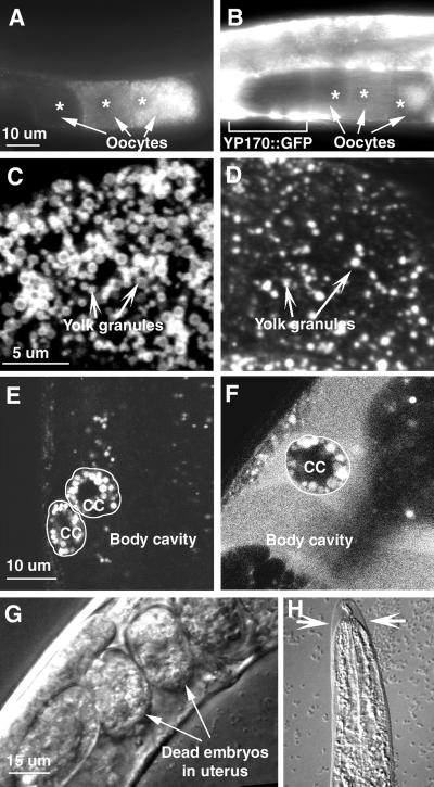
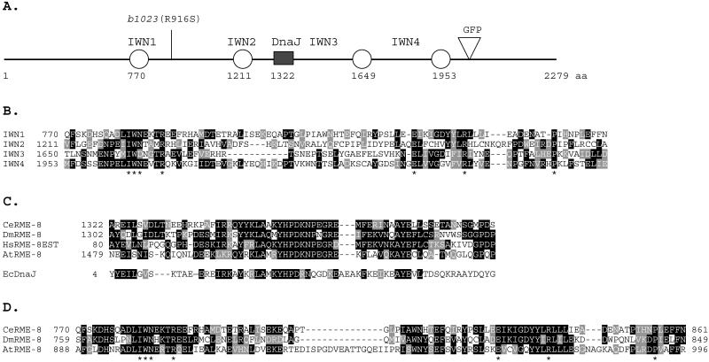
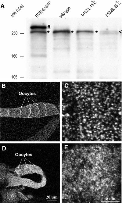
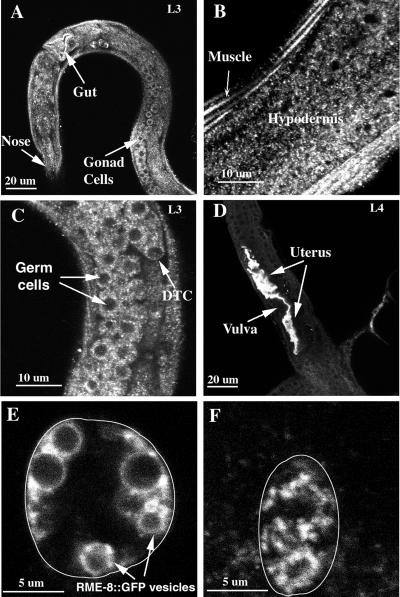
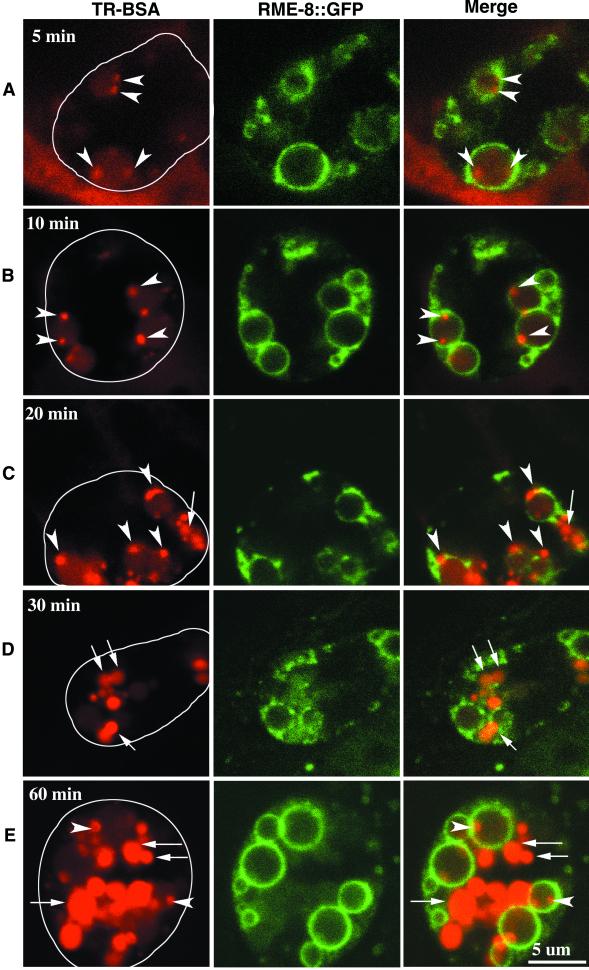
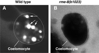
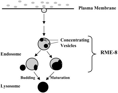
By genetic analysis of Caenorhabditis elegans mutants defective in yolk uptake, we have identified new molecules functioning in the endocytosis pathway. Here we describe a novel J-domain-containing protein, RME-8, identified by such genetic analysis. RME-8 is required for receptor-mediated endocytosis and fluid-phase endocytosis in various cell types and is essential for C. elegans development and viability. In the macrophage-like coelomocytes, RME-8 localizes to the limiting membrane of large endosomes. Endocytosis markers taken up by the coelomocytes rapidly accumulate in these large RME-8-positive endosomes, concentrate in internal subendosomal structures, and later appear in RME-8-negative lysosomes. rme-8 mutant coelomocytes fail to accumulate visible quantities of endocytosis markers. These observations show that RME-8 functions in endosomal trafficking before the lysosome. RME-8 homologues are found in multicellular organisms from plants to humans but not in the yeast Saccharomyces cerevisiae. These sequence homologies suggest that RME-8 fulfills a conserved function in multicellular organisms.


Figures

Figure 1
Figure 1
rme-8 is an essential gene required for endocytosis in multiple cells. rme-8(b1023) mutants display defects in receptor-mediated endocytosis (A–D) and fluid-phase endocytosis (E and F) and show developmental arrest (G and H). In all experiments, worms were increased at the permissive temperature and shifted to the restrictive temperature for 24 h. (A and B) Adult hermaphrodites of wild-type (A) and rme-8(b1023) (B) carrying the YP170::GFP transgene. Arrows point to the three most full-grown oocytes (asterisks labeling the positions of nuclei) with the most mature one on the right. In wild-type, YP170::GFP is efficiently endocytosed and stored in oocytes with the highest level in the most full-grown oocyte. In rme-8(b1023), YP170::GFP has reduced storage in oocytes, and an accumulation in the pseudocoelomic space (e.g., the large white patches indicated by the bracket). (C and D) Confocal micrographs of yolk granules in wild-type (C) and rme-8(b1023) (D) oocytes. Wild-type oocytes have abundant yolk in dense large yolk granules. In contrast, rme-8(b1023) oocytes have much reduced yolk in sparse small vesicles. Yolk proteins were visualized by immunofluorescence staining. Confocal micrographs were taken by focusing on the cortical region of the most mature oocytes. (E and F) Endo cytosis of pseudocoelomic GFP in wild-type (E) and rme-8(b1023) worms (F). In wild-type, the body cavity is dark and void of GFP. Two adjacent coelomocytes (cc, cell boundaries marked with circles) have abundant vesicles containing GFP. In rme-8(b1023), GFP accumulated in the body cavity. The single coelomocyte (cc, cell boundary marked with a circle) in this focal plane has some GFP-containing vesicles. (G and H) Developmental defects in rme-8(b1023). Adult hermaphrodites often contain a few dead embryos (G), and larvae usually arrest in the next molt (H, an arrested L3 larva; arrows point to the unshed cuticle).
Figure 2
Figure 2
RME-8 structure and sequence conservation. Conserved residues for sequence alignments (B–D) are shaded black and similar amino acids are shaded gray. (A) Domain structure of RME-8, showing a central DnaJ-domain (filled box) and four novel IWN repeats (circles). rme-8(b1023) has an amino acid substitution (R916S), as shown on top. In the RME-8::GFP fusion reporter, GFP (inverted triangle) is inserted after the fourth IWN repeat. (B) Alignment of the four IWN repeats in C. elegans RME-8. Invariant amino acids present in all IWN repeats are marked with asterisk at the bottom. These residues are also conserved in IWN repeats of other RME-8 homologues (see D for IWN1). (C) Sequence conservation among the J-domains of RME-8 homologues from C. elegans (CeRME-8), humans (HsRME-8EST), Drosophila (DmRME-8), and Arabidopsis (AtRME-8). The J-domain from E. coli DnaJ protein (EcDnaJ) is shown for comparison at the bottom. (D) Sequence conservation of the IWN1 region of RME-8 homologues.
Figure 3
Figure 3
Detection of RME-8 by Western blot analysis (A) and RME-8 localization in ovary by indirect immunofluorescence staining (B–E). A protein band (*) corresponding to the size of the predicted RME-8 (260 kDa) was detected in wild type (A). This band was present in rme-8(b1023) worms reared at the permissive temperature, but it was greatly reduced (<) in rme-8(b1023) worms that were shifted to the restrictive temperature for 48 h. In addition to the wild-type band, a larger band (No.) corresponding the size of the predicted RME-8::GFP protein (290 kDa) was observed in the RME-8::GFP strain. Equal amounts of total worm proteins were loaded on each lane. (B–E) Confocal images of wild-type (B and C) and rme-8(b1023) (D and E) ovaries stained with anti–RME-8 antibodies. Images were obtained with the same exposure parameters focusing on the middle focal plane at low magnification (B and D) or on the cortical focal plane at high magnification (C and E). Wild-type RME-8 is localized to the oocyte cortex (B) on punctate structures (C). Mutant RME-8 carrying the b1023 mutation shows a diffuse cytoplasmic staining (D) and less localization to the punctate structures (E). Worms are shifted to the restrictive temperature for 24 h.
Figure 4
Figure 4
RME-8 is expressed in many cell types throughout development. RME-8 expression was detected by immunofluorescence antibody staining with affinity-purified antibodies (A–D) or by GFP signal from an RME-8::GFP reporter (E and F). RME-8 staining is present in many cells in an L3 larva (A). Particularly, RME-8 staining is abundant in the hypodermis and muscle (B), the developing hermaphrodite gonad (C), and the developing uterus (D). The exposure for the image in D was ∼10-fold lower than in other images to show the RME-8 staining in the uterus; consequently, staining in other cells was underexposed. RME-8::GFP is localized to heterogeneously sized large vesicles in a coelomocyte in a live animal (E). These RME-8::GFP-labeled large vesicles collapse to small puncta after a standard antibody staining process (F). The cell boundaries of coelomocytes are outlined in E and F. DTC, distal tip cell of the developing gonad.
Figure 5
Figure 5
Endocytic trafficking in wild-type coelomocytes. Endocytosis marker TR-BSA was microinjected into the body cavity (where coelomocytes are located), and TR-BSA taken up by coelomocytes was examined by confocal microscopy at 5, 10, 20, 30, and 60 min after injection (A–E). Cross sections of a single coelomocyte (cell boundary outlined on the left image) at a given time point are shown in each row, with TR-BSA (red) on the left, RME-8::GFP (green) in the middle, and the merged image on the right. Endocytosed TR-BSA appears rapidly inside RME-8::GFP-labeled vesicles (A, 5 min; best viewed in red only). Some TR-BSA fills RME-8::GFP-labeled vesicles at a low level, whereas some TR-BSA is concentrated into small areas (arrowheads; A, 5 min; B, 10 min). These areas containing concentrated TR-BSA increase in size over time and begin to appear in RME-8::GFP-negative vesicles (arrow) by ∼20 min after injection (C). Over a longer time, more TR-BSA appears in RME-8::GFP-negative vesicles (arrows; D, 30 min; E, 60 min). At these time points, newly endocytosed TR-BSA appears in RME-8::GFP-labeled vesicles that have characteristic concentrating vesicles (arrowheads).
Figure 6
Figure 6
Endocytic trafficking is blocked in rme-8(b1023) mutant coelomocytes. Similar trafficking experiments were done as in Figure 5, but in worms that did not carry the RME-8::GFP reporter. Worms were shifted to the restrictive temperature for 48 h before being used for the trafficking experiments. At 30 min after injection, there was little or no uptake of TR-BSA in rme-8(b1023) mutant coelomocytes (B), whereas abundant TR-BSA–containing vesicles (arrows) were present in wild-type coelomocytes (A). Coelomocyte boundary is outlined.
Figure 7
Figure 7
A model illustrating RME-8 function in endosomal trafficking in the C. elegans coelomocyte. Lysosome-bound endocytosis markers such as TR-BSA are internalized and delivered to RME-8–labeled endosomes. TR-BSA is depicted as gray to dark areas with a degree of darkness, indicating relative concentration of TR-BSA. It is unclear how many steps of intracellular trafficking are involved before TR-BSA enters RME-8–labeled endosomes (see DISCUSSION). In the RME-8–labeled endosome, TR-BSA is gradually concentrated into small concentrating vesicles (dark area). These concentrating vesicles are then delivered to the RME-8 negative lysosomes, after either a vesicular transport model (left branch of the pathway) in which the concentrating vesicles bud off and fuse with existing lysosomes or a maturation model (right branch of the pathway) in which the entire RME-8–positive endosomes become lysosomes.

Similar articles

Cited by

References

    1. Adams MD, et al. The genome sequence of Drosophila melanogaster. Science. 2000;287:2185–2195. - PubMed
    1. Bettinger JC, Lee K, Rougvie AE. Stage-specific accumulation of the terminal differentiation factor LIN-29 during Caenorhabditis elegans development. Development. 1996;122:2517–2527. - PubMed
    1. Brenner S. The genetics of Caenorhabditis elegans. Genetics. 1974;77:71–94. - PMC - PubMed
    1. Bryant NJ, Stevens TH. Vacuole biogenesis in Saccharomyces cerevisiae: protein transport pathways to the yeast vacuole. Microbiol Mol Biol Rev. 1998;62:230–247. - PMC - PubMed
    1. Christoforidis S, McBride HM, Burgoyne RD, Zerial M. The Rab5 effector EEA1 is a core component of endosome docking. Nature. 1999;397:621–625. - PubMed

Publication types

MeSH terms

Associated data

LinkOut - more resources