. 2001 Jul;12(7):2031-46.
doi: 10.1091/mbc.12.7.2031.

Vaccinia virus DNA replication occurs in endoplasmic reticulum-enclosed cytoplasmic mini-nuclei

Affiliations

Vaccinia virus DNA replication occurs in endoplasmic reticulum-enclosed cytoplasmic mini-nuclei

N Tolonen et al. Mol Biol Cell. 2001 Jul.

Abstract

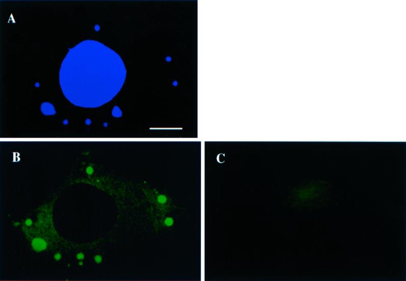
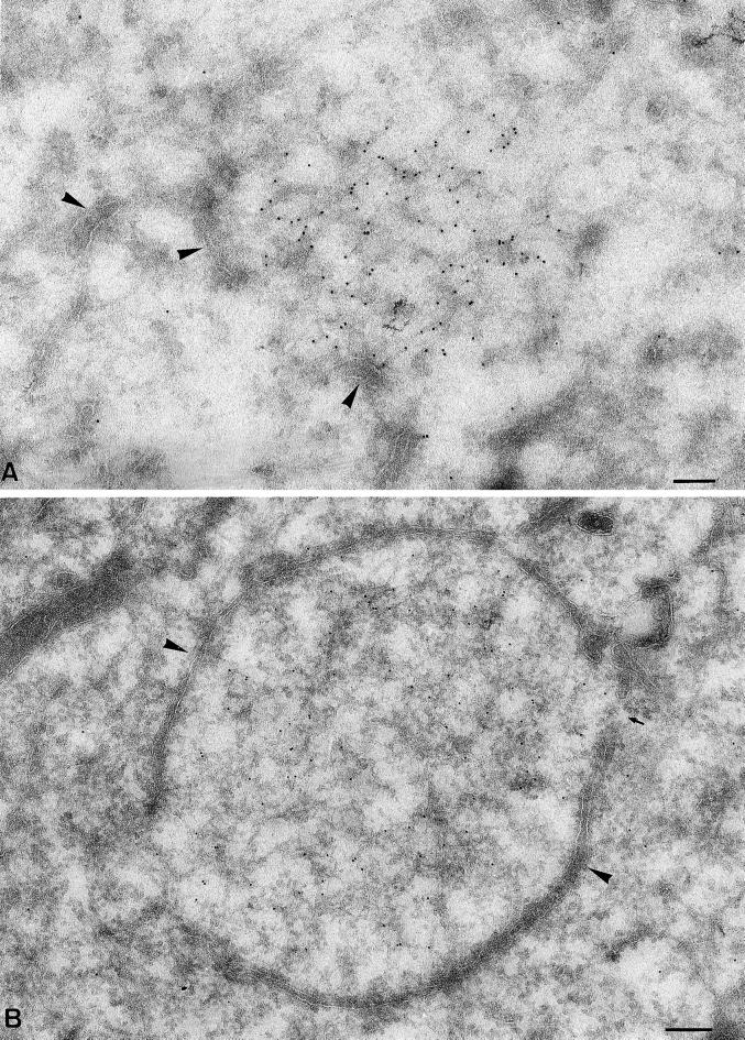
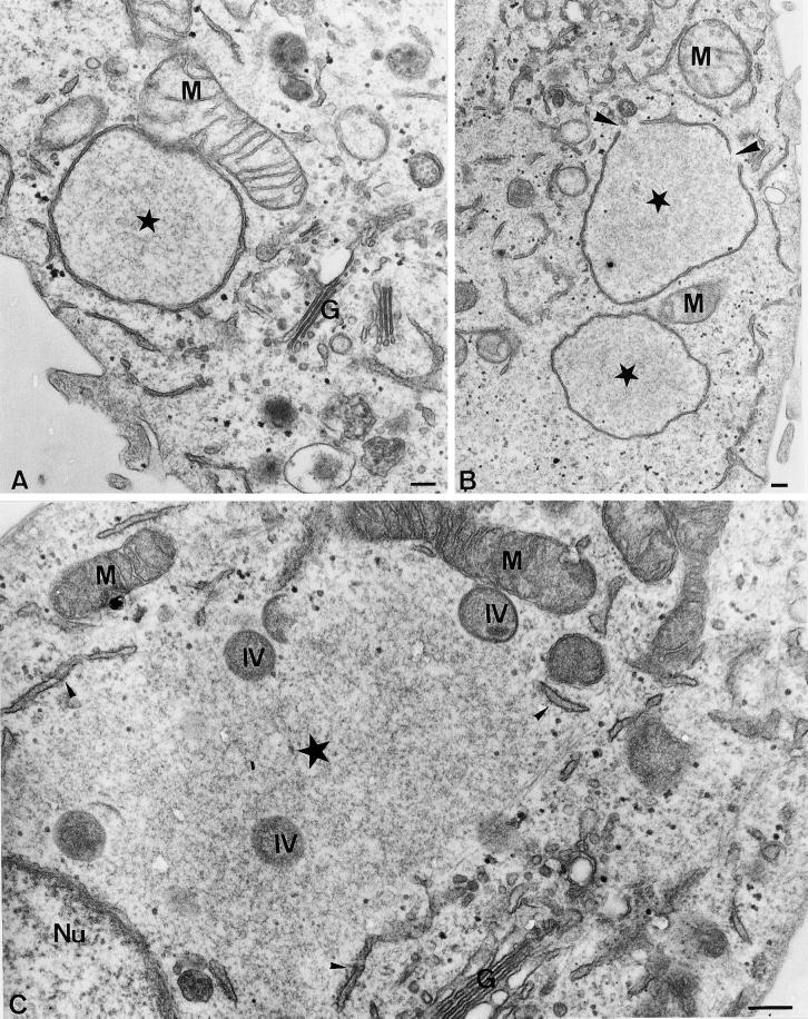
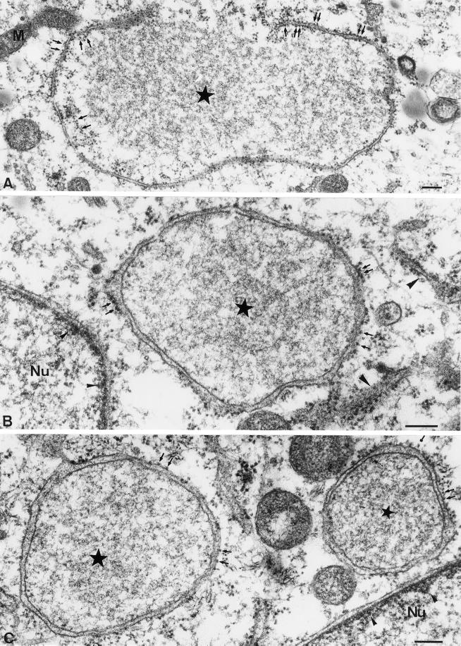
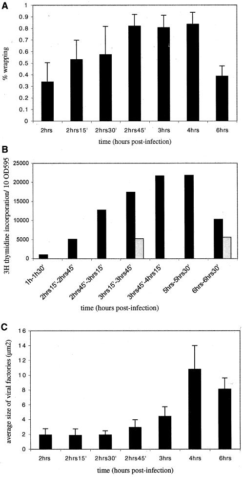
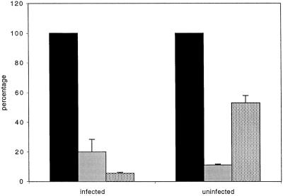
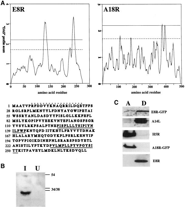
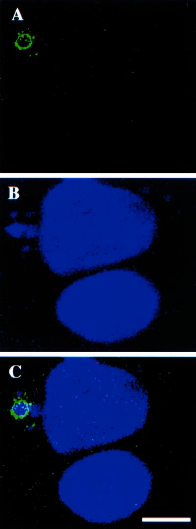
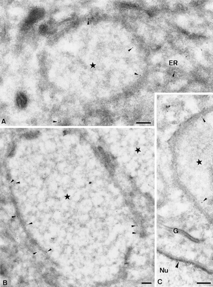
Vaccinia virus (vv), a member of the poxvirus family, is unique among most DNA viruses in that its replication occurs in the cytoplasm of the infected host cell. Although this viral process is known to occur in distinct cytoplasmic sites, little is known about its organization and in particular its relation with cellular membranes. The present study shows by electron microscopy (EM) that soon after initial vv DNA synthesis at 2 h postinfection, the sites become entirely surrounded by membranes of the endoplasmic reticulum (ER). Complete wrapping requires ~45 min and persists until virion assembly is initiated at 6 h postinfection, and the ER dissociates from the replication sites. [(3)H]Thymidine incorporation at different infection times shows that efficient vv DNA synthesis coincides with complete ER wrapping, suggesting that the ER facilitates viral replication. Proteins known to be associated with the nuclear envelope in interphase cells are not targeted to these DNA-surrounding ER membranes, ruling out a role for these molecules in the wrapping process. By random green fluorescent protein-tagging of vv early genes of unknown function with a putative transmembrane domain, a novel vv protein, the gene product of E8R, was identified that is targeted to the ER around the DNA sites. Antibodies raised against this vv early membrane protein showed, by immunofluorescence microscopy, a characteristic ring-like pattern around the replication site. By electron microscopy quantitation the protein concentrated in the ER surrounding the DNA site and was preferentially targeted to membrane facing the inside of this site. These combined data are discussed in relation to nuclear envelope assembly/disassembly as it occurs during the cell cycle.


Figures

Figure 1
Figure 1
p35 colocalizes with the viral replication sites in the cytoplasm of infected cells. Monolayers of HeLa cells were infected (A and B) or mock infected (C) and fixed at 3 h postinfection. A is labeled with Hoechst at 0.5 mg/ml, whereas B and C are labeled with anti-p35 and donkey anti-rabbit-fluorescein isothiocyanate. Infected cells show prominent p35- (B) and Hoechst (A)-positive spots in the cytoplasm that entirely colocalize. Such spots are not present in uninfected cells in C. Bar, 5 μm.
Figure 2
Figure 2
Soon after initial DNA synthesis the sites become enwrapped by membranes typical of the ER. (A) Cryosections were fixed at 2 h postinfection and labeled with anti-p35. The labeling appears associated with a fine electron-dense structure apparently lying “free” in the cytoplasm. The arrowheads indicate putative ER membranes that appear in proximity to the p35-positive site. (B) Cryosections fixed at 3 h postinfection showing that the entire p35-positive area is now surrounded by a membrane typical of the ER (large arrowheads). The small arrow indicates a gap in the surrounding membrane. Bars, 200 nm.
Figure 3
Figure 3
Ultrastructure of the viral replication sites in Epon-embedded cells. (A and B) Cells infected and fixed at 2 h 45 min, postinfection. The stars indicate the factory region. Note in A the close association of a mitochondrium (M) with the ER membrane surrounding the factory site; B shows two DNA replication sites, one of which shows gaps in the surrounding membrane (large arrowheads); and C shows the factory region (large star in the center) at 6 h postinfection. The ER (small arrowheads) does not completely surround the factory region anymore. At this time of infection the DNA replication site also contains IVs (indicated), the precursors of the first infectious form of vv, the IMV. Note the close association of mitochondria with the factory region. G, Golgi complex; Nu, nucleus. Bars, 200 nm.
Figure 4
Figure 4
High-magnification views of the DNA replication sites at 3 h postinfection in Epon-embedded infected HeLa cells. In these examples the fixed cells were treated with 1% tannic acid before Epon embedding to better reveal the ribosomes. The stars indicate the factory region. In B note the resemblance of the electron-dense structure of the viral factory to the interior of the nucleus. In A an incompletely closed ER membrane surrounds the factory region. The small arrows indicate ribosomes that appear present on both the inner and outer side of the ER membrane. In B and C the ER membrane completely encloses the factory region and the ribosomes (small arrows) are mostly on the side of the ER membrane facing the cytoplasm. Large arrowheads in B indicate rough ER studded with ribosomes on both sides of the membrane in the vicinity of the DNA replication site. Small arrowheads in B and C point to the electron-dense meshwork adjacent to the INM. Nu, nucleus. Bars, 200 nm.
Figure 5
Figure 5
Extent of ER wrapping coincides with the extent of viral DNA replication and factory growth. In A ER wrapping around the DNA replication sites was determined as described in MATERIALS AND METHODS for 15 randomly selected replication sites as detected by anti-p35 labeling on cryosections. The percentage of wrapping was determined for each site. The values represent the average and SDs of percentage of ER wrapped around 15 DNA sites at each time point tested. In B the extent of DNA replication during the course of infection. Infected (▪) or uninfected (░⃞) HeLa cells were labeled for 30 min with [3H]thymidine at different times of infection (indicated). At the end of the labeling period the cells were lysed and the protein content of each sample was determined with the use of a Bio-Rad protein assay. The amount of TCA-precipitable counts contained in 10 OD595 units was determined by liquid scintillation counting. In C the average size of the factories during the course of infection is represented. From the values obtained in A the average size of the factory region was calculated for 15 factories per time point. The values represent the average size (surface) and SDs of 15 factories for each time point tested. Note that complete (85%) ER wrapping coincides with a peak in viral DNA synthesis as well as an increase in the average size of the factory region. The decrease in factory size at 6 h postinfection is probably the result of the fact that at that time of infection the factory region is more difficult to define. This is because the p35-positive site is more dispersed and not bounded by ER membrane anymore.
Figure 6
Figure 6
Ionomycin treatment severely affects DNA replication. Infected or uninfected HeLa cells (indicated) were treated for 10 min with 5 μm ionomycin (grey shaded bar), ionomycin and BAPTA-AM (dotted bar), or not treated (solid black bar) at 3 h postinfection. Cells were then metabolically labeled with [3H]thymidine for 30 min in the presence or absence of the drugs. The cells were lysed and equal amounts of OD595 units determined by a Bio-Rad protein assay were TCA precipitated and the extent of [3H]thymidine incorporation determined by liquid scintillation counting. The values obtained from untreated control samples were expressed as 100%. The values of the treated samples are expressed as the percentage of [3H]thymidine incorporation relative to the untreated control. All values show the averages and SDs of duplicate samples.
Figure 7
Figure 7
The gene product of E8R is a membrane protein. In A hydrophobicity plots of the gene products of E8R and A18R with the use of the dense alignment surface method. The dotted line on the graphs indicates the loose and the upper uninterrupted line the strict cut-off for the determination of a putative transmembrane region. The amino acid sequence of the E8R gene is depicted under its respective “dense alignment surface” plot. The putative transmembrane domains are underlined and the peptide sequence used to raise antibodies is indicated with a hatched line above the sequence. In B HeLa cells were infected (I) or mock infected (U) for 3 h. and postnuclear supernatants were prepared. Equal amounts of protein were loaded onto a 12.5% SDS-PAGE, blotted onto polyvinylidene difluoride membrane and the E8R protein detected by enhanced chemiluminescence with the use of the peptide antibody. On the right side the positions of the 34-, 38-, and 54-kDa marker proteins are indicated. In C infected and transfected cells were lysed at 6 h postinfection and the lysate subjected to extraction with TX-114. A is the aqueous phase and D the detergent phase. E8R- and A18R-GFP refers to detection of the transfected GFP-tagged proteins with the use of anti-GFP antibodies, whereas E8R represents the untagged vv protein detected by anti-E8R. The A14L and H5R gene products serve as a control for a membrane (Salmons et al., 1997) and a soluble protein, respectively, and were detected with antibodies raised against the respective proteins.
Figure 8
Figure 8
Localization of E8R by immunofluorescence microscopy. HeLa cells grown on coverslips were infected and fixed at 3 h postinfection. Cells were double-labeled with anti-E8R antibody (A) and Hoechst (B). C shows the merged image. The image shows two cells and only the upper cell is infected. In this cell the E8R antibody labels around the Hoechst-positive replication site. This same cell has also several small DNA-sites that are not surrounded by E8R labeling. Bar, 2.5 μm.
Figure 9
Figure 9
EM localization of E8R. HeLa cells were infected with vv and fixed at 3 h postinfection (B), or infected and lipofected with E8R-GFP and fixed at 4 h postinfection (A and C). Sections were labeled with affinity purified anti-E8R (B) or anti-GFP (A and C; anti-GFP or anti-E8R labeling is indicated with small arrowheads). In all three images the factory region is indicated with a star. In A labeling is observed on the inner ER membrane and in ER membranes in continuity with the membranes enclosing the factory region. In B cells were infected for 3 h and labeled with anti-E8R. The image shows one entire replication site and part of a second site in the top right of the picture. Labeling is found on the ER membrane enclosing the DNA site as well as on the inside of the factory region (see text). C shows a part of a factory region of which the inner membrane is labeled. The image also shows an unlabeled Golgi stack (G) as well as the NE. The large arrowhead indicates the electron-dense structure underlying the INM. Nu, nucleus. Bars, 200 nm.

Similar articles

Cited by

References

    1. Banham AH, Leader DP, Smith GL. Phosphorylation of ribosomal proteins by the vaccinia virus B1R protein kinase. FEBS Lett. 1993;321:27–31. - PubMed
    1. Banham AH, Smith GL. Vaccinia virus gene B1R encodes a 34-kDa serine/threonine protein kinase that localizes in cytoplasmic factories and is packaged into virions. Virology. 1992;191:803–812. - PubMed
    1. Beaud G, Beaud R. Preferential virosomal location of underphosphorylated H5R protein synthesized in vaccinia virus-infected cells. J Gen Virol. 1997;78:3297–3302. - PubMed
    1. Beaud G, Beaud R, Leader DP. Vaccinia virus gene H5R encodes a protein that is phosphorylated by the multisubstrate vaccinia virus B1R protein kinase. J Virol. 1995;69:1819–1826. - PMC - PubMed
    1. Beaud G, Sharif A, Topa-Masse A, Leader DP. Ribosomal protein S2/Sa kinase purified from HeLa cells infected with vaccinia virus corresponds to the B1R kinase and phosphorylates in vitro the viral ssDNA-binding protein. J Gen Virol. 1994;75:283–293. - PubMed

Publication types

MeSH terms

LinkOut - more resources