. 2001 Jul;12(7):2047-60.
doi: 10.1091/mbc.12.7.2047.

The Golgi complex is a microtubule-organizing organelle

Affiliations
Free PMC article

The Golgi complex is a microtubule-organizing organelle

K Chabin-Brion et al. Mol Biol Cell. 2001 Jul.
Free PMC article

Abstract

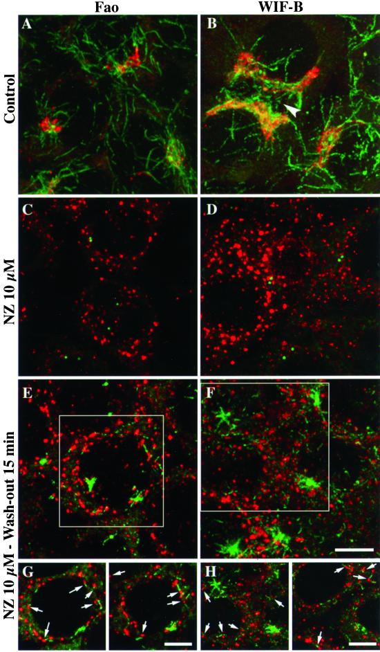
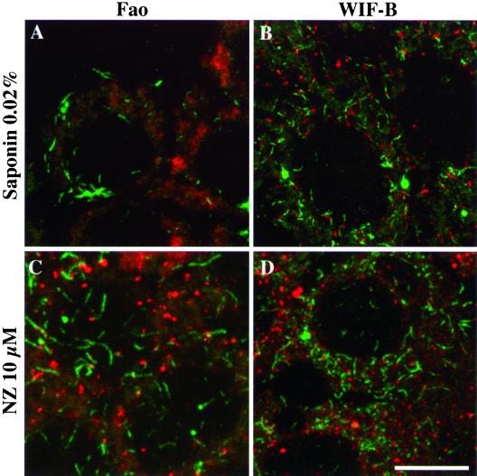
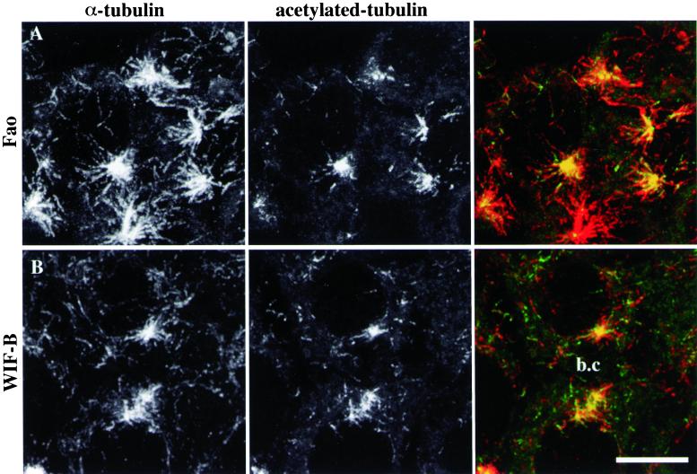
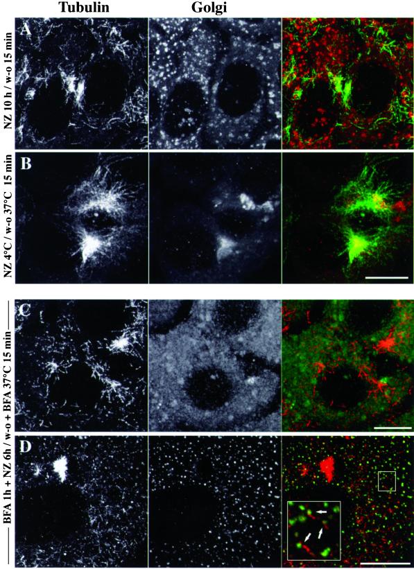
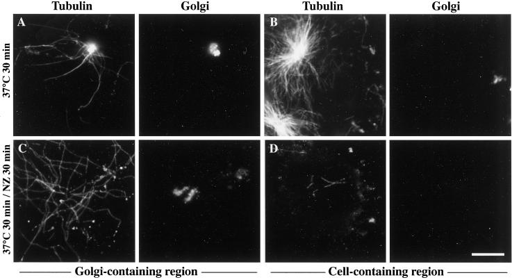
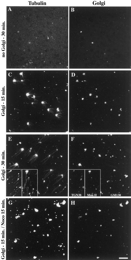
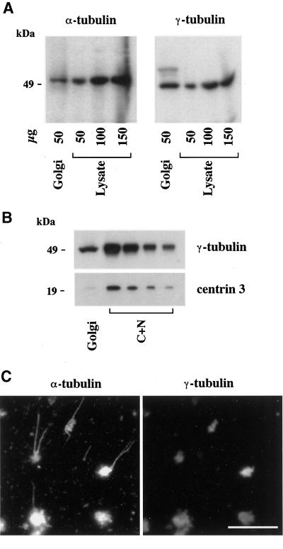
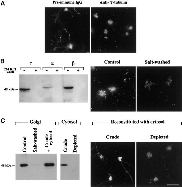
We show that the Golgi complex can directly stimulate microtubule nucleation in vivo and in vitro and thus behaves as a potent microtubule-organizing organelle in interphase cells. With the use of nocodazole wash-out experiments in hepatic cells, we found that the occurrence of noncentrosomal, early stabilized microtubules is highly correlated with the subcellular localization of Golgi membranes. With the use of in vitro reconstituted microtubule assembly systems with or without cytosol, we also found that, in contrast to centrosomally attached microtubules, the distal ends of Golgi-attached microtubules are remotely stabilized in a way that requires additional cytosolic component(s). Finally, we demonstrate that Golgi-based microtubule nucleation is direct and involves a subset of gamma-tubulin bound to the cytoplasmic face of the organelle.


Figures

Figure 1
Figure 1
Acetylated MTs reform in the close vicinity of Golgi fragments. Fao rat hepatoma cells (A, C, E, and G) and WIF-B polarized hepatic cells (B, D, F, and H) were treated without (A and B) or with (C–F) 10 μM nocodazole for 10 h, washed twice with cold culture medium, and incubated at 37°C in nocodazole-free medium for 15 min (E, F, G, and H). Cells were then fixed and processed for double immunofluorescence labeling with the use of antibodies to acetylated tubulin (green) and secretory albumin (red). All cells were examined by confocal microscopy. The images shown in A–F are the superimposition of serial optical sections acquired over the whole cell height (delta z = 0.5 μm). In G and H, two images with a shorter depth of field (made by superimposing 3 consecutive optical sections) were extracted from the indicated regions in E and F, respectively. The arrowhead indicates a bile canaliculus between adjacent WIF-B cells. Scale bar, 10 μm.
Figure 2
Figure 2
Newly formed acetylated MTs associated with Golgi elements are stable. Nocodazole wash-out experiments were conducted as described in Figure 1 on Fao (A and C) and WIF-B cells (B and D). Cells were either permeabilized with HEPES-buffered medium containing 0.2 mg/ml saponin (A, B) or incubated again with 10 μM nocodazole for 20 min at 37°C (C and D). After fixation, cells were subjected to double immunofluorescence labeling of acetylated tubulin (green) and secretory albumin (red). All cells were examined by confocal microscopy. The images are the superimposition of consecutive optical sections taken over the whole cell height (delta z = 0.5 μm). Scale bar, 10 μm.
Figure 3
Figure 3
Scattered acetylated MTs belong to noncentrosomal microtubules. Fao rat hepatoma cells (A) and WIF-B polarized hepatic cells (B) were subjected to the same nocodazole wash-out experiments as described in Figure 1. After fixation, acetylated tubulin (center panels) and α-tubulin (left panels) were visualized by immunofluorescence on a confocal microscope. The images shown are the superimposition of consecutive optical sections acquired over the whole cell height (delta z = 0.5 μm). A bile canaliculus (b.c) is indicated between two adjacent WIF-B cells. Scale bar, 10 μm.
Figure 4
Figure 4
The subcellular localization of newly assembled microtubules is correlated with Golgi membrane location. WIF-B polarized hepatic cells were pretreated (C, D) or not with 10 μM BFA for 1 h at 37°C and then subjected to MT depolymerization by 10 μM nocodazole at 37°C for 6 (C and D) or 10 h (A). In B, MT depolymerization was conducted at 4°C for 2 h. Cells were then allowed to recover from nocodazole treatment at 37°C for 15 min. After fixation, cells were either subjected to the double-immunofluorescence labeling of α-tubulin (left panels) and secretory albumin (middle panels of A–C) or to that of α-tubulin and GM130 (middle panel of D). The images are the superimposition of equidistant optical sections acquired by confocal microscopy over the whole cell height (delta z = 0.5 μm). Scale bars, 10 μm.
Figure 5
Figure 5
Golgi membranes nucleate and stabilize microtubules when added to a permeabilized cell system in which interphase microtubule dynamics were reconstituted. NIH-3T3 fibroblasts cultured on glass coverslips were detergent-extracted with 0.05% Triton X-100 in PEM buffer, rinsed extensively, and kept at 4°C for 3 d. Permeabilized cells were then incubated for 30 min at 37°C with detergent-free, autologous cytosol containing 2.5 μM tubulin, 5 μM okadaic acid, an ATP-regenerating system, and purified Golgi. In C–D, 20 μM nocodazole was added for 30 more min at 37°C. After two rinses with warm PEM buffer, samples were fixed with methanol (−20°C, 4 min) and processed for the double-immunofluorescence labeling of tubulin (left panels) and albumin (right panels). Depending on the distribution of Golgi elements and cells on the coverslip, images of Golgi-containing or control (containing extracted cells) regions are shown as indicated. Scale bar, 10 μm.
Figure 6
Figure 6
Purified Golgi membranes promote the assembly of microtubules in vitro but have no microtubule-stabilizing effect. Purified Golgi membranes were added (C–H) or not (A and B) to 0.1% purified porcine brain tubulin and 1 mM GTP in PEM buffer. After 15- or 30-min incubations at 37°C, followed (G and H) or not (A–F) by the addition of 10 μM nocodazole for 15 more min as indicated, samples were fixed by a 20-fold dilution with 0.25% glutaraldehyde in PEM buffer and sedimented although a 15% sucrose cushion onto glass coverslips. After postfixation in methanol (−20°C, 4 min), samples were processed for the immunofluorescence labeling of α-tubulin (left panels) and GM130 (right panels, B–H), TGN38 or Mannosidase II (F, insets) as indicated. Scale bar, 10 μm
Figure 7
Figure 7
γ-Tubulin is peripherally associated with Golgi membranes. (A) Purified rat liver Golgi membranes (50 μg of total proteins) and increasing amounts of rat hepatocyte lysate were analyzed by 9% SDS-PAGE followed by Western blotting for the presence of α- and γ-tubulin. Band intensities were analyzed by densitometry to estimate the enrichment of Golgi membranes in both markers relative to the lysate (see the text). (B) The amount of centriolar material was measured in Golgi samples compared with a nucleo-centrosomal extract (C+N) by SDS-PAGE (9 and 15% for γ-tubulin and centrin-3, respectively), followed by electrotransfer onto polyvinyl difluoride membranes, postfixation with 0.2% glutaraldehyde (only for centrin), and immunorevelation. Densitometric measurements of band intensities were used to evaluate the ratios of γ-tubulin to centrin signals and to allow a comparison between the Golgi and the C+N extract (see the text). (C) Purified Golgi was incubated for 30 min at 37°C with 0.1% tubulin and 1 mM GTP. After fixation with 0.25% glutaraldehyde, samples were centrifuged onto glass coverslips and postfixed with methanol before the double-labeling of α- and γ-tubulins as indicated. Scale bar, 10 μm.
Figure 8
Figure 8
γ-Tubulin is involved in the nucleation of Golgi-based MTs. (A) As indicated, purified Golgi was preincubated either with anti–γ-tubulin or with preimmune mouse IgGs for 1 h at 37°C and then used in the in vitro MT assembly assay. Samples were processed for the immunofluorescence labeling of α-tubulin. (B) Purified rat liver Golgi was washed as indicated with 2 M KCl and then rinsed three times in PEM buffer before being assayed for the presence of tubulins or to be used in the in vitro MT assembly assay. Samples were analyzed for the presence of tubulins by Western blotting, as described in Figure 7. After the in vitro MT assembly assays, samples were subjected to the immunofluorescence labeling of α-tubulin. (C) Salt-washed Golgi membranes were incubated (1 h, room temperature) with crude WIF-B cytosol or cytosol that was immunodepleted of γ-tubulin and then rinsed twice in PEM buffer and either analyzed by Western blot for the presence of peripherally bound γ-tubulin or used in the in vitro MT assembly assay, followed by the immunofluorescence labeling of α-tubulin. Scale bar, 10 μm.

Similar articles

Cited by

References

    1. Baas PW, Joshi HC. γ-Tubulin distribution in the neuron: implications for the origins of neuritic microtubules. J Cell Biol. 1992;119:171–178. - PMC - PubMed
    1. Biou D, Monnet D, Miller D, Feger J, Durand G. An immunochemical procedure to evaluate the degree of desialylation of α1-acid glycoprotein in rat serum. J Immunol Methods. 1984;74:267–271. - PubMed
    1. Bloom GS, Goldstein LS. Cruising along microtubule highways: how membranes move through the secretory pathway. J Cell Biol. 1998;140:1277–1280. - PMC - PubMed
    1. Bré MH, Kreis TE, Karsenti E. Control of microtubule nucleation and stability in Madin-Darby canine kidney cells: the occurrence of noncentrosomal, stable detyrosinated microtubules. J Cell Biol. 1987;105:1283–1296. - PMC - PubMed
    1. Bulinski JC, Richards JE, Piperno G. Posttranslational modifications of α-tubulin: detyrosination and acetylation differentiate populations of interphase microtubules in cultured cells. J Cell Biol. 1988;106:1213–1220. - PMC - PubMed

Publication types

LinkOut - more resources