. 2001 Jul;12(7):2137-45.
doi: 10.1091/mbc.12.7.2137.

Activation of Rhoa and ROCK are essential for detachment of migrating leukocytes

Affiliations
Free PMC article

Activation of Rhoa and ROCK are essential for detachment of migrating leukocytes

J Alblas et al. Mol Biol Cell. 2001 Jul.
Free PMC article

Abstract

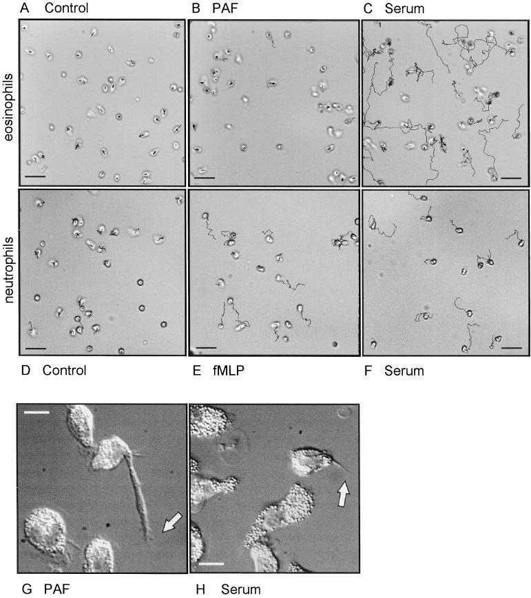
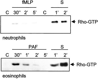
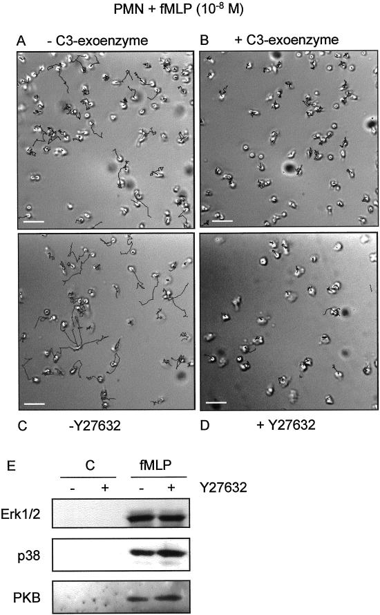
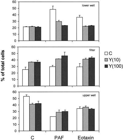
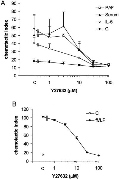
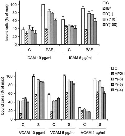
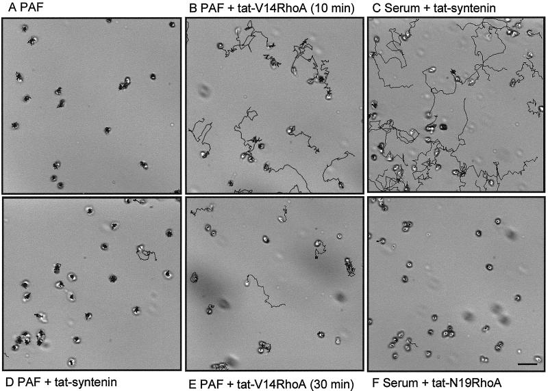
Detachment of the rear of the cell from its substratum is an important aspect of locomotion. The signaling routes involved in this adhesive release are largely unknown. One of the few candidate proteins to play a role is RhoA, because activation of RhoA in many cell types leads to contraction, a mechanism probably involved in detachment. To study the role of RhoA in detachment regulation, we analyzed several subsets of expert migratory leukocytes by video microscopy. In contrast to fast-migrating neutrophils, eosinophils do not detach the rear of the cell unless stimulated with serum. When measuring the amount of active RhoA, with the use of a GST-Rhotekin pulldown assay, we found that serum is an excellent activator of RhoA in granulocytes. Inhibition of RhoA or one of Rho's target proteins, the kinase ROCK, in neutrophils leads to the phenotype seen in eosinophils: the rear of the cell is firmly attached to the substratum, whereas the cell body is highly motile. ROCK-inhibition leads to impaired migration of granulocytes in filters, on glass, and through endothelial monolayers. Also, the ROCK signaling pathway is involved in changes of integrin-mediated adhesion. Eosinophil transduction by a tat-fusion construct containing active RhoA resulted in detachment stimulation in the presence of chemoattractant. From these results we conclude that activation of the RhoA-ROCK pathway is essential for detachment of migratory leukocytes.


Figures

Figure 1
Figure 1
Cell tracks of randomly moving eosinophils (A, B, C, G, and H) and neutrophils (D–F). The cells were attached to albumin-coated glass coverslips in RPMI-HEPES containing 0.5% HSA. Then they were transferred to the same buffer completed as indicated without stimulus (A and D) with PAF (10−7 M; B and G), fMLP (10−8 M; E), or 10% human pooled serum (C, F, and H). The cells were warmed to 37°C and then monitored for 20 min (A–C), 8 min (D–F), or 30 min (G and H). Bar, 30 μm (A–F) or 10 μm (G and H). Video material available for A–H.
Figure 2
Figure 2
RhoA activation assay in neutrophils and eosinophils. The cells were stimulated with fMLP (10−7 M), PAF (10−7 M), or serum for the time-periods indicated, lysed, and subjected to GST-Rhotekin pulldown as described in MATERIALS AND METHODS. Shown are Western blots of active RhoA (arrow).
Figure 3
Figure 3
Migration of neutrophils on albumin-coated glass coverslips. The cells were untreated (A and C) or pretreated with C3-exoenzyme (10 μg/ml; 4 h, B) or Y27632 (10−5 M; 30 min, D) then attached to the coverslips. The coverslips were inverted in assay medium containing fMLP (10−8 M) and embedded. Random movement of the cells was recorded at 20-s intervals. Shown are tracks recorded during an 8-min period. Bar, 30 μm. (E) fMLP-induced activity of Erk1/2, p38, and PKB in neutrophils in the absence or presence of Y27632 (10−5 M; 30 min) with the use of phosphospecific antibodies. Video material available for A–D.
Figure 4
Figure 4
Transendothelial migration of eosinophils in the presence of Y27632. Eosinophils migrating over confluent monolayers of human umbilical vein endothelial cells (details in MATERIALS AND METHODS) were stimulated with PAF (10−7 M) or eotaxin (10−7 M). Pretreatment of the eosinophils with Y27632 was for 30 min as indicated (in μM). After 1 h of migration, the cells were taken out of the lower wells, filters, and upper wells and separately quantified with the use of calcein. The results are expressed as percentage of total cells (mean ± SEM) of a representative experiment performed in duplicate.
Figure 5
Figure 5
Granulocyte chemotaxis in the presence of ROCK-inhibitor Y27632. (A) Eosinophils migrating in the Boyden chamber were stimulated with PAF (10−7 M) present in the lower wells, IL-5 (10−10 M) in the upper well, or with serum (10%) in both wells. (B) Boyden chamber chemotaxis assay of neutrophils stimulated with fMLP (10−8 M). Y27632 pretreatment was in all cases for 30 min. The results are expressed as chemotactic index (mean ± SEM of 2–6 experiments performed in duplicate).
Figure 6
Figure 6
Adhesion of eosinophils to ICAM-1 or VCAM. Eosinophils were calcein-loaded, incubated for 30 min with Y27632 at the indicated concentrations (between brackets, in μM), or with neutralizing antibodies against α4- (HP2/1) or β2- (IB4) integrins. Then the cells were placed in 96-well plates coated with ICAM-1 (5–10 μg/ml) or VCAM (0.1–5 μg/ml) containing control buffer or serum-containing buffer and incubated for 5 min at 37°C. The cells were washed and lysed and fluorescence intensity was measured. The results are expressed as percentage of maximal binding (mean ± SEM of 2–5 experiments performed in duplicate).
Figure 7
Figure 7
Video microscopy of migrating eosinophils in the presence of tat-fusion proteins. Cells were attached to albumin-coated glass coverslips, transferred to medium containing 1 μM of the indicated tat-fusion proteins and/or PAF (10−7 M) or human pooled serum (10%), and embedded at t = 0. Images were recorded with 20-s intervals during 20 min at 37°C. Shown are calculated cell tracks of a representative experiment. Bar, 30 μm. Video material is available for A–F.

Similar articles

Cited by

References

    1. Allen WE, Zicha D, Ridley AJ, Jones GE. A role for cdc42 in macrophage chemotaxis. J Cell Biol. 1998;141:1147–1157. - PMC - PubMed
    1. Amano M, Ito M, Kimura K, Fukata Y, Chihara K, Nakano T, Matsuura Y, Kaibuchi K. Phosphorylation and activation of myosin by Rho-associated kinase (Rho-kinase) J Biol Chem. 1996;271:20246–20249. - PubMed
    1. Aspenström P. Effectors for the Rho GTPases. Curr Opin Cell Biol. 1999;11:95–102. - PubMed
    1. Chrzanowska-Wodnicka M, Burridge K. Rho-stimulated contractility drives the formation of stress fibers and focal adhesions. J Cell Biol. 1996;133:1403–1415. - PMC - PubMed
    1. Essler M, Amano M, Kruse HJ, Kaibuchi K, Weber PC, Aepfelbacher M. Thrombin inactivates myosin light chain phosphatase via Rho and its target Rho kinase in human endothelial cells. J Biol Chem. 1998;273:21867–21874. - PubMed

Publication types

MeSH terms

Substances