. 2001 Jul 15;29(14):2950-62.
doi: 10.1093/nar/29.14.2950.

Cloning and characterization of two guide RNA-binding proteins from mitochondria of Crithidia fasciculata: gBP27, a novel protein, and gBP29, the orthologue of Trypanosoma brucei gBP21

Affiliations

Cloning and characterization of two guide RNA-binding proteins from mitochondria of Crithidia fasciculata: gBP27, a novel protein, and gBP29, the orthologue of Trypanosoma brucei gBP21

D Blom et al. Nucleic Acids Res. .

Abstract

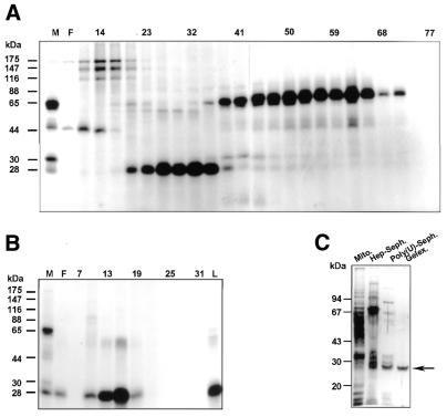
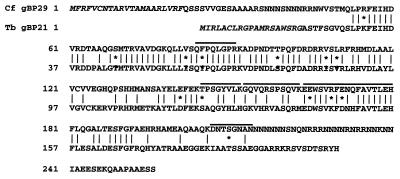
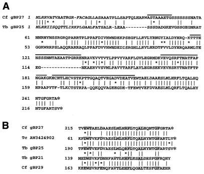
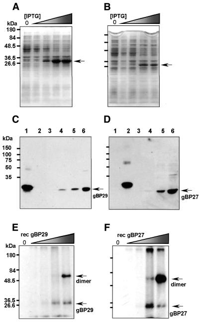
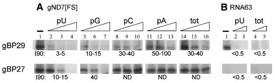
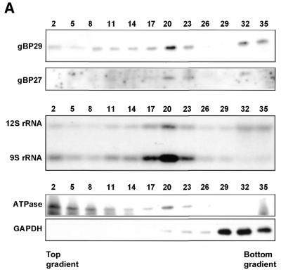
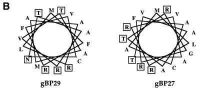
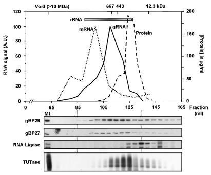
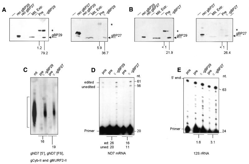
In kinetoplastid protozoa, mitochondrial (mt) mRNAs are post-transcriptionally edited by insertion and deletion of uridylate residues, the information being provided by guide (g)RNAs. Currently popular mechanisms for the editing process envisage a series of consecutive 'cut-and-paste' reactions, carried out by a complex RNP machinery. Here we report on the purification, cloning and functional analysis of two gRNA-binding proteins of 28.8 (gBP29) and 26.8 kDa (gBP27) from mitochondria of the insect trypanosome Crithidia fasciculata. gBP29 and gBP27 proved to be similar, Arg + Ala-rich proteins, with pI values of approximately 10.0. gBP27 has no homology to known proteins, but gBP29 is the C.fasciculata orthologue of gBP21 from Trypanosoma brucei, a gRNA-binding protein that associates with active RNA editing complexes. As measured in UV cross-linking assays, His-tagged recombinant gBP29 and gBP27 bind to radiolabelled poly(U) and synthetic gRNAs, while competition experiments suggest a role for the gRNA 3'-(U)-tail in binding to these proteins. Immunoprecipitates of mt extracts generated with antibodies against gBP29 also contained gBP27 and vice versa. The immunoprecipitates further harbored a large proportion of the cellular content of four different gRNAs and of edited and pre-edited NADH dehydrogenase subunit 7 mRNAs, but only small amounts of mt rRNAs. In addition, the bulk of gBP29 and gBP27 co-eluted with gRNAs from gel filtration columns in the high molecular weight range. Together, these results suggest that the proteins are part of a large macromolecular complex(es). We infer that gBP29 and gBP27 are components of the C.fasciculata editing machinery that may interact with gRNAs.


Figures

Figure 1
Figure 1
Purification of 30/28 kDa proteins that bind to poly(U) and gRNAs. (A) Mitochondrial extracts were loaded onto a heparin–Sepharose column and bound proteins were eluted with a KCl gradient (see Materials and Methods). An aliquot of 7.5 µl of every third heparin–Sepharose fraction, containing 0.1–1.0 µg protein, was tested in a UV cross-linking assay, followed by separation on a 10% (w/v) SDS–PAGE gel. Labelled proteins were then visualised by autoradiography. Lane M, 13 µg mitochondrial extract; lane F, 6.8 µg flow-through. (B) Heparin–Sepharose fractions containing the 28 kDa UV cross-linking activity were pooled, loaded onto a poly(U)–Sepharose column and bound proteins were eluted with a KCl gradient. An aliquot of 7.5 µl of every third poly(U)–Sepharose fraction, containing 10–100 ng protein, was tested in a UV cross-linking assay, followed by separation on a 10% SDS–PAGE gel. Labelled proteins were visualised by autoradiography. Lane M, 13 µg mitochondrial extract; lane F, 1.0 µg flow-through; lane L, 1.0 µg load. (C) Protein samples of different steps in purification of the 28 kDa UV cross-linking activity were separated on a 10% SDS–PAGE gel and silver stained. Mito., 500 ng mitochondrial extract; Hep-Seph., 200 ng pooled heparin–Sepharose fractions containing the 28 kDa UV cross-linking activity; Poly(U)-Seph., 80 ng pooled poly(U)–Sepharose fractions containing the 28 kDa UV cross-linking activity; Gelex., 25 ng PAGE gel-purified 28 kDa UV cross-linking activity. The arrow indicates the position of the 28 kDa protein active in a UV cross-linking assay. Molecular masses of marker proteins are indicated on the left-hand side of each protein gel.
Figure 2
Figure 2
Inferred amino acid sequence of gBP29. Amino acid sequence alignment of C.fasciculata gBP29 (top lines) and T.brucei gBP21 (bottom lines). Identical amino acids are indicated by vertical lines. Asterisks indicate conservative amino acid substitutions: K, R; S, T; D, E; F, Y; V, I, L. Dashes are introduced for optimal alignment. The putative mitochondrial import signals are given in italic and Lys-C generated peptide sequences of gBP29 are indicated above the lines.
Figure 3
Figure 3
Inferred amino acid sequence of gBP27 and T.brucei gBP25. (A) Amino acid sequence alignment of C.fasciculata gBP27 (top lines) and its putative T.brucei orthologue gBP25 (bottom lines). Identical amino acids are indicated by vertical lines. Small asterisks indicate conservative amino acid substitutions: K, R; S, T; D, E; F, Y; V, I, L. Large asterisks represent stop codons. Dashes are introduced for optimal alignment. The putative mitochondrial import signals are given in italic and Lys-C generated peptide sequences of gBP27 are indicated above the lines. (B) Sequence alignment of a C-terminal section of C.fasciculata gBP29 and gBP27, T.brucei gBP21 and gBP25 and a translated T.cruzi EST AW3424902. Identical and conserved amino acids found in all sequences have been highlighted.
Figure 4
Figure 4
Expression of recombinant gBP29 and gBP27, antibody generation and UV cross-linking activity. (A and B) Expression of rec gBP29 (A) and gBP27 (B) in E.coli was induced by addition of increasing concentrations of IPTG (shaded wedge) to the culture medium. After induction, cell extracts were prepared and separated on a 10% SDS–PAGE gel, which was stained with Coomassie Brilliant Blue. Concentrations of IPTG used: lanes 1, 0 mM; lanes 2, 0.01 mM; lanes 3, 0.05 mM; lanes 4, 0.25 mM; lanes 5, 1.25 mM. The arrows indicate the positions of rec gBP29 (A) and rec gBP27 (B). (C and D) rec gBP29, rec gBP27 and C.fasciculata mt extract were run on a 10% SDS–PAGE gel and transferred to PVDF membranes. Proteins were visualized by ECL with polyclonal α-gBP29 (C) and α-gBP27 (D) antibodies. Amounts of protein loaded: lanes 1, 8 ng E.coli extract expressing rec gBP29; lanes 2, 8 ng E.coli extract expressing rec gBP27; lanes 3–6, 0.24, 1.19, 2.38 and 11.9 µg C.fasciculata mt extract, respectively. The arrows indicate the positions of gBP29 (C) and gBP27 (D). (E and F) Increasing amounts of purified native rec gBP29 (E) and gBP27 (F) were cross-linked to radiolabelled poly(U) (see Materials and Methods), separated on a 10% SDS–PAGE gel and visualised by autoradiography. Amounts of protein loaded in each lane: 0, 0.5, 5, 50 and 500 ng purified rec gBP29 (E) or purified rec gBP27 (F). Molecular masses of marker proteins are indicated in kDa on the left-hand side of each protein gel.
Figure 5
Figure 5
RNA binding characteristics of recombinant gBP29 and gBP27. (A) Aliquots of 5 pmol purified gBP29 (upper) or gBP27 (lower) were cross-linked to 0.5 pmol radiolabelled gND7[FS] in the presence of varying amounts of competitor RNAs. The samples were analysed on a 12.5% SDS–PAGE gel, followed by autoradiography. Lane 1, no competitor. Lanes 2–16, before the addition of protein to the mixture, 12, 60 or 300 ng of different competitor RNAs were added: lanes 2–4, poly(U) (pU); lanes 5–7, poly(G) (pG); lanes 8–10, poly(C) (pC); lanes 11–13, poly(A) (pA); lanes 14–16, total RNA from CHP100 cells, a human neuroblastoma cell line (tot). The radioactivity present in cross-linked gBP29 and gBP27 was quantified in a Storm phosphorimager apparatus and the level of binding relative to the experiments without competitor was calculated. The figure shows an autoradiograph of a representative experiment; the numbers given for each competitor RNA represent the mass excess required for ∼90% inhibition of binding (I90), as deduced from a number of different experiments (n = 4 for gBP29; n = 3 for gBP27). The I90 values of gBP27 competition experiments with poly(C), poly(A) and total RNA were not determined (ND); with the highest amounts of these competitors tested (150-fold mass excess) binding levels were still between 20 and 30%. (B) rec gBP29 and gBP27 were cross-linked to 0.5 pmol radiolabelled RNA63, without competitor (lane 1) or with 5 and 20 ng poly(U) (lanes 2 and 3) or 5 and 20 ng total human RNA (lanes 4 and 5). For other details see (A).
Figure 6
Figure 6
Mitochondrial localization of gBP29 and gBP27. (A) Total cell extracts of C.fasciculata were submitted to renografin gradient centrifugation (20–35% w/v) and 36 fractions were collected (fraction 1 was taken from the top of the gradient, etc.). Protein extracts from indicated fractions were submitted to SDS–PAGE (10% polyacrylamide), followed by western blotting with antisera against gBP29 and gBP27 (upper two panels) and mt ATPase and GAPDH (lower two panels). The same fractions were tested for the presence of mitochondrial 9S and 12S rRNA by northern blot hybridization (middle panel). (B) Helical wheel projection of the putative mitochondrial import signal of gBP29 (left) and gBP27 (right). Charged and hydrophilic amino acids are boxed.
Figure 6
Figure 6
Mitochondrial localization of gBP29 and gBP27. (A) Total cell extracts of C.fasciculata were submitted to renografin gradient centrifugation (20–35% w/v) and 36 fractions were collected (fraction 1 was taken from the top of the gradient, etc.). Protein extracts from indicated fractions were submitted to SDS–PAGE (10% polyacrylamide), followed by western blotting with antisera against gBP29 and gBP27 (upper two panels) and mt ATPase and GAPDH (lower two panels). The same fractions were tested for the presence of mitochondrial 9S and 12S rRNA by northern blot hybridization (middle panel). (B) Helical wheel projection of the putative mitochondrial import signal of gBP29 (left) and gBP27 (right). Charged and hydrophilic amino acids are boxed.
Figure 7
Figure 7
Fractionation of mt extract on a Sephacryl S-500 gel filtration column. Mitochondrial extracts were fractionated on a Sephacryl S-500 column as described in Materials and Methods. Fractions were tested by northern blot hybridisation (upper) for the presence of ND7 mRNA (dotted line; A.U. = arbitrary units), the combined presence of ND7[FS], ND7[5′], Cyb-II and MURF2-II gRNAs (solid line) and for 9S + 12S rRNA (inserted bar; the elution profile of 9S + 12S rRNA is indicated by shading). The protein concentration of every fifth fraction was determined by Bradford assay (dashed line). Marker proteins used to calibrate the Sephacryl column were: thyroglobulin (647 kDa), apoferritin (443 kDa) and cytochrome c (12.3 kDa). Every fifth fraction was tested for the presence of gBP29 and gBP27 by western blotting (panels 2 and 3 from the top), for RNA ligase activity by self-adenylylation of two proteins of 43 and 49 kDa, respectively (panel 4), and for TUTase activity by uridylate addition to tRNAs (bottom). Mt, mitochondrial extract.
Figure 8
Figure 8
Co-immunoprecipitation analysis. (A and B) Mt extracts of C.fasciculata were incubated with Staph A coated with α-gBP29 serum (A), α-gBP27 serum (B) or pre-immune serum (Pre) (A and B), followed by washing and centrifugation as described in Materials and Methods. Immunoprecipitates were analysed for the presence of either gBP29 (left-hand panels) or gBP27 (right-hand panels) by PAGE and western blotting. In addition, 8 ng E.coli extracts expressing either gBP29 (rec gBP29) or gBP27 (rec gBP27) and the mt extract used as starting material for the immunoprecipitation reaction (Mit. Extr.) were applied to the PAGE gel. Arrows indicate the positions of gBP29 and gBP27; asterisks mark the positions of IgG heavy chains that are occasionally visible. The numbers under the lanes indicate the amounts of gBP29 or gBP27 found in the immunoprecipitate, as a percentage of the total amount present in the mt extract used in the experiment. (CE) Immunoprecipitates obtained with pre-immune serum (pre), α-gBP29 antibodies and α-gBP27 antibodies were tested for the presence of gND7[FS], gND7[5′], gCyb-II and gMURF2-II by northern blot analysis (C), the presence of ND7 mRNA (edited or unedited at the frameshift position) (D) and 12S rRNA (E) by primer extension analysis. The position of the radiolabelled oligonucleotide (primer) used in the extension reaction is indicated, as is that of the extension products. Primer extension was carried out in the presence of ddGTP instead of dGTP, resulting in a product of 61 nt derived from ND7 RNA edited by insertion of five U residues at the frameshift position and a 56 nt fragment derived from unedited ND7 RNA. Pos., positive control of the primer extension reaction with C.fasciculata total RNA. The numbers under (C)–(E) indicate the ratios of the amounts of RNA present in the immunoprecipitates generated with α-gBP29 and α-gBP27, respectively, and those generated with pre-immune serum.

Similar articles

Cited by

References

    1. Arts G.J. and Benne,R. (1996) Mechanism and evolution of RNA editing in kinetoplastida. Biochim. Biophys. Acta, 1307, 39–54. - PubMed
    1. Alfonzo J.D., Thiemann,O. and Simpson,L. (1997) The mechanism of U insertion/deletion RNA editing in kinetoplastid mitochondria. Nucleic Acids Res., 25, 3751–3759. - PMC - PubMed
    1. Stuart K., Allen,T.E., Heidmann,S. and Seiwert,S.D. (1997) RNA editing in kinetoplastid protozoa. Microbiol. Mol. Biol. Rev., 61, 105–120. - PMC - PubMed
    1. Hajduk S.L. and Sabatini,R.S. (1998) Mitochondrial mRNA editing in kinetoplastid mitochondria. In Grosjean,H. and Benne,R. (eds) Modification and Editing of RNA. ASM press, Washington, DC, pp. 377–393.
    1. Seiwert S.D., Heidmann,S. and Stuart,K. (1996) Direct visualization of uridylate deletion in vitro suggests a mechanism for kinetoplastid RNA editing. Cell, 84, 831–841. - PubMed

Publication types

MeSH terms

Associated data