. 2001 Jul 15;29(14):3030-40.
doi: 10.1093/nar/29.14.3030.

Expression of hsp16 in response to nucleotide depletion is regulated via the spc1 MAPK pathway in Schizosaccharomyces pombe

Affiliations

Expression of hsp16 in response to nucleotide depletion is regulated via the spc1 MAPK pathway in Schizosaccharomyces pombe

L Taricani et al. Nucleic Acids Res. .

Abstract

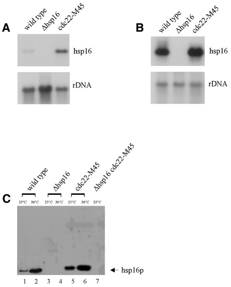
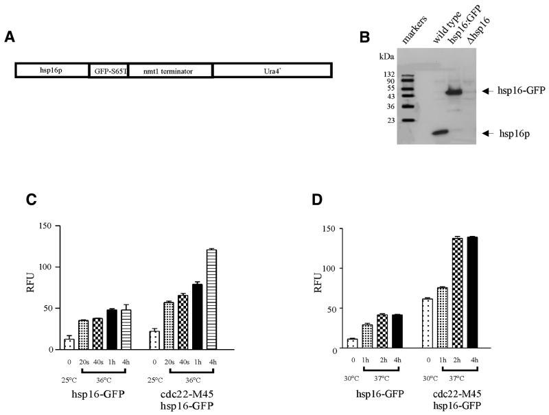
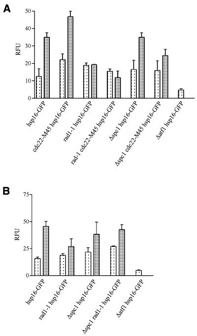
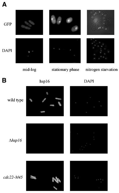
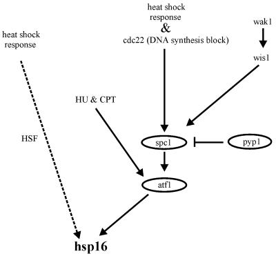
A universal response to elevated temperature and other forms of physiological stress is the induction of heat shock proteins (HSPs). Hsp16 in Schizosaccharomyces pombe encodes a polypeptide of predicted molecular weight 16 kDa that belongs to the HSP20/alpha-crystallin family whose members range in size from 12 to 43 kDa. Heat shock treatment increases expression of the hsp16 gene by 64-fold in wild-type cells and 141-fold in cdc22-M45 (ribonucleotide reductase) mutant cells. Hsp16 expression is mediated by the spc1 MAPK signaling pathway through the transcription factor atf1 and in addition through the HSF pathway. Nucleotide depletion or DNA damage as occurs in cdc22-M45 mutant cells, or during hydroxyurea or camptothecin treatment, is sufficient to activate hsp16 expression through atf1. Our findings suggest a novel role for small HSPs in the stress response following nucleotide depletion and DNA damage. This extends the types of damage that are sensed by the spc1 MAPK pathway via atf1.


Figures

Figure 1
Figure 1
Expression of hsp16. (A and B) Northern blot analysis of hsp16 expression in a cdc22-M45 mutant background. (A) Total RNA was isolated from the strains indicated at 25°C, (B) total RNA was isolated following 4 h heat shock treatment at 36°C. RNA was analyzed by northern hybridization with a probe specific for hsp16 (A and B, upper panels). A ribosomal DNA probe was used as a loading control (A and B, lower panels). (C) hsp16 protein level correlates with RNA expression levels. Hsp16 protein levels are increased in a cdc22-M45 mutant background (lanes 1, 3, 5 and 7 at 25°C; lanes 2, 4 and 6 at 36°C). All strains were cultured at 25°C, then shifted to 36°C for 4 h in YEA medium.
Figure 2
Figure 2
Hsp16–GFP expression. (A) Schematic of hsp16–GFP(S65T)-ura4+ chromosomal fusion placing expression under the control of the native promoter of hsp16. (B) Immunoblot showing that hsp16 and hsp16–GFP levels are similar following heat shock treatment at 36°C for 4 h. The strains were cultured at 25°C then shifted to 36°C for 4 h in YEA media. (C and D) Hsp16 is responsive to heat shock. (C) Expression of hsp16–GFP in wild-type and in a cdc22-M45 mutant background at 25°C and following heat shock treatment at 36°C for various times. (D) Expression of hsp16–GFP in wild-type and in a cdc22-M45 mutant background at 30°C and following heat shock treatment at 37°C for various times. Hsp16–GFP levels were quantitated by fluorimetry
Figure 3
Figure 3
Hsp16–GFP expression is activated following HU or CPT treatment. Analysis of hsp16–GFP levels in cdc22-M45 mutant. (A) Cells were cultured at 25°C (lightly shaded bars) in YEA to mid-exponential phase and then hydroxyurea (heavily shaded bars) was added at a final concentration of 11 mM to one-half of the culture and incubated for 4 h. (B) Cells were cultured at 25°C (lightly shaded bars) in YEA to mid-exponential and then camptothecin (heavily shaded bars) was added at a final concentration of 40 µM to one-half of the culture and incubated for 2 h. Hsp16–GFP levels were quantitated by fluorimetry.
Figure 4
Figure 4
The cellular localization of hsp16 protein. (A) Hsp16–GFP localization in the absence of stress (left panel), upon glucose starvation (middle panel) and nitrogen starvation in spores (right panel). (B) Exponentially growing cells were immunostained with polyclonal anti-GST–hsp16 antibody and Alexa 488 anti-rabbit IgG (H + L) and stained with DAPI. Left panels show Alexa 488 images and the right panels show DAPI images (top panel, wild-type; middle panel, Δhsp16; bottom panel, cdc22-M45). Cells were cultured at 25°C in YEA to a density of 2–5× 10⁶ cells/ml.
Figure 5
Figure 5
A model of the pathway involved in the regulation of hsp16 expression in S.pombe. This model is based on results in this study.

Similar articles

Cited by

References

    1. DeJong W.W., Leunissan,J.A.M. and Voorter,C.E.M. (1993) Evolution of the α-crystallin/small heat shock protein family. Mol. Biol. Evol., 10, 103–126. - PubMed
    1. Carver, J.A. (1999) Probing the structure and interactions of crystallin proteins by NMR spectroscopy. Prog. Retin. Eye Res., 18, 431–462. - PubMed
    1. Boelens W.C., Croes,Y., deRuwe,M., deReu,L. and deJong,W.W. (1998) Negative charges in the C-terminal domain stabilize the αβ-crystallin complex. J. Biol. Chem., 273, 28085–28090. - PubMed
    1. Kim K.K., Kim,R. and Kim,S.H. (1998) Crystal structure of a small heat shock protein. Nature, 394, 595–599. - PubMed
    1. Arrigo A.-P. and Landry,J. (1994) Expression and function of the low-molecular weight heat shock proteins. In Morimoto,R., Tissieres,H. and Georgeopoulos,C. (eds), The Biology of Heat Shock Proteins and Molecular Chaperones. Cold Spring Harbor Laboratory Press, Cold Spring Harbor, NY. pp. 335–373.

Publication types

MeSH terms