. 2001 Jul 15;29(14):3069-79.
doi: 10.1093/nar/29.14.3069.

Tripartite structure of Saccharomyces cerevisiae Dna2 helicase/endonuclease

Affiliations

Tripartite structure of Saccharomyces cerevisiae Dna2 helicase/endonuclease

S H Bae et al. Nucleic Acids Res. .

Abstract

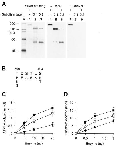
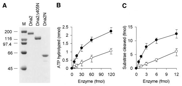
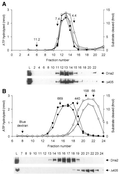
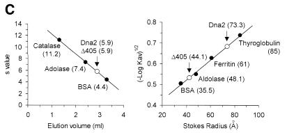
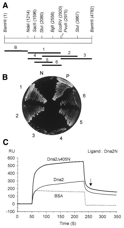
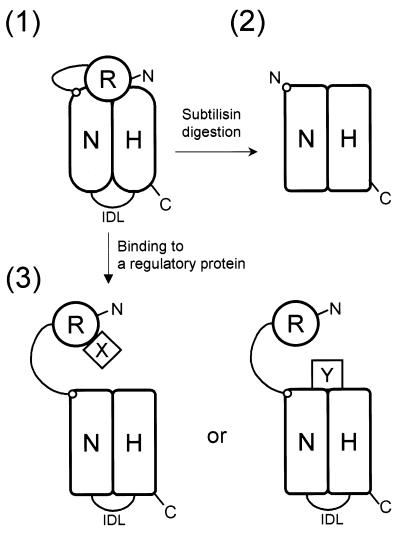
In order to gain insights into the structural basis of the multifunctional Dna2 enzyme involved in Okazaki fragment processing, we performed biochemical, biophysical and genetic studies to dissect the domain structure of Dna2. Proteolytic digestion of Dna2 using subtilisin produced a 127 kDa polypeptide that lacked the 45 kDa N-terminal region of Dna2. Further digestion generated two subtilisin-resistant core fragments of approximately equal size, 58 and 60 kDa. Surprisingly, digestion resulted in a significant (3- to 8-fold) increase in both ATPase and endonuclease activities compared to the intact enzyme. However, cells with a mutant DNA2 allele lacking the corresponding N-terminal region were severely impaired in growth, being unable to grow at 37 degrees C, indicating that the N-terminal region contains a domain critical for a cellular function(s) of Dna2. Analyses of the hydrodynamic properties of and in vivo complex formation by wild-type and/or mutant Dna2 lacking the N-terminal 45 kDa domain revealed that Dna2 is active as the monomer and thus the defect in the mutant Dna2 protein is not due to its inability to multimerize. In addition, we found that the N-terminal 45 kDa domain interacts physically with a central region located between the two catalytic domains. Our results suggest that the N-terminal 45 kDa domain of Dna2 plays a critical role in regulation of the enzymatic activities of Dna2 by serving as a site for intra- and intermolecular interactions essential for optimal function of Dna2 in Okazaki fragment processing. The possible mode of regulation of Dna2 is discussed based upon our recent finding that replication protein A interacts functionally and physically with Dna2 during Okazaki fragment processing.


Figures

Figure 1
Figure 1
Subtilisin selectively degrades the N-terminal region of Dna2. (A) The glycerol gradient fraction of HX-Dna2 enzyme (0.5 mg/ml, 0.2 ml) (7) was incubated with two different concentrations (0.1 and 0.2 µg) of subtilisin at 37°C for 15 min and then analyzed by 10% SDS–PAGE. The glycerol gradient fractions contained 25 mM Tris–HCl, pH 7.8, 1 mM EDTA, 1 mM DTT, 25% glycerol and 0.5 M NaCl. Aliquots containing 0.5 µg Dna2 were subjected to 10% SDS–PAGE and then silver stained or analyzed by western blot analyses using α-Dna2 polyclonal antibodies against full-length HX-Dna2 or α-Dna2N polyclonal antibodies against the N-terminal 405 amino acid fragment. M denotes marker proteins and the numbers on the left indicate the molecular size (kDa). (B) Amino acid sequence of the N-terminus of the 127 kDa polypeptide after subtilisin digestion. The sequence matching amino acids 399–404 in the DNA2 open reading frame is indicated in bold. (C and D) The digested enzymes were assayed for ATPase (C) and endonuclease (D) activities as described in Materials and Methods. Squares denote HX-Dna2 treated with 0.1 (filled) and 0.2 (open) µg subtilisin. Filled circles represent the activity obtained with an untreated HX-Dna2 control incubated in the absence of subtilisin.
Figure 2
Figure 2
Removal of the N-terminal 405 amino acids activates the enzymatic activities of Dna2. (A) Purified HX-Dna2Δ405N was analyzed by 8% SDS–PAGE alongside purified HX-Dna2 and HX-Dna2N. Each lane contained 4 µg purified protein and the gel was subsequently stained with Coomassie brilliant blue. M denotes marker proteins and the numbers on the left of the figure indicate the molecular size (kDa). (B and C) ATPase (B) and endonuclease (C) activities of HX-Dna2Δ405N (filled circle) and HX-Dna2 (open circle). The amount (fmol) of enzyme added is indicated at the bottom of the graph.
Figure 3
Figure 3
Dna2 is active as a monomer and removal of the N-terminal 45 kDa renders the enzyme spherical. (A and B) Purified HX-Dna2 (50 µg) and HX-Dna2Δ405N (50 µg) were subjected to glycerol gradient centrifugation (A) and gel filtration (B) as described in Materials and Methods. ATPase (circles) and endonuclease (squares) activities of HX-Dna2 (closed symbols) and HX-Dna2Δ405N (open symbols) were measured across the glycerol gradient (200 µl) and gel filtration fractions (60 µl) (top of each figure). The material subjected to these steps (indicated by L) and the resulting fractions (2 µl) were subjected to 8% SDS–PAGE and then analyzed by western blotting using α-Dna2 polyclonal antibodies (bottom of each figure). The marker proteins used for glycerol gradients were catalase (11.2 S), aldolase (7.4 S) and BSA (4.4 S) and their sedimentation positions are indicated by arrows. The size markers used for gel filtration were thyroglubulin (669 kDa, 85 Å), ferritin (440 kDa, 61 Å), aldolase (158 kDa, 48.1 Å) and BSA (66 kDa, 35.5 Å). Elution positions of the marker molecules as well as blue dextran were as indicated. (C) Sedimentation values (left) and Stokes radii (right) of the two Dna2 enzymes were determined from (A) and (B), respectively. The Stokes radii of the two Dna2 enzymes were determined by plotting the Stokes radii (Å) of marker molecules against (–logKav)1/2.
Figure 3
Figure 3
Dna2 is active as a monomer and removal of the N-terminal 45 kDa renders the enzyme spherical. (A and B) Purified HX-Dna2 (50 µg) and HX-Dna2Δ405N (50 µg) were subjected to glycerol gradient centrifugation (A) and gel filtration (B) as described in Materials and Methods. ATPase (circles) and endonuclease (squares) activities of HX-Dna2 (closed symbols) and HX-Dna2Δ405N (open symbols) were measured across the glycerol gradient (200 µl) and gel filtration fractions (60 µl) (top of each figure). The material subjected to these steps (indicated by L) and the resulting fractions (2 µl) were subjected to 8% SDS–PAGE and then analyzed by western blotting using α-Dna2 polyclonal antibodies (bottom of each figure). The marker proteins used for glycerol gradients were catalase (11.2 S), aldolase (7.4 S) and BSA (4.4 S) and their sedimentation positions are indicated by arrows. The size markers used for gel filtration were thyroglubulin (669 kDa, 85 Å), ferritin (440 kDa, 61 Å), aldolase (158 kDa, 48.1 Å) and BSA (66 kDa, 35.5 Å). Elution positions of the marker molecules as well as blue dextran were as indicated. (C) Sedimentation values (left) and Stokes radii (right) of the two Dna2 enzymes were determined from (A) and (B), respectively. The Stokes radii of the two Dna2 enzymes were determined by plotting the Stokes radii (Å) of marker molecules against (–logKav)1/2.
Figure 4
Figure 4
Dna2 does not dimerize in vivo. Crude extracts (570 mg) were prepared from insect cells (1 l) co-infected with recombinant baculoviruses expressing either histidine-tagged Dna2 (HX-Dna2) or hemagglutinin-tagged Dna2 (HA-Dna2). Dna2 protein enriched on a heparin–Sepharose column was dialyzed against buffer T (25 mM Tris–HCl, pH 7.5, 10% glycerol, 0.02% Nonidet P-40). The dialysate was adjusted to either 150 (A) or 500 (B) mM NaCl and placed on a Ni2+–NTA–agarose column. The column was eluted with buffer T plus 400 mM imidazole containing the same concentrations of salt [for (A) 150 mM and for (B) 500 mM] as the load materials. The load (L, 10 µg), flow-through (F, 10 µg) and Ni2+ column fractions (1 µl) were subjected to 8% SDS–PAGE and analyzed by western blot using polyclonal antibodies specific for Dna2 (α-Dna2) and monoclonal antibodies specific for the polyhistidine (α-His) and hemagglutinin (α-HA) epitopes.
Figure 5
Figure 5
The dna2Δ405N mutant displays a temperature-sensitive growth phenotype and is moderately sensitive to UV irradiation. (A) Strain YKH12 was transformed with one of pRS314-DNA2 (WT), pRS314-Dna2Δ105N (Δ105), pRS314-Dna2Δ405N (Δ405), pRS314-Dna2Δ550N (Δ550) or pRS314 vector alone (vector). Ten-fold serial dilutions of each transformant were spotted onto complete synthetic medium lacking tryptophan in the absence (–FOA) or presence (+FOA) of 5-FOA. Cells were grown at 30°C for 3 days. (B) Temperature-sensitive growth defect of N-terminal deleted dna2 mutant cells. Strains containing the wild-type DNA2 (WT), dna2Δ105N105N), dna2-Δ405N405N) or dna2-1 (2) allele were streaked onto rich medium in duplicate and grown for 3 days at 25 and 37°C. (C) dna2Δ405N mutants are sensitive to UV irradiation. Cells containing the wild-type DNA2 (WT) (YPH499), dna2Δ105N105) (YJA7) or dna2Δ405N405) (YJA2) alleles were plated onto rich medium and irradiated with UV as indicated. The number of viable colonies formed at 25°C was counted to measure survival.
Figure 6
Figure 6
The defect associated with the N-terminal 45 kDa deletion is rescued by overexpression of the mutant allele itself or a functional RAD27 gene. (A) Temperature sensitivity of the dna2Δ405N mutation is suppressed when growth is retarded. Ten-fold serial dilutions of liquid cultures of wild-type (WT) and dna2Δ405N405) strains were spotted onto solid medium in the absence (no HU) or presence (20 mM HU) of hydroxyurea and the cells were grown for 5 days at 25 and 37°C. (B) Suppression of the temperature-sensitive phenotype of the dna2Δ405N mutant allele. The temperature-sensitive phenotype of the dna2Δ405N mutant strain is suppressed by overexpressing either Dna2Δ405N (Δ405) or Rad27 (RAD27) protein, but not by the N-terminal 45 kDa domain (dna2N). The dna2Δ405N mutant (YJA2) strain was transformed with pYES2 alone (vector) or pYES2-derived plasmids expressing either wild-type Dna2 (WT), Dna2Δ405N (Δ405), the N-terminal 405 amino acid domain (dna2N) or yeast Fen1 (RAD27). Liquid cultures of the resulting transformants were serially diluted and spotted in duplicate onto rich medium containing either dextrose (Dex) or galactose (Gal) and grown for 3 or 5 days, respectively, at 25 and 37°C.
Figure 7
Figure 7
Intramolecular interaction of the N-terminal 405 amino acid fragment with a central region of Dna2 located between the endonuclease and ATPase domains. (A) Restriction enzyme sites in the yeast DNA2 open reading frame that were used to construct plasmids for yeast two-hybrid analyses. The numbers in parentheses indicate the nucleotide position of each restriction enzyme. The bold bars indicate the fragments cloned into yeast two-hybrid vectors. The B fragment containing the N-terminal 405 amino acids was used as bait. The fragments indicated by the numbers 1–6 were used as prey. (B) Yeast two-hybrid analyses. Cells harboring the bait vector were transformed with six different prey vectors (indicated by the numbers 1–6). The transformants obtained were streaked onto synthetic dropout (SD) minimal medium lacking l-histidine and examined for their ability to grow in the presence of 30 mM 3-aminotriazole. Yeast cells harboring the prey vector (pACT2) were used as a negative control (indicated by N). P indicates a positive control provided by the manufacturer to show the interaction between simian virus 40 T antigen and p53. (C) Physical interaction between HX-Dna2N and HX-Dna2Δ405N detected by surface plasmon resonance analysis. The rate of increase in ΔRU, the change in refractory index units (RU), after injection of solutions containing equimolar concentrations of wild-type HX-Dna2 (100 µg/ml; thin line), HX-Dna2 Δ405N (70 µg/ml; thick line) and BSA (250 µg/ml; dotted line) onto a Biosensor chip with immobilized HX-Dna2N is plotted. The arrow indicates the time point (∼25 s after starting washing the chip with a buffer containing no protein) at which stable ΔRU values (200 for Dna2Δ405N and 150 for Dna2) were measured.
Figure 8
Figure 8
A model of the domain structure of Dna2 and its possible mode of regulation. In this model Dna2 consists of three main domains: one potential regulatory domain (indicated by R) and two catalytic domains (indicated by N and H) for endonuclease and ATPase/helicase activities, respectively. The N- and C-termini of Dna2 are indicated by N and C, respectively. IDL denotes the interdomain loop that connects the two catalytic domains. The small circle in the left upper corner of the endonuclease domain denotes the position of amino acid 405 of Dna2. (1) The N-terminal regulatory domain suppresses enzyme activity when it is associated intramolecularly with the region between the two catalytic domains of Dna2. (2) Subtilisin treatment destroys the N-terminal 45 kDa regulatory domain and the interdomain loop. This digestion changes the overall shape of Dna2 from a prolate to a spherical molecule. Removal of the regulatory domain is accompanied by a conformational change in the catalytic domains, increasing the enzymatic activities of Dna2. (3) The N-terminal domain is a target for regulation and is displaced by other proteins (indicated by molecules X and Y) through protein–protein interactions in two different modes, as depicted. This interaction not only activates the enzymatic activities of Dna2 by inducing a conformational change in the catalytic domains, but might also target the enzyme to the location where Okazaki fragment processing occurs.

References

    1. Bae S.H. and Seo,Y.S. (2000) Characterization of the enzymatic properties of the yeast dna2 helicase/endonuclease suggests a new model for Okazaki fragment processing. J. Biol. Chem., 275, 38022–38031. - PubMed
    1. Budd M.E. and Campbell,J.L. (1995) A yeast gene required for DNA replication encodes a protein with homology to DNA helicases. Proc. Natl Acad. Sci. USA, 92, 7642–7646. - PMC - PubMed
    1. Budd M.E. and Campbell,J.L. (2000) The pattern of sensitivity of yeast dna2 mutants to DNA damaging agents suggests a role in DSB and postreplication repair pathways. Mutat. Res., 459, 173–186. - PubMed
    1. Formosa T. and Nittis,T. (1999) Dna2 mutants reveal interactions with DNA polymerase alpha and Ctf4, a Pol alpha accessory factor and show that full Dna2 helicase activity is not essential for growth. Genetics, 151, 1459–1470. - PMC - PubMed
    1. Kang H.Y., Choi,E., Bae,S.H., Lee,K.H., Gim,B.S., Kim,H.D., Park,C., MacNeill,S.A. and Seo,Y.S. (2000) Genetic analyses of Schizosaccharomyces pombe dna2+ reveal that Dna2 plays an essential role in Okazaki fragment metabolism. Genetics, 155, 1055–1067. - PMC - PubMed

Publication types

MeSH terms