Comparative Study
. 2001 Jul 15;29(14):3099-107.
doi: 10.1093/nar/29.14.3099.

Biochemical characterization of a novel hypoxanthine/xanthine dNTP pyrophosphatase from Methanococcus jannaschii

Affiliations
Comparative Study

Biochemical characterization of a novel hypoxanthine/xanthine dNTP pyrophosphatase from Methanococcus jannaschii

J H Chung et al. Nucleic Acids Res. .

Abstract

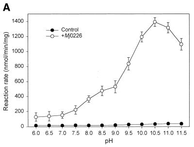
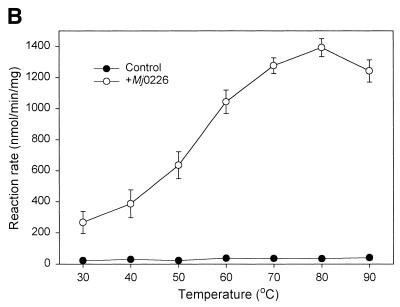
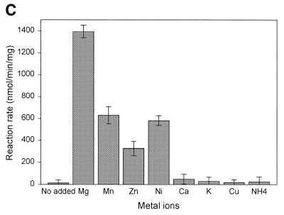
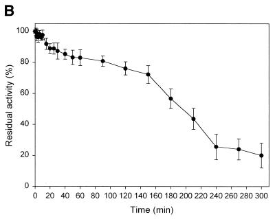
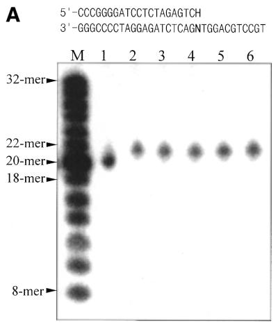
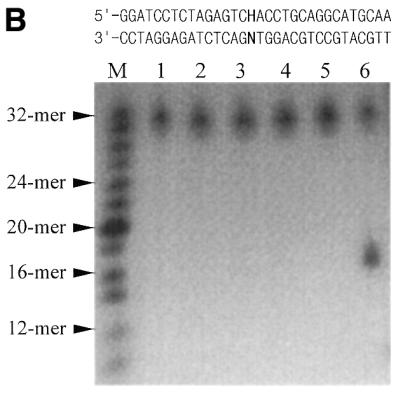
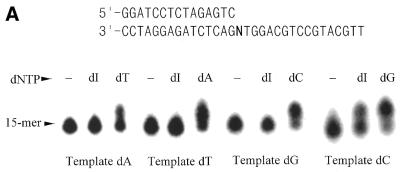
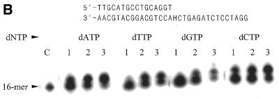
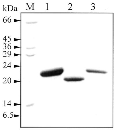
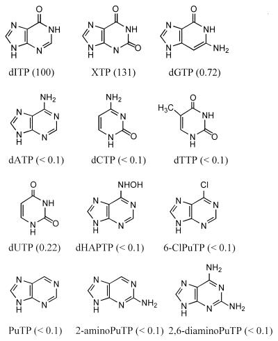
A novel dNTP pyrophosphatase, Mj0226 from Methanococcus jannaschii, which catalyzes the hydrolysis of nucleoside triphosphates to the monophosphate and PPi, has been characterized. Mj0226 protein catalyzes hydrolysis of two major substrates, dITP and XTP, suggesting that the 6-keto group of hypoxanthine and xanthine is critical for interaction with the protein. Under optimal reaction conditions the k(ca)(t) /K(m) value for these substrates was approximately 10 000 times that with dATP. Neither endonuclease nor 3'-exonuclease activities were detected in this protein. Interestingly, dITP was efficiently inserted opposite a dC residue in a DNA template and four dNTPs were also incorporated opposite a hypoxanthine residue in template DNA by DNA polymerase I. Two protein homologs of Mj0226 from Escherichia coli and Archaeoglobus fulgidus were also cloned and purified. These have catalytic activities similar to Mj0226 protein under optimal conditions. The implications of these results have significance in understanding how homologous proteins, including Mj0226, act biologically in many organisms. It seems likely that Mj0226 and its homologs have a major role in preventing mutations caused by incorporation of dITP and XTP formed spontaneously in the nucleotide pool into DNA. This report is the first identification and functional characterization of an enzyme hydrolyzing non-canonical nucleotides, dITP and XTP.


Figures

Figure 1
Figure 1
Optimal conditions for the nucleotide hydrolysis activities of Mj0226 protein. (A) The activity assay in various pH buffers was performed at 80°C. The following buffers were used: MES buffer, pH 6.0–6.5; HEPES buffer, pH 7.0–7.5; Tris–Cl buffer, pH 8.0–8.5; CHES buffer, pH 9.0–9.5; and CAPS buffer, pH 10.0–11.5. (B) The activity assay was performed at various temperatures in CAPS buffer, pH 10.5. (C) The effect of metal ions was assayed at 80°C with each metal ion (4 mM). Control, reaction without Mj0226 protein; +Mj0226, reaction with Mj0226 protein.
Figure 1
Figure 1
Optimal conditions for the nucleotide hydrolysis activities of Mj0226 protein. (A) The activity assay in various pH buffers was performed at 80°C. The following buffers were used: MES buffer, pH 6.0–6.5; HEPES buffer, pH 7.0–7.5; Tris–Cl buffer, pH 8.0–8.5; CHES buffer, pH 9.0–9.5; and CAPS buffer, pH 10.0–11.5. (B) The activity assay was performed at various temperatures in CAPS buffer, pH 10.5. (C) The effect of metal ions was assayed at 80°C with each metal ion (4 mM). Control, reaction without Mj0226 protein; +Mj0226, reaction with Mj0226 protein.
Figure 2
Figure 2
Thermal denaturation and thermostability of Mj0226 protein. (A) Thermal denaturation using CD. Measurements were performed in pH 7.5 buffer containing 20 mM potassium phosphate and 0.1 mg/ml Mj0226 protein at 222 nm. Thermal melting curves were measured at 1°C intervals with an averaging time of 60 s at each temperature and the change in ellipticity was monitored. The raw residue molar ellipticity values were transformed into the fraction of protein unfolded. (B) Thermostability of Mj0226 protein. Mj0226 protein (0.1 mg/ml) was incubated in a pH 7.5 buffer solution containing 20 mM potassium phosphate and 300 mM NaCl at 95°C. Aliquots were withdrawn at periodic intervals and kept on ice and then nucleotide hydrolysis assay was carried out by HPLC. The relative activity is presented as the residual activity of the enzyme compared to the activity of the unheated enzyme control.
Figure 2
Figure 2
Thermal denaturation and thermostability of Mj0226 protein. (A) Thermal denaturation using CD. Measurements were performed in pH 7.5 buffer containing 20 mM potassium phosphate and 0.1 mg/ml Mj0226 protein at 222 nm. Thermal melting curves were measured at 1°C intervals with an averaging time of 60 s at each temperature and the change in ellipticity was monitored. The raw residue molar ellipticity values were transformed into the fraction of protein unfolded. (B) Thermostability of Mj0226 protein. Mj0226 protein (0.1 mg/ml) was incubated in a pH 7.5 buffer solution containing 20 mM potassium phosphate and 300 mM NaCl at 95°C. Aliquots were withdrawn at periodic intervals and kept on ice and then nucleotide hydrolysis assay was carried out by HPLC. The relative activity is presented as the residual activity of the enzyme compared to the activity of the unheated enzyme control.
Figure 3
Figure 3
Exonuclease and endonuclease activity assay of Mj0226 protein. (A) 3′-Exonuclease activity assay of Mj0226. Mj0226 protein was incubated with 100 pmol 5′-labeled HX-containing duplex (top sequences) for 30 min. The products were analyzed by denaturing PAGE using a BAS2000 image analyzer. Lane M, size marker; lane 1, HX:C incubated with Klenow fragment; lane 2, HX:C only; lanes 3–6, HX:A, T, G and C, respectively, incubated with Mj0226. (B) Endonuclease activity assay of Mj0226. Mj0226 protein was incubated with 100 pmol 5′-labeled HX-containing duplex (top sequences) for 30 min. The products were analyzed by denaturing PAGE using a BAS2000 image analyzer. Lane M, size marker; lane 1, HX:T only; lanes 2–5, HX:A, T, G and C, respectively, incubated with Mj0226; lane 6, HX:T incubated with E.coli endonuclease V.
Figure 3
Figure 3
Exonuclease and endonuclease activity assay of Mj0226 protein. (A) 3′-Exonuclease activity assay of Mj0226. Mj0226 protein was incubated with 100 pmol 5′-labeled HX-containing duplex (top sequences) for 30 min. The products were analyzed by denaturing PAGE using a BAS2000 image analyzer. Lane M, size marker; lane 1, HX:C incubated with Klenow fragment; lane 2, HX:C only; lanes 3–6, HX:A, T, G and C, respectively, incubated with Mj0226. (B) Endonuclease activity assay of Mj0226. Mj0226 protein was incubated with 100 pmol 5′-labeled HX-containing duplex (top sequences) for 30 min. The products were analyzed by denaturing PAGE using a BAS2000 image analyzer. Lane M, size marker; lane 1, HX:T only; lanes 2–5, HX:A, T, G and C, respectively, incubated with Mj0226; lane 6, HX:T incubated with E.coli endonuclease V.
Figure 4
Figure 4
DNA replication fidelity of DNA polymerase I. (A) dITP incorporation opposite dN residues of template DNA by DNA polymerase I. 5′-End labeling of 15mer primer DNA annealed to template DNA (top sequences) was used. dITP incorporation into the dN template was with 1 U DNA polymerase (exonuclease) and 100 µM dITP in 20 µl of reaction mixture at 37°C for 15 min. Reaction mixtures were separated by denaturing PAGE and autoradiograms were obtained using a BAS2000 image analyzer. (B) dNTP incorporation opposite HX residues of template DNA by DNA polymerase I. 5′-End-labeling of 16mer primer DNA annealed to template DNAs (top sequences) was used in this experiment. dNTP incorporation into the HX template was with 1 U Klenow fragment and 1–100 µM dNTP in 20 µl of reaction mixture at 37°C for 15 min. Lane C, control; lane 1, 1 µM dNTP; lane 2, 10 µM dNTP; lane 3, 100 µM dNTP.
Figure 4
Figure 4
DNA replication fidelity of DNA polymerase I. (A) dITP incorporation opposite dN residues of template DNA by DNA polymerase I. 5′-End labeling of 15mer primer DNA annealed to template DNA (top sequences) was used. dITP incorporation into the dN template was with 1 U DNA polymerase (exonuclease) and 100 µM dITP in 20 µl of reaction mixture at 37°C for 15 min. Reaction mixtures were separated by denaturing PAGE and autoradiograms were obtained using a BAS2000 image analyzer. (B) dNTP incorporation opposite HX residues of template DNA by DNA polymerase I. 5′-End-labeling of 16mer primer DNA annealed to template DNAs (top sequences) was used in this experiment. dNTP incorporation into the HX template was with 1 U Klenow fragment and 1–100 µM dNTP in 20 µl of reaction mixture at 37°C for 15 min. Lane C, control; lane 1, 1 µM dNTP; lane 2, 10 µM dNTP; lane 3, 100 µM dNTP.
Figure 5
Figure 5
Purification of homologs of Mj0226. Two homologs of Mj0226 from E.coli and A.fulgidus were cloned and purified as described in Materials and Methods. Lane M, molecular weight marker; lane 1, Mj0226 from M.jannaschii; lane 2, Af2237 from A.fulgidus; lane 3, Ec197 from E.coli.
Figure 6
Figure 6
Comparison of base structure of nucleoside triphosphates tested as substrates for Mj0226. The values in parentheses (%) are activity of Mj0226 with the substrates relative to that of dITP. PuTP, purine triphosphate.

Similar articles

Cited by

References

    1. Friedberg E.C., Walker,G.C. and Siede,W. (1995) DNA Repair and Mutagenesis. ASM Press, Washington, DC.
    1. Shapiro R. and Pohl,S.H. (1968) The reaction of ribonucleosides with nitrous acid. Side products and kinetics. Biochemistry, 7, 448–455. - PubMed
    1. Lindahl T. (1979) DNA glycosylases, endonucleases for apurinic/apyrimidinic sites and base excision-repair. Prog. Nucleic Acid Res. Mol. Biol., 22, 135–192. - PubMed
    1. Schuster H. (1960) The reaction of nitrous acid with deoxyribonucleic acid. Biochem. Biophys. Res. Commun., 2, 320–323.
    1. Hill-Perkins M., Jones,M.D. and Karran,P. (1986) Site-specific mutagenesis in vivo by single methylated or deaminated purine bases. Mutat. Res., 162, 153–163. - PubMed

Publication types

MeSH terms