. 2001 Jul;103(3):319-31.
doi: 10.1046/j.1365-2567.2001.01238.x.

Endocytosis and recycling of the complex between CD23 and HLA-DR in human B cells

Affiliations

Endocytosis and recycling of the complex between CD23 and HLA-DR in human B cells

S N Karagiannis et al. Immunology. 2001 Jul.

Abstract

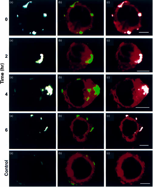
The presentation of extremely low doses of antigen to T cells is enhanced by immunoglobulin E (IgE)-dependent antigen focusing to CD23, the low-affinity receptor for IgE, expressed on activated B cells. CD23 contains a C-type lectin domain in its extracellular sequence and a targeting signal for coated pits, required for endocytosis, in its cytoplasmic sequence. CD23 is non-covalently associated with the major histocompatibility complex class II antigen, human leucocyte antigen HLA-DR, on the surface of human B cells, but the fate of this complex following endocytosis is unknown. To answer this question we have labelled these proteins on the surface of RPMI 8866 B cells and traced their route through the cytoplasm. Endocytosis mediated by anti-CD23 antibodies (BU38 and MHM6) led to the loss of CD23 from the cells. Endocytosis mediated by an antibody to HLA-DR (CR3/43) or an antigen-IgE complex (NP-BSA-anti-NP IgE), however, led to recycling of the HLA-DR-CD23 complex to the cell surface on a time scale (3-6 hr) consistent with the recycling of HLA-DR in antigen presentation. Along the latter pathway CD23 label was observed in cytoplasmic organelles that resembled the 'compartments for peptide loading' or 'class II vesicles' described by previous authors. Two features of the recycling process may contribute to the efficiency of antigen presentation. Peptide exchange may be facilitated by the proximity of HLA-DR and antigen in peptide loading compartments of the endosomal network. The return of CD23 with HLA-DR to the cell surface may then help to stabilize specific B-cell-T-cell interactions, contributing to T-cell activation.


Figures

Figure 1
Figure 1
Flow cytometric comparison of the induction of endocytosis in RPMI 8866 cells by various anti-CD23 antibodies. The percentage of internalized CD23 with the various antibodies are indicated after 3 and 5 hr. Each measurement was the average of at least two experiments. Errors were typically ± 5%.
Figure 2
Figure 2
Confocal optical sections revealing the location of CD23 in RPMI 8866 cells, incubated with the anti-CD23 antibody, BU38. Samples were incubated with mouse anti-human antibody BU38, followed by goat anti-mouse IgG (Fab′)2-FITC (green). Cells were labelled with propidium iodide (red), a nuclear stain, here used for the purposes of highlighting the cell structure. The thickness of optical sections was ∼1 µm. A sample was kept in ice for 8 hr (a), while others were incubated at 37° for (b) 2 hr, (c) 4 hr, (d) 6 hr and (e) 8 hr, to reveal capping (a, b) and internalization (b–e) of CD23. A control (f) shows the lack of non-specific surface labelling in cells incubated with the goat anti-mouse IgG (Fab′)2-FITC but without BU38, and kept on ice for 8 hr. Furthermore, negative controls using mouse anti-human antibodies to proteins not expressed by RPMI 8866 cells such as LFA-1 mouse anti-human antibody to CD21 (Serotec, UK) followed by anti-mouse IgG (Fab′)2-FITC showed lack of non-specific binding to cells (not shown).
Figure 3
Figure 3
Electron microscopic visualization of CD23 domains, on antibody-induced endocytosis in RPMI 8866 cells. RPMI 8866 cells were incubated with the anti-CD23 lectin domain MHM6, followed by anti-mouse IgG, conjugated to 5 nm gold (a,b). Samples were incubated at 4° (a) and at 37° for 10 hr (b). Gold is accumulated within plasmalemmal pits (a, small arrows) and multivesicular cytoplasmic bodies, located in the region of the nucleus after 10 hr (b, small arrows). In another experiment, cells were incubated with the anti-CD23 stalk antibody, EBVCS1, followed by anti-mouse IgG Fab, conjugated to 5 nm gold to label the stalk, and with biotinylated BU38 followed by anti-biotin mAb, conjugated with 10 nm gold, to label the lectin domain (c–e). Cells were incubated at 37° for 3·5 hr (c,d) and 10 hr (e). Cell membrane indentations are seen after 3·5 hr (c,d small arrows), in close proximity to early endosomes (c, large arrow). Early vesicles contain 5 (d small arrowhead) and 10 (d, large arrowhead) nm gold. Following 10 hr at 37° (e) gold is accumulated mostly in aggregates (e, long thin arrows), within multivescicular cytoplasmic bodies (e, small arrows). The 5 nm gold particles representing the stalk domain (e, small arrowheads) are more prevalent than the 10 nm gold particles representing the lectin (e, large arrowheads). Cell sections in (a), (c) and (d) were labelled with uranyl acetate and lead citrate. Cell sections in (b) and (e) were unstained to confirm the presence of gold in the vesicles. C, cytoplasm; N, nucleus; G, golgi apparatus; PM, plasma membrane: scale bars=250 nm.
Figure 4
Figure 4
Co-internalization of HLA-DR and CD23, observed by flow cytometry of acid-washed RPMI 8866 cells. Cells were incubated with CD23 antibody, IOB8, and anti-mouse IgG Fab-R-PE, followed by anti-HLA-DR antibody CR3/43-FITC. Dot plots are divided into four quadrants, defined by the acid-washed controls (b). Sample incubated on ice (a,b) and at 37° for 3 hr (c,d) or 6 hr (e,f).
Figure 5
Figure 5
Three-colour confocal optical section displaying CD23 and HLA-DR in RPMI 8866 cells. Cells were incubated with the mouse anti-human CD23 antibody, EBVCS1, and anti-mouse IgG (Fab′)2-Cy5, followed by anti-HLA-DR CR3/43-FITC. DiBAC4(5) was used to highlight surface and cytoplasmic membranes but does not label the cell nuclei. Optical section thickness: ∼1 µm. Cells were kept on ice or incubated at 37° for 2, 4, or 6 hr. A negative control (c, marking the bottom row), kept on ice, contained the secondary antibody anti-mouse IgG (Fab′)2-Cy5. Vertical panels represent (a) HLA-DR (green) and CD23 (white), (b) HLA-DR (green) and cytoplasmic membranes (red) and (c) CD23 (white) and surface and cytoplamic membranes (red).
Figure 6
Figure 6
Electron microscopic visualization of anti-HLA-DR endocytosis of CD23. RPMI 8866 cells were labelled with IOB8 and goat anti-mouse IgG Fab, conjugated to 5 nm gold, followed by the anti-HLA-DR antibody CR3/43. Sections in (a), (b), (c), (e) and (f) were stained with uranyl acetate and lead citrate to enhance vesicle structure. The unstained section in (d) was used to confirm the presence of aggregated gold in the vesicles. (a) Sample kept on ice: small arrows point to surface molecules and to plasmalemmal pits. Samples incubated at 37° for 3·5 hr (b,c) or 10 hr (d–f). (b) Gold in plasmalemmal pits (small arrow) and an early endosome (large arrow); (c) gold-filled vesicle (large arrow) budding from plasmalemmal pit (small arrow); (d) aggregated gold particles (long thin arrows) in multivesicular cytoplasmic bodies, located in groups within the cell cytoplasm; (e,f) groups of multivesicular bodies containing aggregated gold (arrowheads). Some vesicles have a coiled rope motif (large arrowheads) and others do not (small arrowheads). C, cytoplasm; PM, plasma membrane: scale bars=250 nm.
Figure 7
Figure 7
Endocytosis induced by an antigen–IgE complex in RPMI 8866 cells. Cells were incubated with EBVCS1 and anti-mouse IgG (Fab′)2-FITC, followed by NP-IgE and NP-BSA. Propidium iodide was used to label nuclei and highlight the cell structure. Cells were kept on ice (a) or incubated at 37° for 2 hr (b), 4 hr (c), or 6 hr (d). Controls included a sample incubated with IgE without NP-BSA (e) and NP-BSA without IgE (f) that were incubated at 37° for 2 hr. Optical section thickness ∼ 1 µm.
Figure 8
Figure 8
Electrophoretic analysis of intracellular and extracellular 125I-labelled CD23 from iodinated RPMI 8866 cells following endocytosis induced by anti-HLA DR. Cells were incubated with or without CR3/43 antibody for 18 hr at 37°. (a) Lane 1, CD23 from untreated control cells; lane 2, cells treated with CR3/43; lane 3, untreated control cells after acid washing; lane 4, cells treated with CR3/43 and subjected to acid washing. (b) Lane 1, CD23 from untreated control cells; lane 2, cells treated with CR3/43 antibody; lane 3, untreated control cells after chymotrypsin treatment; lane 4, cells incubated with CR3/43 after chymotrypsin treatment.

Similar articles

Cited by

References

    1. Kikutani H, Suemura M, Owaki H, et al. Fc epsilon receptor, a specific differentiation marker transiently expressed on mature B cells before isotype switching. J Exp Med. 1986;164:1455–69. - PMC - PubMed
    1. Yokota A, Kikutani H, Tanaka T, Sato R, Barsumian EL, Suemura M, Kishimoto T. Two species of human Fcε receptor II (FcεRII/CD23): tissue-specific and IL-4 specific regulation of gene expression. Cell. 1988;55:611–18. - PubMed
    1. Yokota A, Yukawa K, Yamamoto A, Sugiyama K, Suemura M, Tashiro Y, Kishimoto T, Kikutani H. Two forms of the low-affinity Fc receptor for the IgE differentially mediate endocytosis and phagocytosis: Identification of the critical cytoplasmic domains. Proc Natl Acad Sci USA. 1992;89:5030–4. - PMC - PubMed
    1. Beavil AJ, Edmeades RL, Gould HJ, Sutton BJ. α-helical coiled-coil stalks in the low-affinity receptor for IgE (FcεRII/CD23) and related C-type lectins. Proc Natl Acad Sci USA. 1992;809:753–7. - PMC - PubMed
    1. Beavil RL, Graber P, Aubonney N, Bonnefoy J-Y, Gould HJ. CD23/FcεRII and its soluble fragments can form oligomers on the cell surface and in solution. Immunology. 1995;84:202–6. - PMC - PubMed

Publication types

MeSH terms