

Intralesional isotype profiles in human localized cutaneous leishmaniasis lesions
- PMID: 11454104
- PMCID: PMC2517705
- DOI: 10.1111/j.1365-2613.2001.iep0082-0129-x
Intralesional isotype profiles in human localized cutaneous leishmaniasis lesions
Abstract
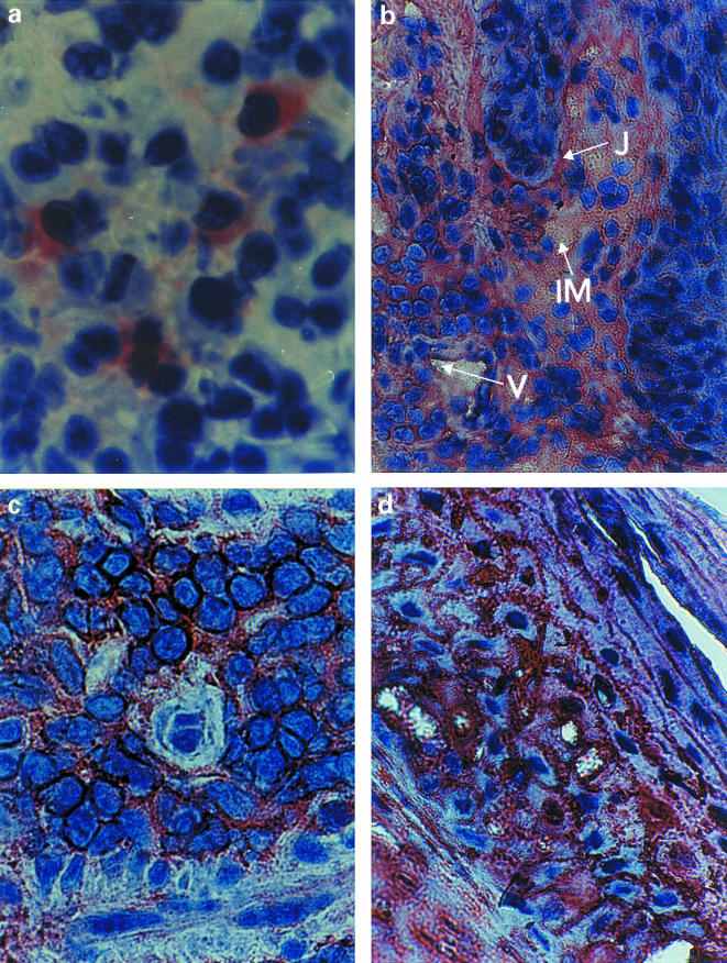
This immunocytochemical study evaluates the presence of IgG1-4, IgA and IgE immunoglobulins in active lesions of 25 localized cutaneous leishmaniasis patients from three bioclimatic areas (Awa, Afa and Bsha) in Mérida State, Venezuela. All immunoglobulin isotypes except IgE were detected, with variable intensity, in one or more of the epidermal or dermal components of skin lesions. IgG1 and IgG2 were detected significantly more frequently than IgG3, IgG4 and IgA. The ranking of the isotypes according to frequency of detection was the same in all areas: IgG1 = IgG2 > IgG3 = IgG4 = IgA, but considered as whole, all isotypes were detected significantly more frequently in patients from the Awa area than in those from the Bsha area. The predominant expression of isotypes IgG1 and IgG2 suggests a preferential Th1 like immune response. Anti-Leishmania immunoserum stained only parasites and their debris, suggesting that most of the immunostaining was nonspecific.
Figures

Similar articles
-
Antibody isotype responses to Schistosoma japonicum antigens in subjects from a schistosomiasis area with repeated praziquantel chemotherapy compared with a new endemic zone in Hunan Province, P.R. China.Trans R Soc Trop Med Hyg. 2002 Mar-Apr;96(2):210-5. doi: 10.1016/s0035-9203(02)90310-x. Trans R Soc Trop Med Hyg. 2002. PMID: 12055818 Clinical Trial.
-
[Study on immune status of patients with schistosomiasis japonica in Poyang Lake region I characterization of antigen-specific antibody isotype responses to Schistosoma japonicum].Zhongguo Xue Xi Chong Bing Fang Zhi Za Zhi. 2012 Oct;24(5):510-3, 521. Zhongguo Xue Xi Chong Bing Fang Zhi Za Zhi. 2012. PMID: 23373252 Chinese.
-
[Immunoglobulin isotype and IgG subclass profiles in American tegumentary leishmaniasis].Rev Soc Bras Med Trop. 2005 Mar-Apr;38(2):137-41. doi: 10.1590/s0037-86822005000200002. Epub 2005 Mar 30. Rev Soc Bras Med Trop. 2005. PMID: 15821787 Portuguese.
-
IgE and IgG subclass regulation by IL-4 and IFN-gamma in human helminth infections. Assessment by B cell precursor frequencies.J Immunol. 1993 Jul 1;151(1):458-65. J Immunol. 1993. PMID: 8326137
-
Case of pemphigus with immunoglobulin G and A antibodies, binding to both the intercellular spaces and basement membrane zone.J Dermatol. 2016 Feb;43(2):194-6. doi: 10.1111/1346-8138.13041. Epub 2015 Jul 29. J Dermatol. 2016. PMID: 26219735
Cited by
-
Identification of novel Leishmania major antigens that elicit IgG2a response in resistant and susceptible mice.Korean J Parasitol. 2006 Mar;44(1):43-8. doi: 10.3347/kjp.2006.44.1.43. Korean J Parasitol. 2006. PMID: 16514281 Free PMC article.
References
-
- Boenisch T. Staining Methods. In: Naish SJ, editor. Handbook of Immunochemicals Staining Methods. Carpinteria: CA. Dako Corporation; 1989. p. 13.
-
- Carvalho EM, Johnson WD, Barreto E, et al. Cell mediated immunity in American cutaneous and mucosal leishmaniasis. J. Immunol. 1985;135:4144–4148. - PubMed
-
- Castés M, Agnelli A, Verde O, Rondón AJ. Characterization of the cellular immune response in American cutaneous leishmaniasis. Clin. Immunol. Immunopath. 1983;27:176–186. - PubMed
-
- Finkelman FD, Holmes J, Katona IM, et al. Lymphokine control of in vivo immunoglobulin isotype selection. Ann. Rev. Immunol. 1990;8:303–333. - PubMed
Publication types
MeSH terms
Substances
LinkOut - more resources
Full Text Sources
Miscellaneous
