. 2001 Aug;49(2):176-81.
doi: 10.1136/gut.49.2.176.

Keratinocyte growth factor and coeliac disease

Affiliations

Keratinocyte growth factor and coeliac disease

V M Salvati et al. Gut. 2001 Aug.

Abstract

Background: Coeliac disease is characterised by increased epithelial renewal associated with a mucosal T cell response to gliadin. Keratinocyte growth factor (KGF) is produced by cytokine activated gut stromal cells and may be a link between mucosal T cell activation in untreated coeliac disease and epithelial hyperplasia.

Aims: To characterise expression of KGF in coeliac disease.

Methods: KGF transcripts in coeliac disease were measured by quantitative competitive reverse transcription-polymerase chain reaction (RT-PCR) and localised using in situ hybridisation. KGF production by gluten reactive CD4+ T cell clones was examined. In addition, KGF transcripts were measured following ex vivo challenge of coeliac biopsies with a peptic-tryptic digest of gliadin.

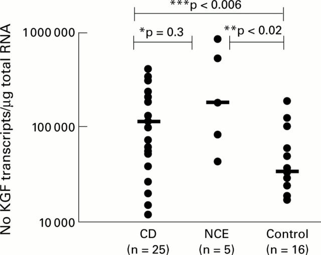
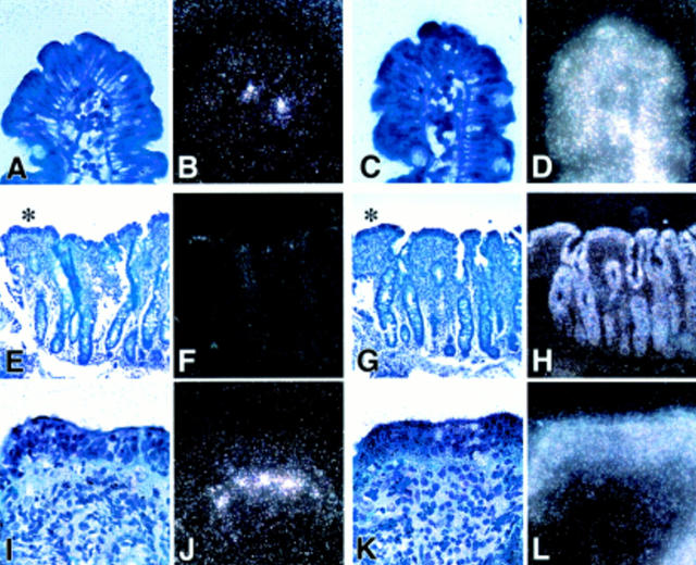
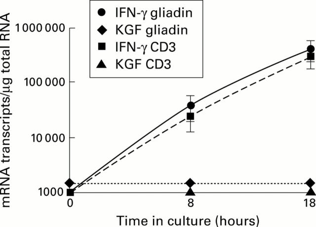
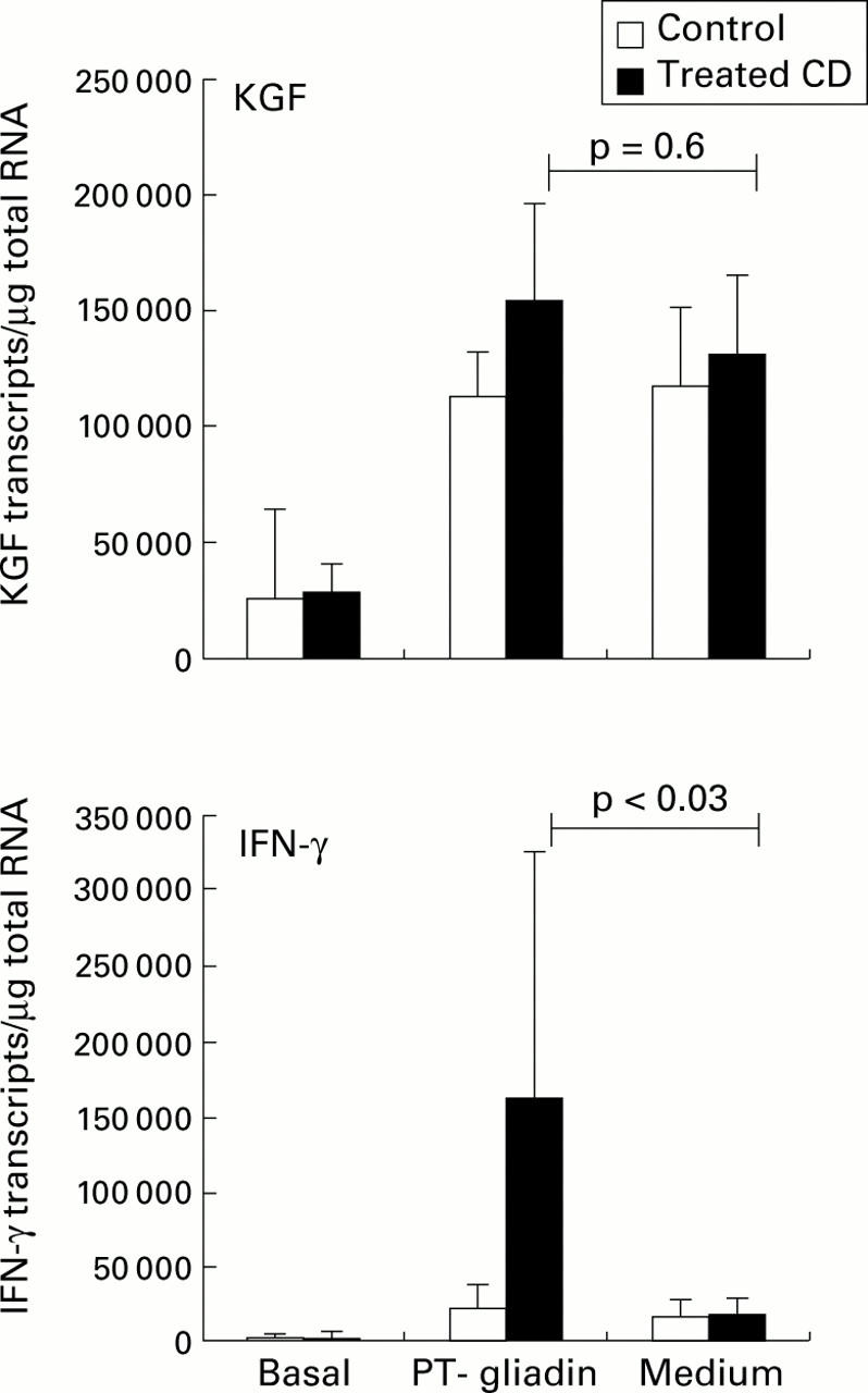
Results: KGF transcripts were elevated in coeliac biopsies compared with normal controls but were not different from non-coeliac disease controls. By in situ hybridisation, KGF mRNA containing cells were present in the upper half of the lamina propria, most abundantly just under the epithelium. There was no signal from cells within the epithelium. Gluten reactive T cell clones did not make KGF. In vitro challenge of coeliac biopsies generated a strong interferon gamma response but a specific KGF response could not be detected because of an extremely high number of KGF transcripts in all cultured biopsies.

Conclusions: KGF is overexpressed in coeliac biopsies and in tissues with non-coeliac enteropathy. No evidence was found for KGF production by intraepithelial lymphocytes or lamina propria T cells.


Figures

Figure 1
Figure 1
Keratinocyte growth factor (KGF) mRNA transcripts in whole small intestinal biopsies from patients with coeliac disease (CD), non-coeliac enteropathy (NCE), and normal controls. Bars represent median KGF mRNA transcripts. ***CD versus controls, p<0.006; **NCE versus controls, p<0.02; *CD versus NCE, p=0.3
Figure 2
Figure 2
Detection of keratinocyte growth factor (KGF) mRNA by in situ hybridisation. (A, B) and (C, D) represent light/dark images of a control section hybridised with antisense probes to KGF mRNA (A, B) and to β-actin (C, D) as a positive control at a magnification of 50×. Very few KGF transcripts were detectable in control sections below the tip of the villi. (E) and (F) show light and dark field images, respectively, at 20× magnification stained for KGF. In (I) and (J) at 50× magnification the flat villi indicated by an asterisk in (E) are shown in more detail. KGF positive hybridising cells are distributed in the subepithelial region of the lamina propria below the tip of the flattened villi. (G, H) (20×) and (K, L) (50×) represent light (G, K) and dark field (H, L) images of the corresponding β-actin staining as a positive control.
Figure 3
Figure 3
Number of keratinocyte growth factor (KGF) and interferon γ (IFN-γ) mRNA transcripts in four gut derived gluten reactive CD4+ T cell clones and lines challenged in vitro with a peptic-tryptic digest of gliadin or anti-CD3 antibodies for eight and 18 hours. Each point at the indicated time represents mean (SEM) values. KGF mRNA expression was below the detection limit (1000 transcripts/µg total RNA) while IFN-γ mRNA transcripts were increased after eight and 18 hours in the presence of gliadin or anti-CD3 antibodies (positive control).
Figure 4
Figure 4
Keratinocyte growth factor (KGF) and interferon γ (IFN-γ) mRNA transcripts measured by competitive quantitative RT-PCR in treated coeliac disease (CD) (n=6) and control biopsies (n=8) challenged in vitro with gliadin. Small intestinal biopsies were cultured for eight hours in the presence or absence of a peptic-tryptic (PT) digest of gliadin (1 mg/ml). Bars represent median (SEM). IFN-γ mRNA transcripts were markedly induced by gliadin in coeliac mucosa compared with biopsies cultured in medium alone (p<0.03). KGF mRNA transcripts were increased in both coeliacs and controls, in the presence and absence of gliadin.

Similar articles

Cited by

References

    1. J Clin Invest. 1995 Jan;95(1):55-65 - PubMed
    1. Science. 1994 Nov 18;266(5188):1253-5 - PubMed
    1. Am J Pathol. 1996 Aug;149(2):521-9 - PubMed
    1. Gut. 1996 Dec;39(6):787-94 - PubMed
    1. Am J Pathol. 1997 Nov;151(5):1469-76 - PubMed

Publication types

MeSH terms