. 2001 Aug;49(2):190-8.
doi: 10.1136/gut.49.2.190.

Loss of transforming growth factor beta signalling in the intestine contributes to tissue injury in inflammatory bowel disease

Affiliations

Loss of transforming growth factor beta signalling in the intestine contributes to tissue injury in inflammatory bowel disease

K B Hahm et al. Gut. 2001 Aug.

Abstract

Background: Inflammatory bowel disease (IBD) is a chronic inflammation of the gastrointestinal tract caused by an abnormal and uncontrolled immune response to one or more normally occurring gut constituents.

Aim: Given the effects of transforming growth factor beta1 (TGF-beta1) on both the immune system and extracellular matrix, we postulated that alterations in TGF-beta signalling in intestinal epithelial cells may play an important role in the development of IBD.

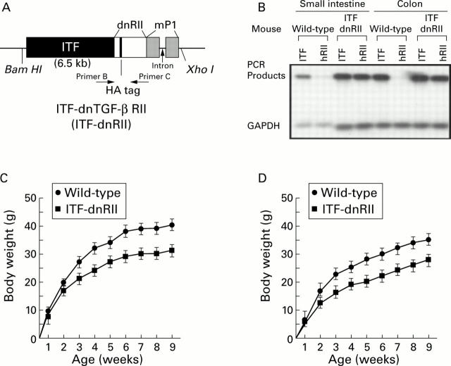
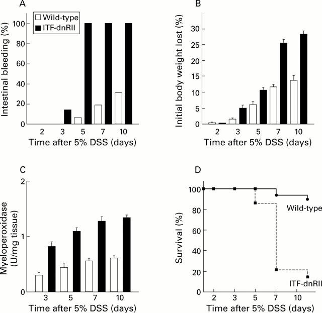
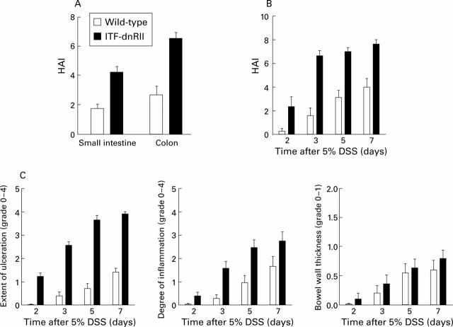
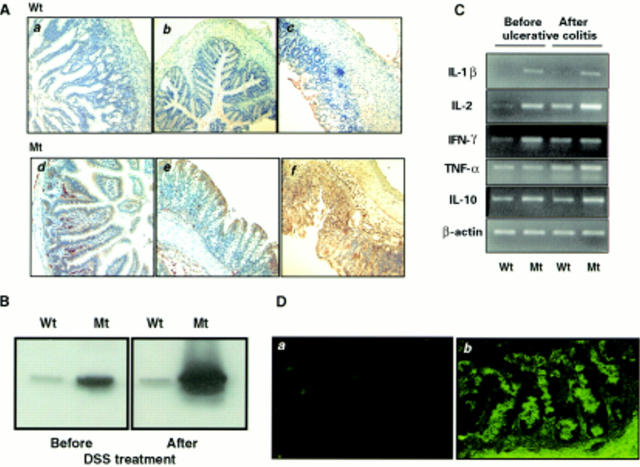
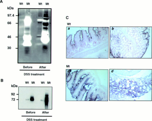
Methods: TGF-beta signalling was inactivated in mouse intestine by expressing a dominant negative mutant form of the TGF-beta type II receptor under the control of the mouse intestinal trefoil peptide (ITF)/TFF3 promoter. Transgenic mice (ITF-dnRII) developed spontaneous colitis presenting with diarrhoea, haematochezia, and anal prolapse when not maintained under specific pathogen free (SPF) conditions. Under SPF conditions we induced colitis by mixing dextran sodium sulphate (DSS) in drinking water to examine the significance of loss of TGF-beta signalling in the pathogenesis of IBD.

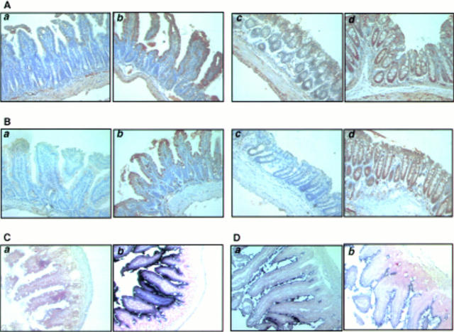
Results: Transgenic mice showed increased susceptibility to DSS induced IBD, and elicited increased expression of major histocompatibility complex class II, generation of autoantibodies against intestinal goblet cells, and increased activity of matrix metalloproteinase in intestinal epithelial cells compared with wild-type littermates challenged with DSS.

Conclusions: Deficiency of TGF-beta signalling specifically in the intestine contributes to the development of IBD. Maintenance of TGF-beta signalling may be important in regulating immune homeostasis in the intestine


Figures

Figure 1
Figure 1
Generation of dominant negative mutant transforming growth factor β receptor II (TGF-β RII) mice—ITF-TGF-β dnRII transgenic mice. (A) Schematic representation of the transgene. The mouse intestinal trefoil factor (ITF) fragment spans −6500 bp to +35 bp. The 0.6 kb human TGF-β RII fragment spans +322 bp to + 911 bp and contains a haemagglutinin (HA) tag sequence and a segment of the mouse protamine that provides an intron and a polyadenylation site. Transgenic mice were generated using inbred FVB/N zygotes. Of the 15 mice born, six were positive for ITF-dnRII, bred into lines, and designated ITF1 to ITF6. Pups from ITF1, ITF2, and ITF3 were used for the current experiments. (B) TGF-β dnRII expression. Using total RNA from the intestines of wild-type mice and ITF-dnRII, reverse transcription-polymerase chain reaction was performed using the specific primer sets for human TGF-β RII, mouse ITF, and mouse GAPDH. (C, D) Body weights of wild-type littermates and ITF-dnRII transgenic mice. Body weight was measured every week for nine weeks in males (C) and females (D).
Figure 2
Figure 2
Tissue distribution of transforming growth factor β receptor II (TGF-β RII), intestinal trefoil factor (ITF), and dnRII. (A) Immunohistochemical staining of TGF-β RII using anti-TGF-β RII antibody and (B) TGF-β dnRII using anti-haemagglutinin (HA) tag antibody in the small intestine of wild-type littermates (a), small intestine of ITF-dnRII mice (b), colon of wild-type littermates (c), and colon of ITF-dnRII transgenic mice (d). Slightly increased expression of TGF-β type II was observed in the intestine of ITF-dnRII mice compared with wild-type littermates. Because we inserted the HA sequence in transgene construct, the HA tag was identified only in the intestine of ITF-dnRII mice by immunohistochemical staining with HA antibody; none was stained in the intestine of wild-type littermates. (C) In situ hybridisation was performed for the presence of human TGF-β RII mRNA using a biotinylated TGF-β RII probe (206 bp) in the colon of wild-type littermates (a) and the colon of ITF-dnRII transgenic mice (b). (D) In situ hybridisation of mouse ITF mRNA. Expression pattern of mouse ITF gene was similarly expressed in transgenic mice (a) and wild-type littermates (b) (×200 magnification)
Figure 3
Figure 3
Dextran sodium sulphate (DSS) administration resulted in an increased incidence of intestinal bleeding, weight loss, colitis, and death in ITF-dnRII transgenic mice. (A) Prevalence of haematochezia. Intestinal bleeding (% incidence) was more common in ITF-dnRII mice. Earlier and more severe anal bleeding developed in mice lacking TGF-β signalling after administration of DSS (p<0.001). (B) Changes in body weight. Weight loss (expressed as per cent of initial body weight) was more prominent in ITF-dnRII mice than in wild-type mice. (C) Mucosal myeloperoxidase (MPO) activity. Changes in mucosal MPO activity in the colon of wild-type littermates and ITF-dnRII mice. ITF-dnRII mice showed significantly higher levels of mucosal MPO than wild-type mice (p<0.01). (D) Survival. Survival was significantly decreased in ITF-dnRII mice compared wild-type littermates using Kaplan-Meyer analysis. (ITF-dnRII transgenic mice were from ITF1-3 lines.)
Figure 4
Figure 4
ITF-dnRII transgenic mice showed increased pathological scores Score criteria for extent of ulceration are as follows: 0, no architectural change; 1, focal superficial ulceration; 2, diffuse superficial ulceration; 3, focal gland dropout or focal deep ulceration; 4, extensive glandular dropout or deep ulceration. Scores for inflammation are as follows: 0, no increased inflammatory infiltrates; 1, focal mild inflammation; 2, diffuse mild inflammation; 3, cryptic abscess formation; 4, diffuse dense inflammation. Presence of oedema: 0, no significant oedema; 1, broad zone of oedema. Based on these criteria, comparison of histological activity index (HAI) is shown according to site of inflammation (A), days after 5% dextran sodium sulphate (DSS) administration (B), and criteria of each score (C). The extent of mucosal ulceration was significantly different between transgenic mice and wild-type littermates (p<0.001). (ITF-dnRII transgenic mice were from ITF1-3 lines.)
Figure 5
Figure 5
Increased expression of major histocompatibility complex (MHC) class II, cytokines, and autoantibodies in the intestine of ITF-dnRII transgenic mice. (A) Immunohistochemical staining of MHC class II. Scant MHC class II expression was observed in epithelial cells of the small intestine (a) and colon (b) of wild-type (Wt) FVB/N mice but increased expression of MHC class II was observed in epithelial cells of the small intestine (d) and colon (e) of ITF-dnRII transgenic mice (Mt). After induction of inflammatory bowel disease (IBD), MHC class II antigen expression was more markedly increased in transgenic mice (f) compared with wild-type mice (c) (×100 magnification). (B) Western blotting of MHC class II. Western blotting showed similar findings, with ITF-dnRII mice (Mt) showing increased expression of MHC class II antigens in intestine compared with wild-type littermates (Wt) (DSS, dextran sodium sulphate). (C) Changes in cytokines. Interleukin (IL)-1β, IL-2, interferon γ (IFN-γ), and IL-10 were all expressed at higher levels in the colons of transgenic mice (Mt) compared with those of wild-type mice (Wt). Expression of tumour necrosis factor α (TNF-α) was increased after induction of ulcerative colitis. (D) Autoantibodies in serum of ITF-dnRII transgenic mice. Autoantibodies were identified by indirect immunofluorescence using monkey ileum sections and FITC coupled rat antimouse immunoglobulin polyclonal antibody. Autoantibodies were detected in the serum of ITF-dnRII mice (b) whereas none was seen after applying sera obtained from wild-type littermates (a). Positive sera containing IgG autoantibodies showed blurry, drop-like staining (×200 magnification).
Figure 6
Figure 6
Matrix metalloproteinase (MMP) expression in mucosal homogenates of wild-type and transgenic mice. (A) Even before induction of ulcerative colitis, colon extracts of transgenic mice showed increased MMP gelatinolytic bands (52, 72, and 92 kDa) which become more prominent after induction of ulcerative colitis. Even applying only one tenth of the amount of protein compared with wild-type (Wt) littermates onto gelatin zymography gel, MMP activity was significantly increased in ITF-dnRII (Mt) mice. (B) MMP expression was confirmed by western blot analysis using a mixture of MMP-2, MMP-3, and MMP-9 antibodies. (C) In situ hybridisation of MMP-9 showed increased mRNA expression in small intestine (c) and colon (d) of ITF-dnRII transgenic mice (Mt) compared with wild-type littermates (Wt) (a, b) (×200 magnification).

Comment in

  • Signals on the immune tract.
    Neurath MF. Neurath MF. Gut. 2001 Aug;49(2):164-5. doi: 10.1136/gut.49.2.164. Gut. 2001. PMID: 11454787 Free PMC article. No abstract available.

References

    1. Hum Cell. 1996 Sep;9(3):229-36 - PubMed
    1. J Gastroenterol. 1996 Dec;31(6):907-16 - PubMed
    1. Gastroenterology. 1997 Sep;113(3):825-32 - PubMed
    1. Am J Physiol. 1997 Oct;273(4 Pt 1):G769-75 - PubMed
    1. Gastroenterology. 1997 Dec;113(6):1828-35 - PubMed

MeSH terms