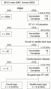
Prevalence of asymptomatic ST segment elevation in right precordial leads with right bundle branch block (Brugada-type ST shift) among the general Japanese population
- PMID: 11454832
- PMCID: PMC1729874
- DOI: 10.1136/heart.86.2.161
Prevalence of asymptomatic ST segment elevation in right precordial leads with right bundle branch block (Brugada-type ST shift) among the general Japanese population
Abstract
Objective: To examine the modality and morbidity of asymptomatic ST segment elevation in leads V1 to V3 with right bundle branch block (Brugada-type ST shift).
Methods: 8612 Japanese subjects (5987 men and 2625 women, mean age 49.2 years) who underwent a health check up in 1997 were investigated. Those with Brugada-type ST shift underwent the following further examinations over a two year period after the initial check up: ECG, echocardiogram, 24 hour Holter monitoring, treadmill exercise testing, signal averaged ECG, and slow kinetic sodium channel blocker loading test (cibenzoline, 1.4 mg/kg).
Results: Asymptomatic Brugada-type ST shift was found in 12 of 8612 (0.14%) subjects. Eleven of these 12 subjects were followed up. Follow up ECG exhibited persistent Brugada-type ST shift in seven of 11 (63.6%) subjects. ST shift was transformed from a saddle back to a coved type in three subjects. None of the subjects had morphological abnormalities or abnormal tachyarrhythmias. Positive late potentials were found in seven of 11 (63.6%) subjects. Augmentation of ST shift was shown by both submaximal exercise and drug administration in one of the 11 subjects (9.1%).
Conclusions: Asymptomatic subjects with Brugada-type ST shift were not unusual, at a rate of 0.14% in the general Japanese population. Almost all of the subjects had some abnormalities in non-invasive secondary examinations. Additional and prospective studies are needed to confirm the clinical significance and the prognosis of asymptomatic Brugada-type ST shift.
Figures


Similar articles
-
[Doubts of the cardiologist regarding an electrocardiogram presenting QRS V1-V2 complexes with positive terminal wave and ST segment elevation. Consensus Conference promoted by the Italian Cardiology Society].G Ital Cardiol (Rome). 2010 Nov;11(11 Suppl 2):3S-22S. G Ital Cardiol (Rome). 2010. PMID: 21361048 Italian.
-
Prevalence and clinical course of the juveniles with Brugada-type ECG in Japanese population.Pacing Clin Electrophysiol. 2005 Jun;28(6):549-54. doi: 10.1111/j.1540-8159.2005.40020.x. Pacing Clin Electrophysiol. 2005. PMID: 15955188
-
Three-year follow-up of patients with right bundle branch block and ST segment elevation in the right precordial leads: Japanese Registry of Brugada Syndrome. Idiopathic Ventricular Fibrillation Investigators.J Am Coll Cardiol. 2001 Jun 1;37(7):1916-20. doi: 10.1016/s0735-1097(01)01239-6. J Am Coll Cardiol. 2001. PMID: 11401132
-
The prevalence, incidence and prognostic value of the Brugada-type electrocardiogram: a population-based study of four decades.J Am Coll Cardiol. 2001 Sep;38(3):765-70. doi: 10.1016/s0735-1097(01)01421-8. J Am Coll Cardiol. 2001. PMID: 11527630
-
Right bundle branch block, intermittent ST segment elevation and inducible ventricular tachycardia in an asymptomatic patient: an unusual presentation of the Brugada syndrome?G Ital Cardiol. 1998 Aug;28(8):893-8. G Ital Cardiol. 1998. PMID: 9773315 Review.
Cited by
-
The 10-Year Prognosis and Prevalence of Brugada-Type Electrocardiograms in Elderly Women: A Longitudinal Nationwide Community-Based Prospective Study.J Cardiovasc Nurs. 2020 Nov/Dec;35(6):E25-E32. doi: 10.1097/JCN.0000000000000722. J Cardiovasc Nurs. 2020. PMID: 32609463 Free PMC article.
-
Recent advances in the treatment of Brugada syndrome.Expert Rev Cardiovasc Ther. 2018 Jun;16(6):387-404. doi: 10.1080/14779072.2018.1475230. Epub 2018 May 18. Expert Rev Cardiovasc Ther. 2018. PMID: 29757020 Free PMC article. Review.
-
Long-Term Prognosis of Brugada-Type ECG and ECG With Atypical ST-Segment Elevation in the Right Precordial Leads Over 20 Years: Results From the Circulatory Risk in Communities Study (CIRCS).J Am Heart Assoc. 2016 Aug 8;5(8):e002899. doi: 10.1161/JAHA.115.002899. J Am Heart Assoc. 2016. PMID: 27503848 Free PMC article.
-
Prevalence and associated factors of ECG abnormality patterns indicative of cardiac channelopathies among adult general population of Tehran, Iran: a report from the Tehran Cohort Study (TeCS).BMC Cardiovasc Disord. 2024 Oct 17;24(1):566. doi: 10.1186/s12872-024-04235-w. BMC Cardiovasc Disord. 2024. PMID: 39415094 Free PMC article.
-
Brugada syndrome.Int J Cardiol. 2005 May 25;101(2):173-8. doi: 10.1016/j.ijcard.2004.03.068. Int J Cardiol. 2005. PMID: 15882659 Free PMC article. Review.
References
MeSH terms
LinkOut - more resources
Full Text Sources
