. 2001 Jul 15;534(Pt. 2):381-93.
doi: 10.1111/j.1469-7793.2001.00381.x.

Mechanism of Ba(2+) block of a mouse inwardly rectifying K+ channel: differential contribution by two discrete residues

Affiliations

Mechanism of Ba(2+) block of a mouse inwardly rectifying K+ channel: differential contribution by two discrete residues

N Alagem et al. J Physiol. .

Abstract

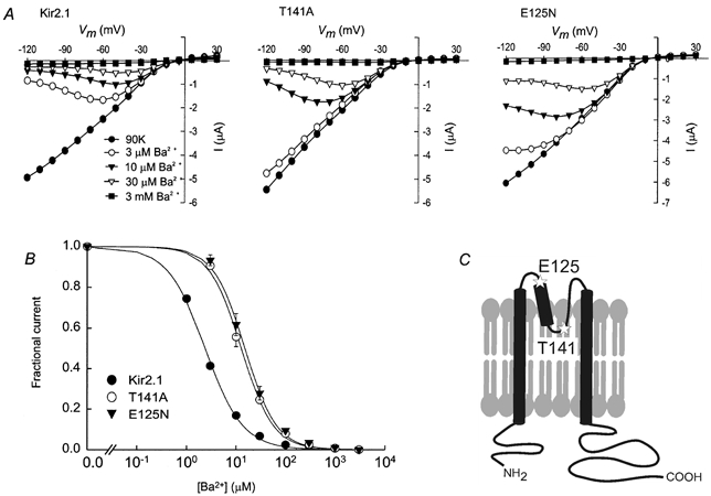
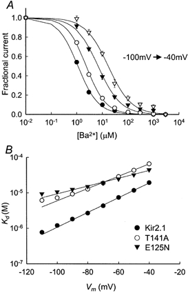
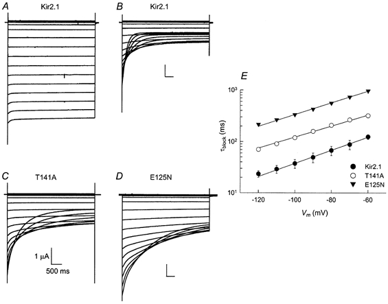
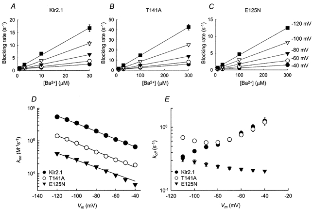
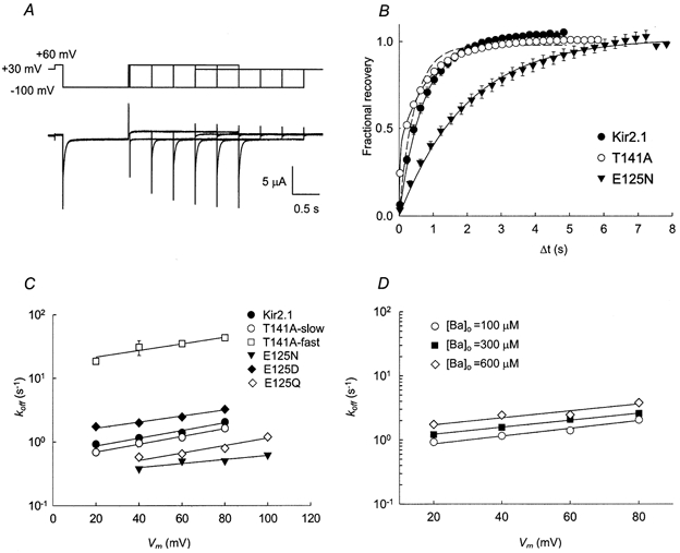
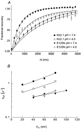
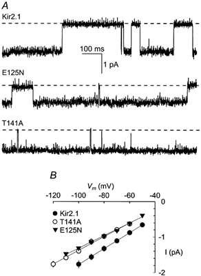
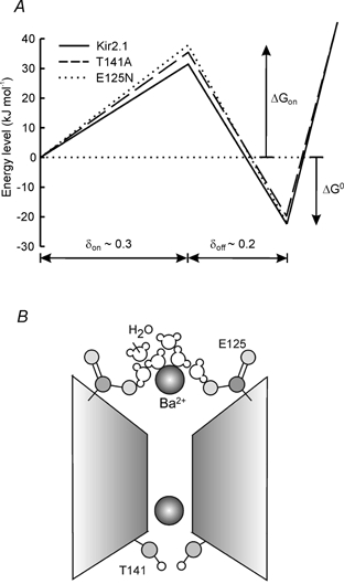
1. The block of the IRK1/Kir2.1 inwardly rectifying K+ channel by a Ba(2+) ion is highly voltage dependent, where the ion binds approximately half-way within the membrane electrical field. The mechanism by which two distinct mutations, E125N and T141A, affect Ba(2+) block of Kir2.1 was investigated using heterologous expression in Xenopus oocytes. 2. Analysis of the blocking kinetics showed that E125 and T141 affect the entry and binding of Ba(2+) to the channel, respectively. Replacing the glutamate at position 125 with an asparagine greatly decreased the rate at which the Ba(2+) ions enter and leave the pore. In contrast, replacing the polar threonine at position 141 with an alanine affected the entry rate of the Ba(2+) ions while leaving the exit rate unchanged. 3. Acidification of the extracellular solution slowed the exit rate of the Ba(2+) from the wild-type channel, but had no such effect on the Kir2.1(E125N) mutant. 4. These results thus reveal two unique roles for the amino acids at positions 125 and 141 in aiding the interaction of Ba(2+) with the channel. Their possible roles in K+ permeation are discussed.


Figures

Figure 1
Figure 1. Steady-state Ba2+ block of Kir2.1 and the two mutants Kir2.1(T141A) and Kir2.1(E125N)
A, typical steady-state current vs. voltage plots measured for one representative oocyte in the presence of 90K with varying concentrations of extracellular Ba2+ for Kir2.1 (left), Kir2.1(T141A) (centre) and Kir2.1(E125N) (right). B, fractional current vs. Ba2+ concentration at steady state, at a holding potential of −80 mV. The smooth lines are fits to the Hill equation (eqn (2)). C, a simplified two-dimensional cartoon showing the approximate positions of E125 and T141 (stars) in the Kir2.1 channel.
Figure 2
Figure 2. Voltage dependence of the steady-state Ba2+ block
A, Ba2+ concentration dependence of the steady-state block of Kir2.1 at different holding potentials (−100 to −40 mV, indicated by different symbols) in 20 mV increments. The data points at -80 mV are the same as in Fig. 1B. The smooth lines are fits to the Hill equation (eqn (2)). B, voltage dependence of Kd. The smooth lines are fits to eqn (3).
Figure 3
Figure 3. Time dependence of Ba2+ block of the Kir2.1 channel and the Kir2.1(T141A) and Kir2.1(E125N) mutants
A–D, current traces elicited by voltage steps ranging from +30 to −120 mV in 10 mV decrements. Each family of traces was recorded from a representative oocyte expressing Kir2.1, Kir2.1(E125N) or Kir2.1(T141A) channels. The bath solution contained 90K (a) or 90K and 10 μm Ba2+ (B–D). The Kir2.1 currents shown in A and B were recorded from the same oocyte. E, voltage dependence of the blocking time constant. The data points were obtained in the presence of 100 μm Ba2+.
Figure 4
Figure 4. Voltage dependence of Ba2+ blocking kinetics
A–C, the blocking rate (the reciprocal of the blocking time constant, τblock) as a function of the extracellular Ba2+ concentration at various membrane potentials. The continuous lines are linear regressions. D, voltage dependence of the blocking rate constants (kon). Data points are the slopes of the linear regression as shown in A–C. The error bars are the standard deviation of the fit (where error bars are not visible, they are smaller than the symbols). Continuous lines are fits to eqn (6). E, voltage dependence of the unblocking rate constant (koff). Data points were obtained by multiplying Kd by kon for each voltage.
Figure 5
Figure 5. Recovery from Ba2+ block at positive potentials
A, illustration of the double pulse protocol used to measure recovery from block at +60 mV (top) and the associated Kir2.1 current traces (bottom). The current traces are superimposed. Channel block was initiated by a hyperpolarizing pulse to −100 mV for 1.2 s. B, time course of current recovery at +60 mV. In the case of Kir2.1 and Kir2.1(E125N) channels, the continuous lines are fits to eqn (8). In the case of Kir2.1(T141A), the data were fitted using eqn (8) (dashed line) and eqn (9) (continuous line). C, voltage dependence of the unblocking rate. The continuous lines are fits using eqn (10). D, recovery of Kir2.1 from the block at various external Ba2+ concentrations. The data points at [Ba2+]o= 100 μm are the same as in C.
Figure 6
Figure 6. Effect of lowering the extracellular pH on the unblocking rates in Kir2.1 and Kir2.1(E125N) at positive potentials
A, time course for current recovery at +60 mV in external solutions at either pH 7.4 (filled symbols) or pH 4.0 (open symbols) for the Kir2.1 (squares) and Kir2.1(E125N) (triangles) channels. The continuous lines are fits to eqn (8). B, voltage dependence of the unblocking rates. The data points at pH 7.4 are the same as in Fig. 5C. The continuous lines are fits to eqn (10).
Figure 7
Figure 7. Single-channel recordings of Kir2.1, Kir2.1(E125N) and Kir2.1(T141A) channels
A, single-channel currents of oocytes expressing Kir2.1 (top), Kir2.1(E125N) (middle) and Kir2.1(T141A) (bottom) channels. The currents were recorded from cell-attached patches at -80 mV. The dashed lines denote closed channel current levels. B, current vs. voltage plot of the single-channel current amplitudes. The continuous lines are linear regressions.
Figure 8
Figure 8. Energetic profile of Ba2+ blocking interaction with the Kir2.1 pore
A, energy profile for Ba2+ binding to the Kir2.1 channel and the two mutants Kir2.1(T141A) and Kir(E125N). The ΔGon values were calculated from the blocking rates at 0 mV using the Eyring rate theory model (see Discussion): 31.4 kJ mol−1 for Kir2.1 (continuous line), 35.6 kJ mol−1 for Kir2.1(T141A) (dashed line) and 37.7 kJ mol−1 for Kir2.1(E125N) (dotted line). The Δ values were calculated from the steady-state dissociation constants (Kd) at 0 mV (see Discussion): −22.6 kJ mol−1 for Kir2.1, −19.7 kJ mol−1 for Kir2.1(T141A) and −22.2 kJ mol−1 for Kir2.1(E125N). B, a cartoon model describing the role of E125 and T141 residues in barium block. The negatively charged E125 side-chain presumably participates in the interaction with the Ba2+ ion and its associated water molecules. The polar hydroxyl group (light grey) of T141 stabilizes the interaction of the blocking ion with its binding site.

Similar articles

Cited by

References

    1. Biermans G, Vereecke J, Carmeliet E. The mechanism of the inactivation of the inward-rectifying K current during hyperpolarizing steps in guinea-pig ventricular myocytes. Pflügers Archiv. 1987;410:604–613. - PubMed
    1. Carmeliet E, Mubagwa K. Characterization of the acetylcholine-induced potassium current in rabbit cardiac Purkinje fibres. Journal of Physiology. 1986;371:219–237. - PMC - PubMed
    1. Choe H, Sackin H, Palmer LG. Permeation properties of inward-rectifier potassium channels and their molecular determinants. Journal of General Physiology. 2000;115:391–404. - PMC - PubMed
    1. Dart C, Leyland ML, Spencer PJ, Stanfield PR, Sutcliffe MJ. The selectivity filter of a potassium channel, murine Kir2. 1, investigated using scanning cysteine mutagenesis. Journal of Physiology. 1998;511:25–32. - PMC - PubMed
    1. Doring F, Derst C, Wischmeyer E, Karschin C, Schneggenburger R, Daut J, Karschin A. The epithelial inward rectifier channel Kir7. 1 displays unusual K+ permeation properties. Journal of Neuroscience. 1998;18:8625–8636. - PMC - PubMed

Publication types

MeSH terms

LinkOut - more resources