

In vivo mechanisms of vascular endothelial growth factor-mediated increased hydraulic conductivity of Rana capillaries
- PMID: 11454965
- PMCID: PMC2278710
- DOI: 10.1111/j.1469-7793.2001.00479.x
In vivo mechanisms of vascular endothelial growth factor-mediated increased hydraulic conductivity of Rana capillaries
Abstract
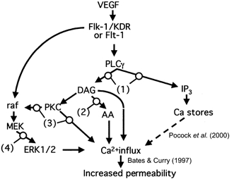
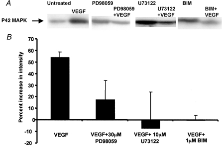
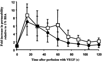
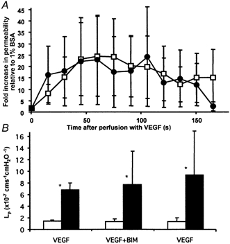
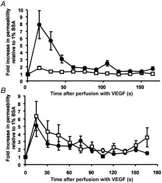
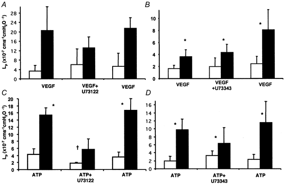
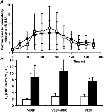
1. Vascular endothelial growth factor (VEGF) increases hydraulic conductivity (L(p)) in vivo. To determine the signal transduction cascade through which this is mediated, we measured the effect of inhibition of various signalling pathways on VEGF-mediated acute increases in L(p) in individually perfused frog mesenteric microvessels. 2. VEGF receptors have previously been shown to activate phospholipase C-gamma (PLCgamma), protein kinase C (PKC) and MEK, the mitogen-activated and extracellular signal-related kinase (ERK) kinase. To determine the role of these signalling pathways we measured the effects of inhibitors of each on the VEGF-mediated increase in L(p). 3. VEGF-mediated increases in L(p) were attenuated by pre-treatment with the PLC inhibitor U73122, but not affected by treatment with the inactive enantiomer U73343. The PLC inhibitor was also able to attenuate the increase in L(p) mediated by the inflammatory mediator ATP. 4. Inhibition of either PKC or MEK activation using the selective inhibitors bisindolylmaleimide (BIM, 1 microM) and PD98059 (30 microM), respectively, did not change the VEGF-mediated increase in L(p). However, PD98059, BIM and U73122 all reduced phosphorylation of ERK1/2 determined by Western blot analysis with anti-phospho-ERK1/2 antibodies. 5. Furthermore, inhibition of the conversion of diacyl glycerol (DAG) to arachidonic acid, by perfusion with the DAG lipase inhibitor RHC80267 (50 microM), did not attenuate the increase in L(p) brought about by VEGF. 6. These data suggest that VEGF acutely increases microvascular permeability in vivo through a mechanism that is dependent on PLC stimulation, but is independent of PKC or MEK activation or production of arachidonic acid from DAG. We therefore propose that VEGF acutely acts to increase L(p) through the direct actions of DAG, independently of PKC or arachidonic acid.
Figures
Similar articles
-
Vascular endothelial growth factors and vascular permeability.Cardiovasc Res. 2010 Jul 15;87(2):262-71. doi: 10.1093/cvr/cvq105. Epub 2010 Apr 16. Cardiovasc Res. 2010. PMID: 20400620 Free PMC article. Review.
-
Vascular endothelial growth factor increases Rana vascular permeability and compliance by different signalling pathways.J Physiol. 2001 May 15;533(Pt 1):263-72. doi: 10.1111/j.1469-7793.2001.0263b.x. J Physiol. 2001. PMID: 11351033 Free PMC article.
-
Hyperosmolality induces activation of cPKC and nPKC, a requirement for ERK1/2 activation in NIH/3T3 cells.Am J Physiol Cell Physiol. 2000 Jan;278(1):C102-9. doi: 10.1152/ajpcell.2000.278.1.C102. Am J Physiol Cell Physiol. 2000. PMID: 10644517
-
Role of phospholipase C, protein kinase C, and calcium in VEGF-induced venular hyperpermeability.Am J Physiol. 1999 Feb;276(2):H535-42. doi: 10.1152/ajpheart.1999.276.2.H535. Am J Physiol. 1999. PMID: 9950855
-
Regulation of VEGF-induced endothelial cell PAF synthesis: role of p42/44 MAPK, p38 MAPK and PI3K pathways.Br J Pharmacol. 2001 Nov;134(6):1253-62. doi: 10.1038/sj.bjp.0704367. Br J Pharmacol. 2001. PMID: 11704645 Free PMC article.
Cited by
-
Endothelial progenitor cells in the host defense response.Pharmacol Ther. 2023 Jan;241:108315. doi: 10.1016/j.pharmthera.2022.108315. Epub 2022 Nov 24. Pharmacol Ther. 2023. PMID: 36436689 Free PMC article. Review.
-
Vascular endothelial growth factors and vascular permeability.Cardiovasc Res. 2010 Jul 15;87(2):262-71. doi: 10.1093/cvr/cvq105. Epub 2010 Apr 16. Cardiovasc Res. 2010. PMID: 20400620 Free PMC article. Review.
-
Effects of methyl p-hydroxybenzoate (methyl paraben) on Ca2+ concentration and histamine release in rat peritoneal mast cells.Br J Pharmacol. 2003 May;139(2):381-7. doi: 10.1038/sj.bjp.0705248. Br J Pharmacol. 2003. PMID: 12770943 Free PMC article.
-
Lipid Peroxidation Drives Gasdermin D-Mediated Pyroptosis in Lethal Polymicrobial Sepsis.Cell Host Microbe. 2018 Jul 11;24(1):97-108.e4. doi: 10.1016/j.chom.2018.05.009. Epub 2018 Jun 21. Cell Host Microbe. 2018. PMID: 29937272 Free PMC article.
-
Effect of antiangiogenic therapy on tumor growth, vasculature and kinase activity in basal- and luminal-like breast cancer xenografts.Mol Oncol. 2012 Aug;6(4):418-27. doi: 10.1016/j.molonc.2012.03.006. Epub 2012 Mar 31. Mol Oncol. 2012. PMID: 22521242 Free PMC article.
References
-
- Aiello LP, Bursell SE, Clermont A, Duh E, Ishii H, Takagi C, Mori F, Ciulla TA, Ways K, Jirousek M, Smith LE, King GL. Vascular endothelial growth factor-induced retinal permeability is mediated by protein kinase C in vivo and suppressed by an orally effective beta-isoform-selective inhibitor. Diabetes. 1997;46:1473–1480. - PubMed
-
- Albelda SM, Sampson PM, Haselton FR, McNiff JM, Mueller SN, Williams SK, Fishman AP, Levine EM. Permeability characteristics of cultured endothelial cell monolayers. Journal of Applied Physiology: Respiratory Environmental and Exercise Physiology. 1988;64:308–322. - PubMed
-
- Bates DO, Curry FE. Vascular endothelial growth factor increases hydraulic conductivity of isolated perfused microvessels. American Journal of Physiology. 1996;271:H2520–2528. - PubMed
-
- Bates DO, Curry FE. Vascular endothelial growth factor increases microvascular permeability via a Ca2+-dependent pathway. American Journal of Physiology. 1997;273:H687–694. - PubMed
Publication types
MeSH terms
Substances
Grants and funding
LinkOut - more resources
Full Text Sources
Miscellaneous