. 2001 Jul 15;534(Pt. 2):565-81.
doi: 10.1111/j.1469-7793.2001.t01-1-00565.x.

Medullary respiratory neurones and control of laryngeal motoneurones during fictive eupnoea and cough in the cat

Affiliations

Medullary respiratory neurones and control of laryngeal motoneurones during fictive eupnoea and cough in the cat

D M Baekey et al. J Physiol. .

Abstract

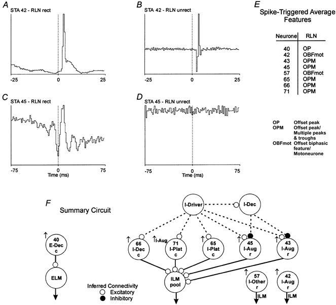
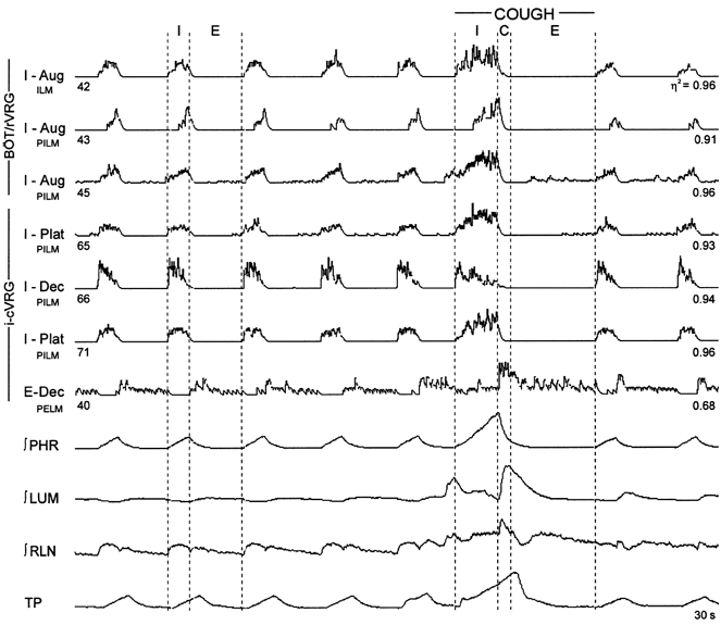
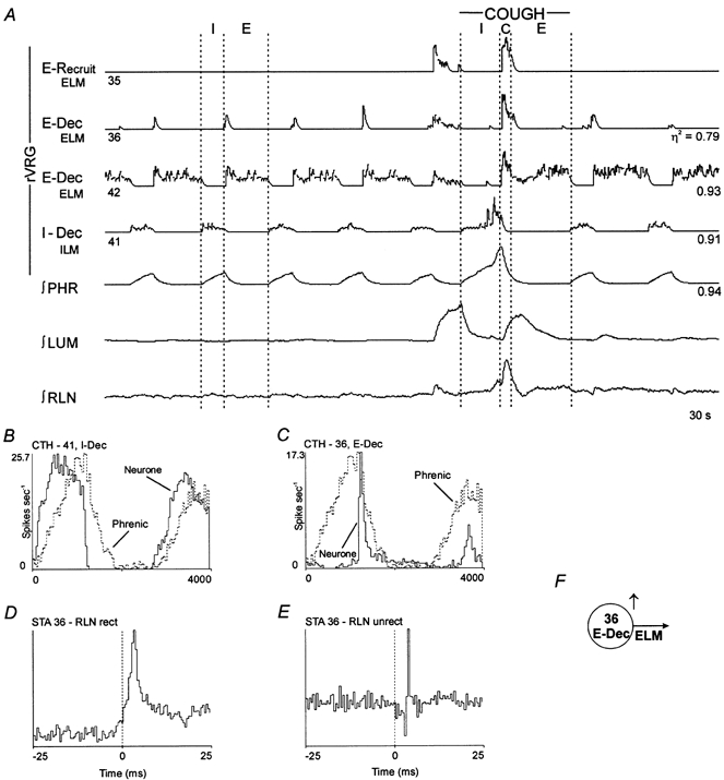
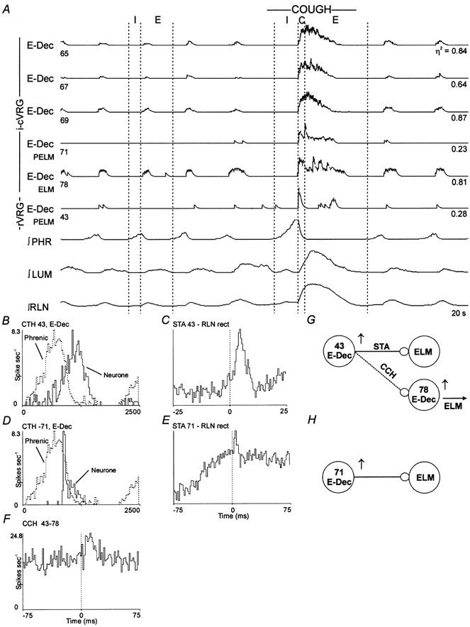
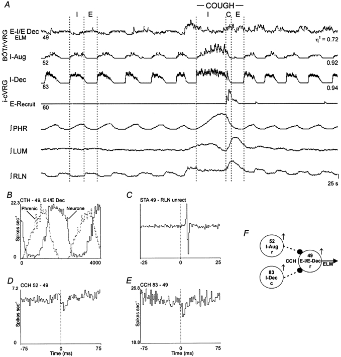
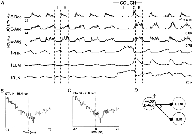
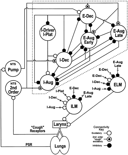
1. This study addressed the hypothesis that ventrolateral medullary respiratory neurones participate in the control of laryngeal motoneurones during both eupnoea and coughing. 2. Data were obtained from 28 mid-collicular decerebrated, artificially ventilated cats. Cough-like motor patterns (fictive cough) in phrenic, lumbar and recurrent laryngeal nerves were elicited by mechanical stimulation of the intrathoracic trachea. Microelectrode arrays were used to monitor simultaneously several neurones in the ventral respiratory group, including the Bötzinger and pre-Bötzinger complexes. Spike trains were evaluated for responses during fictive cough and evidence of functional connectivity with spike-triggered averages of efferent recurrent laryngeal nerve activity. 3. Primary features were observed in averages triggered by 94 of 332 (28 %) neurones. An offset biphasic wave with a positive time lag was present in the unrectified average for 10 inspiratory and 13 expiratory neurones. These trigger neurones were respectively identified as inspiratory laryngeal motoneurones with augmenting, decrementing, plateau and "other" discharge patterns, and expiratory laryngeal motoneurones with decrementing firing patterns. 4. Rectified averages triggered by inspiratory neurones included 37 offset peaks, 11 central peaks and one offset trough. Averages triggered by expiratory neurones had 12 offset peaks, six central peaks and four offset troughs. Relationships inferred from these features included premotor actions of inspiratory neurones with augmenting, decrementing, plateau and "other" patterns on inspiratory laryngeal motoneurones, and premotor actions of decrementing and "other" expiratory neurones on expiratory laryngeal motoneurones. Corresponding changes in neuronal firing patterns during fictive cough supported these inferences. 5. The data confirm and extend previous results on the control of laryngeal motoneurones during eupnoea and support the hypothesis that the same premotor neurones help to shape motoneurone firing patterns during both eupnoea and coughing.


Figures

Figure 1
Figure 1. Spike-triggered averages with features identifying inspiratory neurones as laryngeal motoneurones and putative premotor neurones
An offset biphasic feature in an unrectified (unrect) average of recurrent laryngeal nerve (RLN) efferent activity indicates the trigger neurone was a motoneurone (B). An offset peak (primary feature) in a full-wave rectified (rect) average (C) and a flat unrectified average (D) suggests the neurone was excitatory to motoneurones. A, spike-triggered average (STA); offset peak with a lag of 2.0 ms; half-width, 1.0 ms; number of trigger events, 16763. B, STA; offset biphasic feature. C, STA; offset peak with a lag of 3.0 ms; half-width, 4.5 ms; number of trigger events, 41235. D, STA; flat. E, spike-triggered average feature table summarizing primary features in spike-triggered average histograms in the data set. F, summary circuit showing inferred functional connections detected in the data set in Fig. 2. Changes in peak firing rate (↑, increase) and pattern (Aug, augmenting) during cough. c, intermediate-caudal VRG; r, rostral VRG, which contains the Bötzinger and pre-Bötzinger complexes; ELM, expiratory laryngeal motoneurone; ILM, inspiratory laryngeal motoneurone. Continuous lines represent inferred connectivity from the STA. Dotted lines are hypothesized connections.
Figure 2
Figure 2. Simultaneous responses of inspiratory laryngeal motoneurones and putative premotor inspiratory neurones during fictive cough
Integrated activities of six inspiratory and one expiratory neurone, phrenic (∫PHR), lumbar (∫LUM) and recurrent laryngeal (∫RLN) nerves, and tracheal pressure (TP). C, estimated neural correlate of the compressive phase; E, expiratory phase; I, inspiratory phase; BÖT/rVRG, rostral VRG, which contains the Bötzinger and pre-Bötzinger complexes; i-cVRG, intermediate-caudal VRG; η2, statistical measure of respiratory modulation; PELM, premotor to expiratory laryngeal motoneurones; ILM, inspiratory laryngeal motoneurone; PILM, premotor to inspiratory laryngeal motoneurones.
Figure 3
Figure 3. Laryngeal expiratory and inspiratory motoneurones
Spike-triggered averages and concurrent responses during the neural compressive phase of fictive cough were consistent with E-Recruit neurone 35 and E-Dec neurones 36 and 42 being motoneurones; note the similarity with the ∫RLN. The I-Dec cell is another example of an inspiratory motoneurone. A, simltaneous activity of expiratory and inspiratory laryngeal motoneurones. B and C, cycle-triggered histograms (CTHs); 100 cycles averaged. D, STA; offset peak with a lag of 1.5 ms; half-width, 1.5 ms; number of trigger events, 5291. E, STA; offset biphasic feature. F, model of inference from STA.
Figure 4
Figure 4. Responses during fictive cough and functional connectivity of a set of decrementing expiratory neurones
A, simultaneous activity of six E-Dec neurones. Data were consistent with E-Dec neurones 43 and 71 exciting expiratory motoneurones. B and D, CTHs; 100 cycles averaged. C, STA; offset peak with a lag of 2.0 ms; half-width, 4.0 ms; number of trigger events, 1771. E, STA; offset peak with a lag of 1.5 ms; half-width, 3.0 ms; number of trigger events, 885. F, cross-correlogram (CCH); offset peak with a lag of 7.5 ms; half-width, 10.0 ms; detectability index, 5.6; correlation strength, 0.58; 2424 reference and 10591 target spikes. G and H, summary models representing inferred connectivity.
Figure 5
Figure 5. Connectivity among inspiratory neurones and an expiratory motoneurone, and discharge patterns during fictive cough
A, discharge patterns of two inspiratory and two expiratory neurones. Results suggest I-Aug neurone 52 and I-Dec neurone 83 inhibit expiratory motoneurone 49. B, CTH; 100 cycles averaged. C, STA; offset biphasic feature with a lag of 3.0 ms; number of trigger events, 39081. D, CCH; offset trough with a lag of 6.0 ms; half-width, 3.0 ms; detectability index, 5.2; correlation strength, 0.27; 39098 reference and 38274 target spikes. E, CCH; offset trough with a lag of 6.0 ms; half-width 4.5 ms; detectability index, 3.7; correlation strength, 0.14; 18590 reference and 63998 target spikes. Correlogram was scaled-up to show significant primary trough by subtraction of 70 % of the counts in the minimum bin from each bin. F, summary circuit of inferred connectivity.
Figure 6
Figure 6. Changes in firing pattern during fictive cough and functional connectivity of augmenting expiratory neurones
A, discharge patterns of the three expiratory neurones. Results suggest E-Aug neurones 44 and 56 inhibited either inspiratory or expiratory laryngeal motoneurones. B, STA; offset trough with a lag of 12.0 ms; half-width, 3.0 ms; number of trigger events, 29626. C, STA; offset trough with a lag of 9.0 ms; half-width, 6.0 ms: number of trigger events, 19798. D, summary model representing possible inferred connectivity. Question mark indicates that the troughs in the averages could represent inhibition of ELM or ILM (see text for further explanation).
Figure 7
Figure 7. Network model for the control of laryngeal motoneurones
Schematic diagram of ventrolateral medullary respiratory neuronal network connections and hypothesized inputs from nucleus tractus solitarius (NTS) cough receptor second-order neurones and pulmonary stretch receptor (PSR) Pump cells. Neurone connections onto ILM and ELM arise from the ‘core’ network (enclosed by dashed-line box). E-Aug Early and E-Aug Late, neurones that begin discharging prior to and during the latter part of the expiratory phase, respectively. I-Driver, inspiratory neurone also active before the expiratory-inspiratory phase transition and with a relatively constant discharge rate throughout the inspiratory phase (I-Plat); definition specifically limited to BÖT/rVRG neurones with previously identified excitatory functional links to other inspiratory neurones (Balis et al. 1994). Other abbreviations are described in detail in the text. For a detailed description of the ‘core’ (excluding ILM and ELM) of the model, see Shannon et al. (1998, 2000). The connections with asterisks and question marks represent those inferred from the results of this study.

Similar articles

Cited by

References

    1. Balis UJ, Morris KF, Koleski J, Lindsey BG. Simulation of ventrolateral medullary neural network for respiratory rhythmogenesis inferred from spike train cross-correlation. Biological Cybernetics. 1994;70:311–327. - PubMed
    1. Barillot JC, Bianchi AL. Activité des motoneurones laryngés pendant le réflexe de Hering-Breuer. Journal de Physiologie. 1971;63:783–792. - PubMed
    1. Barillot JC, Bianchi AL, Gogan P. Laryngeal expiratory motoneurones: morphology and electrophysiological evidence of separate sites for excitatory and inhibitory synaptic events. Neuroscience Letters. 1984;47:107–112. - PubMed
    1. Barillot JC, Dussardier M. Activité des motoneurones laryngés expiratoires. Journal de Physiologie. 1976;72:311–343. - PubMed
    1. Barillot JC, Grélot L, Reddad S, Bianchi AL. Discharge patterns of laryngeal motoneurones in the cat: an intracellular study. Brain Research. 1990;509:99–106. - PubMed

Publication types

LinkOut - more resources