

Simvastatin induces regression of cardiac hypertrophy and fibrosis and improves cardiac function in a transgenic rabbit model of human hypertrophic cardiomyopathy
- PMID: 11457751
- PMCID: PMC2768618
- DOI: 10.1161/hc2801.094031
Simvastatin induces regression of cardiac hypertrophy and fibrosis and improves cardiac function in a transgenic rabbit model of human hypertrophic cardiomyopathy
Abstract
Background: Hypertrophic cardiomyopathy is a genetic disease characterized by cardiac hypertrophy, myocyte disarray, interstitial fibrosis, and left ventricular (LV) dysfunction. We have proposed that hypertrophy and fibrosis, the major determinants of mortality and morbidity, are potentially reversible. We tested this hypothesis in beta-myosin heavy chain-Q(403) transgenic rabbits.
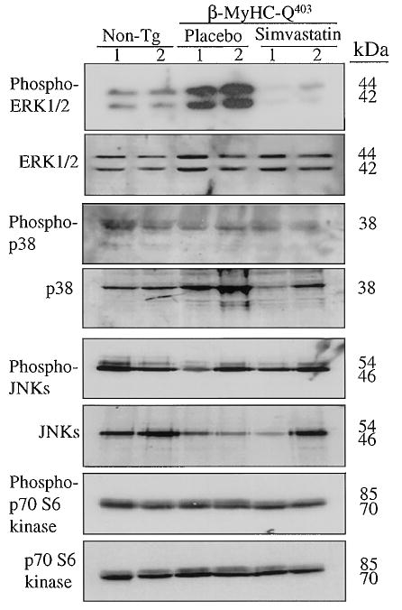
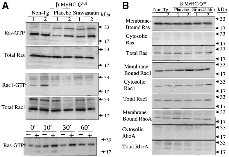
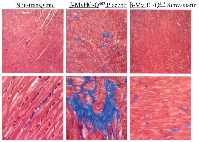
Methods and results: We randomized 24 beta-myosin heavy chain-Q(403) rabbits to treatment with either a placebo or simvastatin (5 mg. kg(-1). d(-1)) for 12 weeks and included 12 nontransgenic controls. We performed 2D and Doppler echocardiography and tissue Doppler imaging before and after treatment. Demographic data were similar among the groups. Baseline mean LV mass and interventricular septal thickness in nontransgenic, placebo, and simvastatin groups were 3.9+/-0.7, 6.2+/-2.0, and 7.5+/-2.1 g (P<0.001) and 2.2+/-0.2, 3.1+/-0.5, and 3.3+/-0.5 mm (P=0.002), respectively. Simvastatin reduced LV mass by 37%, interventricular septal thickness by 21%, and posterior wall thickness by 13%. Doppler indices of LV filling pressure were improved. Collagen volume fraction was reduced by 44% (P<0.001). Disarray was unchanged. Levels of activated extracellular signal-regulated kinase (ERK) 1/2 were increased in the placebo group and were less than normal in the simvastatin group. Levels of activated and total p38, Jun N-terminal kinase, p70S6 kinase, Ras, Rac, and RhoA and the membrane association of Ras, RhoA, and Rac1 were unchanged.
Conclusions: Simvastatin induced the regression of hypertrophy and fibrosis, improved cardiac function, and reduced ERK1/2 activity in the beta-myosin heavy chain-Q(403) rabbits. These findings highlight the need for clinical trials to determine the effects of simvastatin on cardiac hypertrophy, fibrosis, and dysfunction in humans with hypertrophic cardiomyopathy and heart failure.
Figures
References
-
- Heart and Stroke Statistical Update. Dallas, Tex: American Heart Association; 2001. [April 1, 2001]. Available at: http://www.americanheart.org/statistics/pdf/HSSTATS2001_1.0.pdf.
-
- Haider AW, Larson MG, Benjamin EJ, et al. Increased left ventricular mass and hypertrophy are associated with increased risk for sudden death. J Am Coll Cardiol. 1998;32:1454–1459. - PubMed
-
- Assayag P, Carre F, Chevalier B, et al. Compensated cardiac hypertrophy: arrhythmogenicity and the new myocardial phenotype, I: fibrosis. Cardiovasc Res. 1997;34:439–444. - PubMed
-
- Maron BJ, Shirani J, Poliac LC, et al. Sudden death in young competitive athletes: clinical, demographic, and pathological profiles. JAMA. 1996;276:199–204. - PubMed
Publication types
MeSH terms
Substances
Grants and funding
LinkOut - more resources
Full Text Sources
Other Literature Sources
Medical
Research Materials
Miscellaneous
