. 2001 Jul;108(2):189-201.
doi: 10.1172/JCI12694.

Progressive atrioventricular conduction defects and heart failure in mice expressing a mutant Csx/Nkx2.5 homeoprotein

Affiliations

Progressive atrioventricular conduction defects and heart failure in mice expressing a mutant Csx/Nkx2.5 homeoprotein

H Kasahara et al. J Clin Invest. 2001 Jul.

Abstract

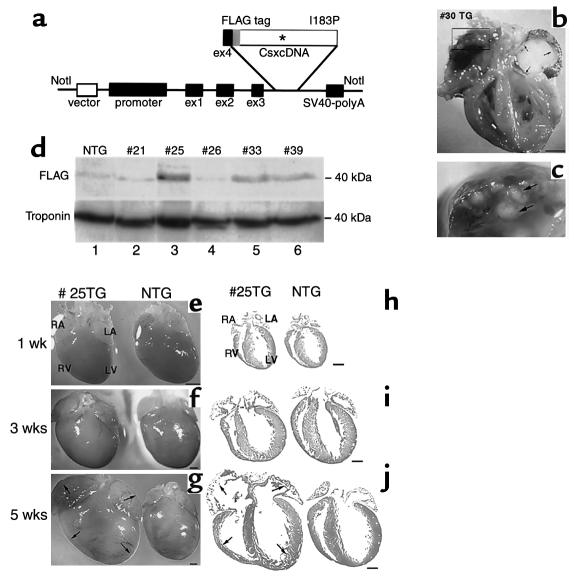
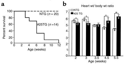
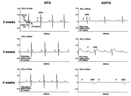
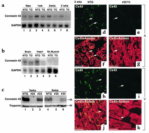
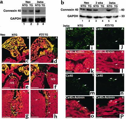
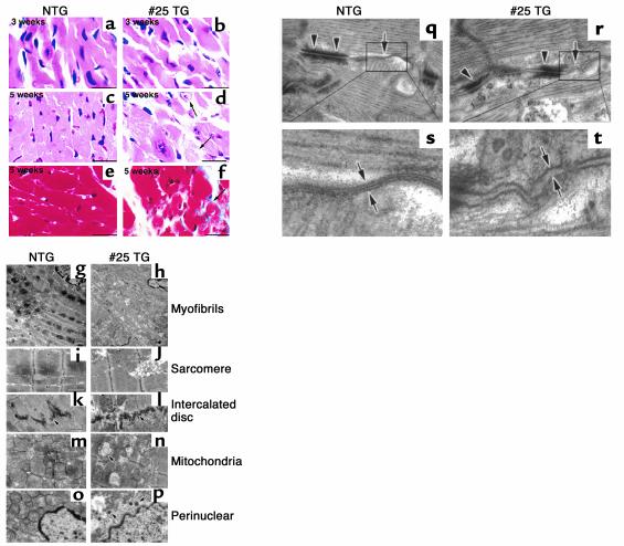
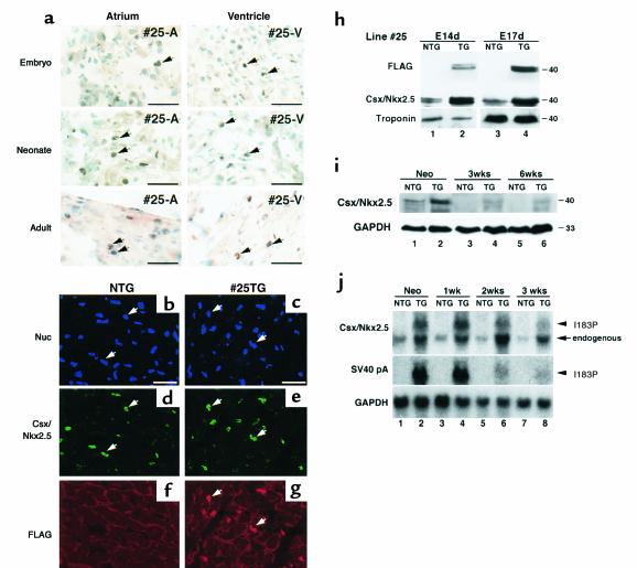
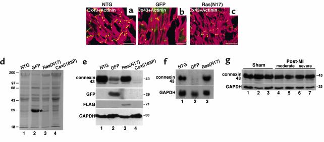
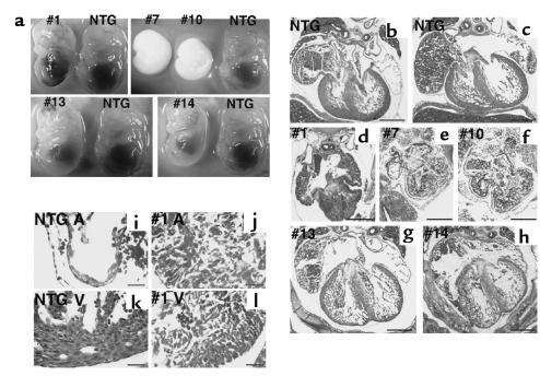
A DNA nonbinding mutant of the NK2 class homeoprotein Nkx2.5 dominantly inhibits cardiogenesis in Xenopus embryos, causing a small heart to develop or blocking heart formation entirely. Recently, ten heterozygous CSX/NKX2.5 homeoprotein mutations were identified in patients with congenital atrioventricular (AV) conduction defects. All four missense mutations identified in the human homeodomain led to markedly reduced DNA binding. To examine the effect of a DNA binding-impaired mutant of mouse Csx/Nkx2.5 in the embryonic heart, we generated transgenic mice expressing one such allele, I183P, under the beta-myosin heavy chain promoter. Unexpectedly, transgenic mice were born apparently normal, but the accumulation of Csx/Nkx2.5(I183P) mutant protein in the embryo, neonate, and adult myocardium resulted in progressive and profound cardiac conduction defects and heart failure. P-R prolongation observed at 2 weeks of age rapidly progressed into complete AV block as early as 4 weeks of age. Expression of connexins 40 and 43 was dramatically decreased in the transgenic heart, which may contribute to the conduction defects in the transgenic mice. This transgenic mouse model may be useful in the study of the pathogenesis of cardiac dysfunction associated with CSX/NKX2.5 mutations in humans.


Figures

Figure 1
Figure 1
Generation of Csx/Nkx2.5(I183P) TG mice driven by β-MHC promoter. (a) Schematic of TG vector showing inserted FLAG-tagged mouse wild-type and Csx/Nkx2.5(I183P) under the β-MHC promoter and 4 exons prior to the first coding ATG site. (b and c) Heart failure in a Csx/Nkx2.5(I183P) TG founder mouse no. 30 showing four cardiac-chamber enlargement with a large organized (white) thrombus in the left atrium (b) and multiple small thrombi in right atrium (c). Bar, 2 mm. (d) Mutant protein expression in five TG mouse lines (nos. 21, 25, 26, 33, and 39). Numbers 25, 33, and 39 mice expressed FLAG-tagged mutant protein around 40 kDa (lanes 3, 5, and 6). The mutant protein was barely detectable in line no. 21 (lane 2) and line no. 26 (lane 4). (ej) Hearts dissected from 1-week-old (e), 3-week-old (f), and 5-week-old (g) TG and NTG mice showed no apparent difference between TG and NTG at 1 week old, slight atrial dilation and spherical ventricle at 3 weeks old, and significant dilation at 5 weeks old. Hematoxylin/eosin–stained heart sections at 1 week (h), 3 weeks (i), and 5 weeks (j) are shown. Bars, 1 mm.
Figure 2
Figure 2
Analysis of heart failure. (a) Survival analysis of Csx/Nkx2.5(I183P) TG mice from line no. 25 mice. Heterozygous offspring were generated by crossing the TG male with NTG female. Percentage of survival of NTG mice (n = 20; dotted line) and no. 25 TG mice (n = 14; continuous line). (b) Heart weight/body weight (mg/g) of NTG and TG from 2 weeks to 5.5 weeks of age. Heart weight/body weight was higher in NTG mice until 3 weeks of age, almost comparative at 3.5 weeks, but was significantly increased in the TG heart at 4.5 weeks, and was further increased at 5.5 weeks. The number of mice examined is indicated in each bar. *P < 0.05.
Figure 3
Figure 3
Selected surface ECG from TG mice 2, 3, and 4 weeks old showed rapidly progressive conduction defects. At 2 weeks, NTG mice showed a regular ECG trace with P-QRS-T waves with SCL 121 ms (HR = 495 bpm), PR interval 31 ms, QRS 12 ms, QT 30 ms. TG also showed regular P-QRS-T waves, with prolonged SCL (180 ms; HR = 333), PR (41 ms), QRS (14 ms), and QT (40 ms) intervals. At 3 weeks, PR, QRS, and QT intervals were markedly prolonged in TG mice. At 4 weeks of age, P and QRS waves were independently recorded (complete AV block) in TG mice. QRS voltage was progressively decreased from 2 weeks of age. See Tables 3 and 4.
Figure 4
Figure 4
Reduced expression of connexin 43 in the TG heart. (a) Northern blot analysis of connexin 43 from neonatal stage to 3 weeks of age. Connexin 43 mRNA expression was similar between NTG (lane 1) and TG (lane 2) at the neonatal stage, but gradually decreased in the TG mouse heart (compare lane 3 vs. 4, lane 5 vs. 6, and lane 7 vs. 8). Connexin 43 transcript in TG mice was barely detectable at 3 weeks of age (lane 8). GAPDH expression is also shown. (b) Connexin 43 downregulation was heart specific. Northern blot analysis of connexin 43 in brain, heart, and skeletal muscle. GAPDH expression is also shown. (c) Western blot analysis of connexin 43 in the TG ventricle at 3 and 6 weeks from two different TG lines (numbers 25 and 33), compared with NTG ventricles. Connexin 43 protein expression was markedly reduced in no. 25 line and moderately reduced in no. 33 line. Troponin T expression was also shown. (dk) Immunofluorescent staining of connexin 43 (FITC) and sarcomeric actinin (rhodamine; merged image in f, g, j, k) in atrium (A; dg) and ventricle (V; hk) at 3 weeks, and neonates of NTG and TG mice. (d and f) Connexin 43 staining (FITC) was observed in the NTG atrium (arrows) localized in cell-cell junctions, whereas connexin 43 expression in the TG atrium was occasionally detectable (e and g). Note ventricular expression of connexin 43 (FITC) in NTG heart (h and j) compared with TG heart (i and k). Bars, 50 μm.
Figure 5
Figure 5
Reduced expression of connexin 40 in TG heart. (a) Northern blot analysis of connexin 40 in neonates (lanes 1 and 2) and mice 3 weeks of age (lanes 3 and 4). Connexin 40 mRNA expression is similar between NTG (lane 1) and TG (lane 2) at the neonatal stage, but was barely detectable in the TG heart at 3 weeks of age (lane 4), compared with the NTG heart (lane 3). RNA was extracted from the whole neonatal heart and from the atria of 3-week-old mice. GAPDH expression is also shown. (b) Western blot of connexin 40 for atria at neonates (lanes 1 and 2), and at 2 weeks (lanes 3 and 4) and 3 weeks of age (lanes 5 and 6). Connexin 40 protein expression in TG atria is markedly reduced at 2 weeks (lane 4) and 3 weeks of age (lane 6). (ch) Immunofluorescent staining of connexin 40 (FITC) and sarcomeric actinin (rhodamine) on the neonatal heart. Only merged images are shown. Connexin 40 was expressed in atria (c and d), intraventricular septum (IVS; e and f), and Purkinje fiber in the ventricle (g and h). Bars, 300 μm. (ip) Connexin 40 (FITC) and sarcomeric actinin (rhodamine) staining of 3-week-old NTG (i, k, m, and o) and TG heart (j, l, n, and p). Connexin 40 (FITC) was observed in NTG atrium (arrows) localized in cell-cell junctions, whereas connexin 40 expression in TG atrium was occasionally detectable (j and l). Ventricular expression of connexin 40 (FITC) in NTG heart (m and o) compared with TG heart (n and p). Bars, 50 μm.
Figure 6
Figure 6
Histopathological analysis of heart failure. (af) Heart sections from 3- and 5-week-old mice stained with hematoxylin/eosin (ad) and Masson’s trichrome (e and f). Hematoxylin/eosin–stained sections showed no apparent differences in 3-week-old TG mouse heart (b) and NTG (a). Degenerated cardiomyocytes evidenced by swelling of nucleus and myofibrils (arrows in d) were detected at 5 weeks of age. Intercellular space of TG heart is wider than NTG heart (d). Masson’s trichrome–stained heart sections were obtained from NTG (e) and TG (f). Arrowheads (f) point to interstitial fibrosis (blue). Bars, 50 μm. (gp) Electron micrographs of the left ventricles from NTG (g, i, k, m, and o) mice and TG (h, j, l, n, and p) at 8 weeks of age. (g and h) Myofibrils of the TG heart were sparsely distributed compared with NTG heart. Sarcomere (i and j) and intercalated discs (k and l) were well-organized in the TG hearts similar to the NTG heart. Mitochondria were swollen and mitochondrial cristae were slightly broken. Mitochondria showed oval structures suggestive of mitochondrial deformation (m and n). The location of electron-dense small vesicles were perinuclear as well as cytoplasmic (p). Bars, 1.66 μm in g and h; 0.97 μm in ip. (qt) The longitudinal zones of the intercellular junction occupied by desmosome (arrowheads) and gap junction (arrows) in NTG (q and s) and TG heart (r and t). Higher magnification of gap junction in NTG heart demonstrates three electron-dense lines indicating two membranes contacting at the middle (s). In the TG heart, intercellular contact at the gap junction was barely detected (t). Bars, 0.15 μm.
Figure 7
Figure 7
Expression of Csx/Nkx2.5(I183P) and endogenous Csx/Nkx2.5 in TG mice. (a) Immunohistochemistry showed Csx/Nkx2.5(I183P) mutant protein expression in atria and ventricle in embryonic, neonatal, and adult atria (#25-A) and ventricles (#25-V). Bars = 50 mm. (bg) Adult heart from NTG (b, d, f) and TG (c, e, g) mice were coimmunostained with anti-Csx/Nkx2.5 Ab (d, e, FITC) and anti-FLAG Ab (f, g, rhodamine). Nuclear staining was shown in Blue (b, c). Most of the FITC and rhodamine stainings in e and g were colocalized. Bars = 50 mm. (h) Mutant protein expression at 14 (lane 2) and 17 dpc (lane 4) by Western blotting using anti-FLAG pAb (upper panels) and anti-Csx/Nkx2.5 mAb (lower panels). In lane 2 and 4, both endogenous and mutant proteins were recognized with anti-Csx/Nkx2.5 Ab and showed approximately twofold higher Csx/Nkx2.5 protein expression than NTG (lane 1 vs. lane 2, lane 3 vs. lane 4). (i) Western blot analysis of heart lysate from neonate, 3 and 6 weeks of NTG and TG mice with anti-Csx/Nkx2.5 mAb detected endogenous protein in NTG hearts (lanes 1, 3, 5) as well as the endogenous plus the mutant protein in TG heart (lanes 2, 4, 6). (j) Northern blot analysis of Csx/Nkx2.5 and SV40 poly A. At neonatal stage, Csx/Nkx2.5 mRNA was detected as a major single band in NTG heart (Csx/Nkx2.5, lane 1), and two bands in TG heart (lane 2). The slower migrating ban hybridized with SV40 poly A probe indicating the transcript of I183P mutant (SV40 pA, lane 2). In NTG hearts, Csx/Nkx2.5 mRNA level was downregulated after birth (compare lane 1 vs. lanes 3, 5, 7). However, the downregulation of the endogenous Csx/Nkx2.5 was not observed in TG hearts (lanes 2, 4, 6, 8), indicating the upregulation of endogenous Csx/Nkx2.5 in TG hearts.
Figure 8
Figure 8
Connexin 43 expression in other heart failure mouse models. (ac) Connexin 43 expression in two other heart failure TG mouse models expressing either GFP protein (b) or dominant negative H-Ras[Ras(N17)] (c) were analyzed and compared with the NTG mouse (a). Heart sections were coimmunostained for connexin 43 (FITC) and α-actinin (rhodamine). Connexin 43 downregulation was observed in GFP-TG (b), which showed additional green fluorescence in both cytosol and nuclei of the GFP signal. Bars, 30 μm. (d) SDS-PAGE– and Coomassie blue–stained heart lysate from NTG (lane 1), GFP-TG (lane 2), Ras(N17)-TG (lane 3), and Csx/Nkx2.5(I183P)-TG (lane 4) mice detected high GFP expression around 29 kDa (lane 2, marked with an asterisk). (e) Western blot analysis from these lysates revealed connexin 43 downregulation in GFP-TG (lane 2) and Csx/Nkx2.5(I183P)-TG mice (lane 4). The membrane was blotted with anti-GFP and anti-FLAG for Ras(N17). Csx/Nkx2.5(I183P) expression was under detection levels (data not shown). GAPDH expression is also shown. (f) Northern blot analysis from NTG (lane 1), GFP-TG (lane 2), or Ras(N17)-TG (lane 3) showed downregulation of connexin 43 mRNA in GFP-TG the mouse (lane 2). (g) Connexin 43 expression in post-myocardial infarction heart failure mice. Noninfarcted upper septum dissected from three sham-operated (lanes 1–3), two moderate (lanes 4 and 5), and two severe (lanes 6 and 7) heart failure mice were analyzed for connexin 43 expression. No significant differences were detected among these mice.
Figure 9
Figure 9
TG mice expressing wild-type Csx/Nkx2.5 showed embryonic lethal phenotype with various heart defects. (a) F0 embryos of wild-type Csx/Nkx2.5 TG mice at 14.5 dpc. Five TG-positive (nos. 1, 7, 10, 13, and 14) and control NTG embryos are compared. Number 1 embryo showed generalized hemorrhage, and no. 7 and no. 10 embryos were almost absorbed. Numbers 13 and 14 embryos were relatively normal, except they had slight growth retardation compared with NTG embryos. (bh) Hematoxylin/eosin–stained sections of the embryos. Compared to the two NTG hearts (b and c), no. 1 (d), no. 7 (e), and no. 10 (f) embryos showed thick myocardium accompanied with small atrial and ventricular cavity. Numbers 13 (g) and 14 (h) embryos showed relatively normal heart formation, but the ventricular wall was thinner than the NTG heart. Bars, 0.4 mm. (il) Higher magnification of NTG (i and k) and no. 1 TG mouse hearts (j and l). NTG heart clearly showed differentiated cardiomyocytes in atria (i) and ventricle (k). In the TG atria (j) and the TG ventricle (l), cardiomyocytes did contact each other. Bars, 50 μm.

Similar articles

Cited by

References

    1. Bodmer R. The gene tinman is required for specification of the heart and visceral muscles in Drosophila. Development. 1993;118:719–729. - PubMed
    1. Azpiazu N, Frasch M. Tinman and bagpipe: two homeo box genes that determine cell fates in the dorsal mesoderm of Drosophila. Genes Dev. 1993;7:1325–1340. - PubMed
    1. Lints TJ, Parsons LM, Hartley L, Lyons I, Harvey RP. Nkx-2.5: a novel murine homeobox gene expressed in early heart progenitor cells and their myogenic descendants. Development. 1993;119:419–431. - PubMed
    1. Komuro I, Izumo S. Csx: a murine homeobox-containing gene specifically expressed in the developing heart. Proc Natl Acad Sci USA. 1993;90:8145–8149. - PMC - PubMed
    1. Tonissen KF, Drysdale TA, Lints TJ, Harvey RP, Krieg PA. XNkx-2.5, a Xenopus gene related to Nkx-2.5 and tinman: evidence for a conserved role in cardiac development. Dev Biol. 1994;162:325–328. - PubMed

Publication types

MeSH terms