Comparative Study
. 2001 Jul;108(2):223-32.
doi: 10.1172/JCI10716.

Bcl-2-dependent oxidation of pyruvate dehydrogenase-E2, a primary biliary cirrhosis autoantigen, during apoptosis

Affiliations
Comparative Study

Bcl-2-dependent oxidation of pyruvate dehydrogenase-E2, a primary biliary cirrhosis autoantigen, during apoptosis

J A Odin et al. J Clin Invest. 2001 Jul.

Abstract

The close association between autoantibodies against pyruvate dehydrogenase-E2 (PDC-E2), a ubiquitous mitochondrial protein, and primary biliary cirrhosis (PBC) is unexplained. Many autoantigens are selectively modified during apoptosis, which has focused attention on apoptotic cells as a potential source of "neo-antigens" responsible for activating autoreactive lymphocytes. Since increased apoptosis of bile duct epithelial cells (cholangiocytes) is evident in patients with PBC, we evaluated the effect of apoptosis on PDC-E2. Autoantibody recognition of PDC-E2 by immunofluorescence persisted in apoptotic cholangiocytes and appeared unchanged by immunoblot analysis. PDC-E2 was neither cleaved by caspases nor concentrated into surface blebs in apoptotic cells. In other cell types, autoantibody recognition of PDC-E2, as assessed by immunofluorescence, was abrogated after apoptosis, although expression levels of PDC-E2 appeared unchanged when examined by immunoblot analysis. Both overexpression of Bcl-2 and depletion of glutathione before inducing apoptosis prevented this loss of autoantibody recognition, suggesting that glutathiolation, rather than degradation or loss, of PDC-E2 was responsible for the loss of immunofluorescence signal. We postulate that apoptotic cholangiocytes, unlike other apoptotic cell types, are a potential source of immunogenic PDC-E2 in patients with PBC.


Figures
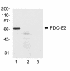
Figure 1
Figure 1
PBC patient sera immunoblot PDC-E2. To identify PBC sera monospecific for PDC-E2 (66 kDa), reduced NRC lysate (80 μg per gel lane) was immunoblotted with PBC patient sera (diluted 1:2500). Two of twenty-five patient sera examined detected only a 66-kDa band (lane 1), and preincubation of these sera with purified PDC (lane 2) blocked detection of this protein band. No protein band was detected when blotting only with the secondary Ab (lane 3). Molecular size markers (Mr × 10–3) are shown on the left. Blotting by each monospecific serum was examined three times with identical results. A representative blot is shown.
Figure 2
Figure 2
PDC-E2 is not cleaved during apoptosis. Reduced NRC lysates from control cells (C) and apoptotic (A) cells were immunoblotted with three different PBC patient sera (lanes 1–6) and SLE patient sera monospecific for NuMA (lanes 7 and 8, top panel), PARP (lanes 7 and 8, middle panel), and U1 70K (lanes 7 and 8, bottom panel). Apoptosis was induced by UV-B irradiation. Equal amounts of protein were electrophoresed in each gel lane. Neither loss of intact PDC-E2 (66 kDa) nor any PDC-E2 cleavage fragments were seen in the apoptotic lysates (lanes 2, 4, and 6) compared with control lysates (lanes 1, 3, and 5, respectively). Likewise, no cleavage of any other PBC autoantigen was detected. Blotting of NuMA, PARP, and U1 70K in the apoptotic lysate showed generation of their expected caspase cleavage fragments (lane 8), confirming induction of apoptosis. Molecular size markers (Mr × 10–3) are shown on the left. Blotting by each serum was examined at least three times with identical results. A representative blot is shown.
Figure 3
Figure 3
PDC-E2 staining by PBC patient sera in apoptotic cells does not localize to cell membrane blebs or apoptotic bodies. Both control cells and cells treated with UV-B to induce apoptosis were examined by confocal, immunofluorescence microscopy. Cells were stained with DAPI (blue) (ah) to distinguish apoptotic cells (cells labeled with stars) with characteristic condensed, fragmented chromatin from nonapoptotic cells (unlabeled cells). Control NRC were costained with PBC patient serum monospecific for PDC-E2 (green) (a) and a mAb specific for COX-1 (red) (e). Immunoreactivity against PDC-E2 and COX-1 colocalized in mitochondria. In apoptotic NRCs, immunoreactivity against PDC-E2 (green) (b) remained perinuclear and colocalized with COX-1 (red) (f). Preincubation of PBC patient sera with purified PDC blocked staining of PDC-E2 (green) in both nonapoptotic and apoptotic NRCs (g). Cells were stained with PI (red) to distinguish cell membrane blebs or apoptotic bodies in apoptotic cells (c and d). Staining of PDC-E2 (green) in apoptotic NRCs did not localize to cell membrane blebs or apoptotic bodies (c). In contrast, immunoreactivity against PDC-E2 (green) was not detected in apoptotic HeLa cells (d), although immunoreactivity against COX-1 (red) was detected (h). Each experiment was repeated twice with each of the monospecific PBC patient sera with identical results. Bar, 20 μm. Representative images are shown.
Figure 4
Figure 4
Persistence of PDC-E2 staining by PBC patient sera in apoptotic cells is cell-type dependent and independent of the stimulus used to induce apoptosis. Both control cells and cells treated with UV-B or staurosporine (STS) to induce apoptosis were examined by confocal, immunofluorescence microscopy. Cells were stained with DAPI (ah) to distinguish apoptotic cells (cells labeled with stars) from nonapoptotic cells (unlabeled cells). After UV-B treatment, PBC patient sera staining of PDC-E2 was detected in apoptotic HSGs (j), but not in apoptotic Jurkat T cells (l). When inducing apoptosis with STS, PDC-E2 immunoreactivity again was undetectable in apoptotic HeLa (n), while persisting in apoptotic NRCs (p), and was undetectable in apoptotic HeLa (n). Bars, 20 μm. All cell types were stained independently with two different PDC-E2 monospecific sera at least three times with similar results each time. Representative images are shown.
Figure 5
Figure 5
PDC-E2 does not leak out of mitochondria during apoptosis. Reduced lysates of subcellular fractions of control (C) and UV-B–irradiated, apoptotic (A) HeLa cells were immunoblotted with SLE and PBC patient sera monospecific for PARP (lanes 1 and 2) and PDC-E2 (lanes 3–6), respectively. Intact PARP predominated in the control nuclear (Nucl) fraction (lane 1), while its caspase cleavage fragment was the predominant form detected in the apoptotic nuclear fraction (lane 2), confirming induction of a high degree of caspase activity in the UV-B–treated cells. PDC-E2 was strongly detected in both control and apoptotic mitochondrial (Mito) fractions (lanes 5 and 6), but not in either cytosolic (Cyto) fraction (lanes 3 and 4). Molecular size markers (Mr × 10–3) are shown on the left. The experiment was repeated twice with identical results. A representative blot is shown.
Figure 6
Figure 6
Depletion of cellular glutathione by BSO treatment before UV-B irradiation prevents the loss of the PDC-E2 staining in apoptotic HeLa cells. UV-B–irradiated cells were stained with DAPI (a and b) to differentiate nonapoptotic from apoptotic cells and with monospecific PBC patient serum against PDC-E2 (c and d). Apoptotic cells are labeled with stars. Staining was analyzed by confocal, immunofluorescence microscopy. In the absence of pretreatment with BSO, staining of PDC-E2 was again not detected in apoptotic HeLa cells after UV-B irradiation (c). However, staining of PDC-E2 persisted in apoptotic HeLa cells pretreated with BSO before UV-B irradiation (d). Bars, 20 μm. Each experiment was repeated at least three times with identical results. Representative images are shown.
Figure 7
Figure 7
Direct oxidation of PDC-E2 by oxidized glutathione abrogates recognition of PDC-E2 by PBC patient sera. (a) Apoptotic NRC lysate was treated with SDS, boiled, and then sequentially treated with 5 mM DTT, 10 mM GSSG, and 25 mM DTT. Strong blotting of PDC-E2 was detected in lysate treated with 5 mM DTT (lane 1). Blotting of PDC-E2 was absent after addition of GSSG to the DTT-treated lysate (lane 2), but subsequent treatment with additional DTT restored detection of PDC-E2 (lane 3). (b) The difference between recognition of oxidized versus reduced PDC-E2 by PBC patient autoantibodies was quantified by immunoblotting lysate treated with 10 mM GSSG vs. 5 mM DTT. A representative blot is shown. (c) The oxidative state of PDC-E2 does not significantly affect its transfer to nitrocellulose. PDC-E2 was immunoprecipitated using PBC patient sera from lysate of HeLa cells incubated with [35S]-methionine to label proteins. Half the immunoprecipitate was treated with DTT and the other half with GSSG before SDS-PAGE was performed. The autoradiogram performed after transfer to nitrocellulose showed that similar amounts of both reduced and oxidized PDC-E2 were transferred. Results using two different PBC patient sera are shown (lanes 1 and 2). (d) The expression level of Bcl-2 correlates with persistence of PDC-E2 staining following apoptosis. Eighty micrograms of lysate protein was loaded per lane and immunoblotted with an mAb specific for human Bcl-2. High levels of Bcl-2 were only detected in the Bcl-2–transfected HeLa (lane 1) and HSG cell lysates (lane 2).
Figure 8
Figure 8
Oxidation of PDC-E2 during apoptosis is Bcl-2 dependent. Control and UV-B–irradiated cells were stained with DAPI (af) to differentiate nonapoptotic from apoptotic cells and with monospecific PBC patient serum against PDC-E2 (gl). Apoptotic cells are labeled with stars. Staining was analyzed by confocal, immunofluorescence microscopy. Staining of PDC-E2 was present in apoptotic, Bcl-2–transfected HeLa cells (h) and apoptotic, freshly isolated IBDECs (j), though not in apoptotic, primary fibroblasts (l). Bars, 20 μm. Representative images are shown.

Comment in

Similar articles

Cited by

References

    1. Kaplan MM. Primary biliary cirrhosis. N Engl J Med. 1996;335:1570–1580. - PubMed
    1. Culp KS, Fleming CR, Duffy J, Baldus WP, Dickson ER. Autoimmune associations in primary biliary cirrhosis. Mayo Clin Proc. 1982;57:365–370. - PubMed
    1. Poupon RE, et al. Combined analysis of randomized controlled trials of ursodeoxycholic acid in primary biliary cirrhosis. Gastroenterology. 1997;113:884–890. - PubMed
    1. Angulo P, et al. Long-term ursodeoxycholic acid delays histological progression in primary biliary cirrhosis. Hepatology. 1999;29:644–647. - PubMed
    1. Pares A, et al. Long-term effects of ursodeoxycholic acid in primary biliary cirrhosis: results of a double-blind controlled multicentric trial. UDCA- Cooperative Group from the Spanish Association for the Study of the Liver. J Hepatol. 2000;32:561–566. - PubMed

Publication types

MeSH terms