Case Reports
. 2001 Jul;108(2):233-40.
doi: 10.1172/JCI12023.

A family with complement factor D deficiency

Affiliations
Case Reports

A family with complement factor D deficiency

D H Biesma et al. J Clin Invest. 2001 Jul.

Abstract

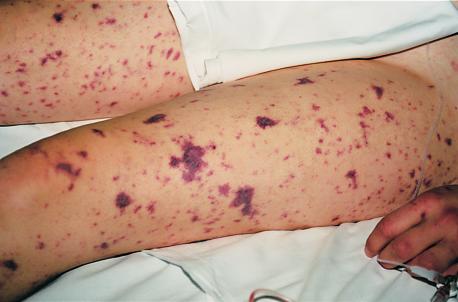
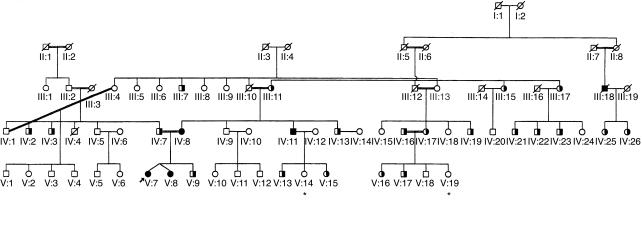
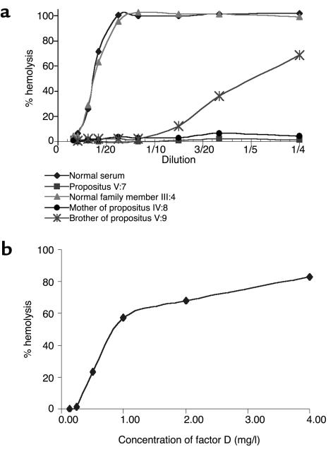
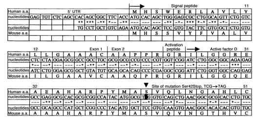
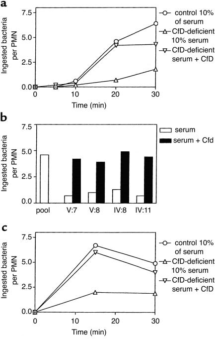
A complement factor D deficiency was found in a young woman who had experienced a serious Neisseria meningitidis infection, in a deceased family member with a history of meningitis, and in three relatives without a history of serious infections. The patient and these three relatives showed a normal activity of the classical complement pathway, but a very low activity of the alternative complement pathway and a very low capacity to opsonize Escherichia coli and N. meningitidis (isolated from the patient) for phagocytosis by normal human neutrophils. The alternative pathway-dependent hemolytic activity and the opsonizing capacity of these sera were restored by addition of purified factor D. The family had a high degree of consanguinity, and several other family members exhibited decreased levels of factor D. The gene encoding factor D was found to contain a point mutation that changed the TCG codon for serine 42 into a TAG stop codon. This mutation was found in both alleles of the five completely factor D-deficient family members and in one allele of 21 other members of the same family who had decreased or low-normal factor D levels in their serum. The gene sequence of the signal peptide of human factor D was also identified. Our report is the first, to our knowledge, to document a Factor D gene mutation. The mode of inheritance of factor D deficiency is autosomal recessive, in accordance with the localization of the Factor D gene on chromosome 19. Increased susceptibility for infections in individuals with a partial factor D deficiency is unlikely.


Figures

Figure 1
Figure 1
Skin changes in the propositus.
Figure 2
Figure 2
Pedigree of the family with factor D deficiency. Heterozygotes and homozygotes for the C125A (Ser42→stop) mutation are indicated by half-filled and filled symbols, respectively. Open symbols indicate family members with wild-type C125 configuration. Symbols with a slash indicate deceased and (except for III:18) noninvestigated family members; symbols with an asterisk indicate living, noninvestigated family members. Double lines indicate consanguineous marriages. The propositus is indicated by an arrow.
Figure 3
Figure 3
Factor D activity of normal serum and partially or completely factor D–deficient serum. (a) Depiction of the factor D activity of the various sera diluted with different amounts of factor D–depleted serum (dilutions from 1:50 to 1:4). Diamonds, normal serum; squares, propositus V:7; triangles, normal family member III:4; circles, mother of the propositus IV:8; asterisks, brother V:9 of the propositus. (b) The restoration of factor D activity in the serum of the propositus by addition of different amounts of purified factor D. The results shown in these figures are representative of three independent experiments.
Figure 4
Figure 4
The 5′ sequence of human and mouse factor D cDNA. The mouse sequence is from ref. . Identity between mouse nucleotides and human nucleotides are indicated by dashes, differences by asterisks. 5′ UTR, 5′ untranslated region; first arrow, start of protein synthesis; second arrow, start of circulating protein sequence; arrowhead, position of mutation. The signal peptide runs from amino acid 1 (methionine) to amino acid 23 (arginine); the activation peptide comprises amino acids 24 (glycine) and 25 (arginine).
Figure 5
Figure 5
Opsonization of E. coli or N. meningitidis by factor D–deficient serum. (a) The kinetics of E. coli phagocytosis: circles, normal pool serum; upward triangles, serum of propositus V:7; downward triangles, serum of propositus with added factor D. Means ± SEM of three independent experiments. ANOVA showed P values less than 0.0001 between all means. Differences between results obtained at 20 and 30 minutes with factor D–deficient serum and either normal serum or factor D–deficient serum with added factor D are significant (P < 0.05, two-tailed P value). (b) The restoration by factor D of the E. coli opsonizing defect in all four factor D–deficient sera measured at 10% serum and 20 minutes of incubation. Open columns, without added factor D; filled columns, with added factor D (2 μg/ml). V:7, propositus; V:8, sister of the propositus; IV:8, mother of the propositus; IV:11, maternal uncle of the propositus. (c) The kinetics of N. meningitidis phagocytosis: circles, normal pool serum; upward triangles, serum of propositus V:7; downward triangles, serum of propositus with added factor D. Means ± SEM of five independent experiments. ANOVA showed P = 0.0015 between all means. Differences between results obtained at 15 and 30 minutes with factor D–deficient serum and either normal or factor D–deficient serum with added factor D are significant (P < 0.02; two-tailed P value).

Similar articles

Cited by

References

    1. Ochs HD, Wedgwood RJ, Frank MM, Heller SR, Hosea SW. The role of complement in the induction of antibody responses. Clin Exp Immunol. 1983;53:208–216. - PMC - PubMed
    1. White RT, et al. Human adipsin is identical to complement factor D and is expressed at high levels in adipose tissue. J Biol Chem. 1992;267:9210–9213. - PubMed
    1. Hiemstra PS, et al. Complete and partial deficiencies of complement factor D in a Dutch family. J Clin Invest. 1989;84:1957–1961. - PMC - PubMed
    1. Figueroa J, Andreoni J, Densen P. Complement deficiency states and meningococcal disease. Immunol Res. 1993;12:295–311. - PubMed
    1. Meyer, M.M. 1961. In Experimental immunochemistry. 2nd edition. Kabat, E.A., and Meyer, M.M., editors. Charles C. Thomas. Springfield, Illinois, USA. 133–240