. 2001 Jul 16;194(2):127-34.
doi: 10.1084/jem.194.2.127.

Mapping precursor movement through the postnatal thymus reveals specific microenvironments supporting defined stages of early lymphoid development

Affiliations

Mapping precursor movement through the postnatal thymus reveals specific microenvironments supporting defined stages of early lymphoid development

E F Lind et al. J Exp Med. .

Abstract

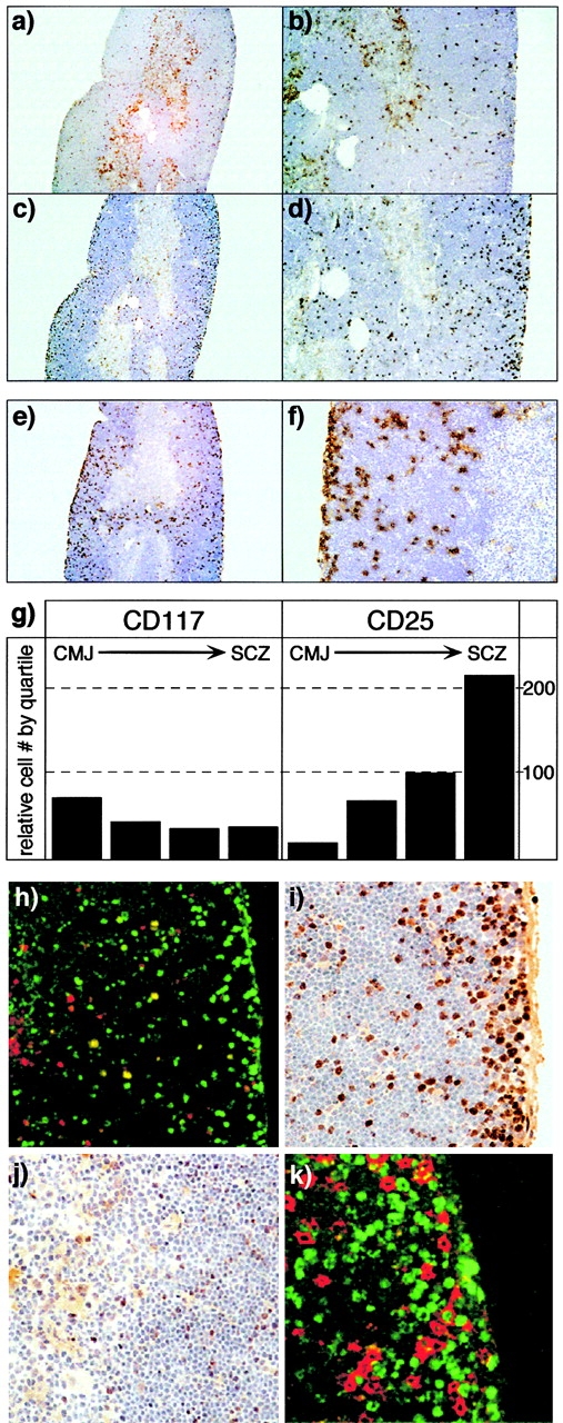
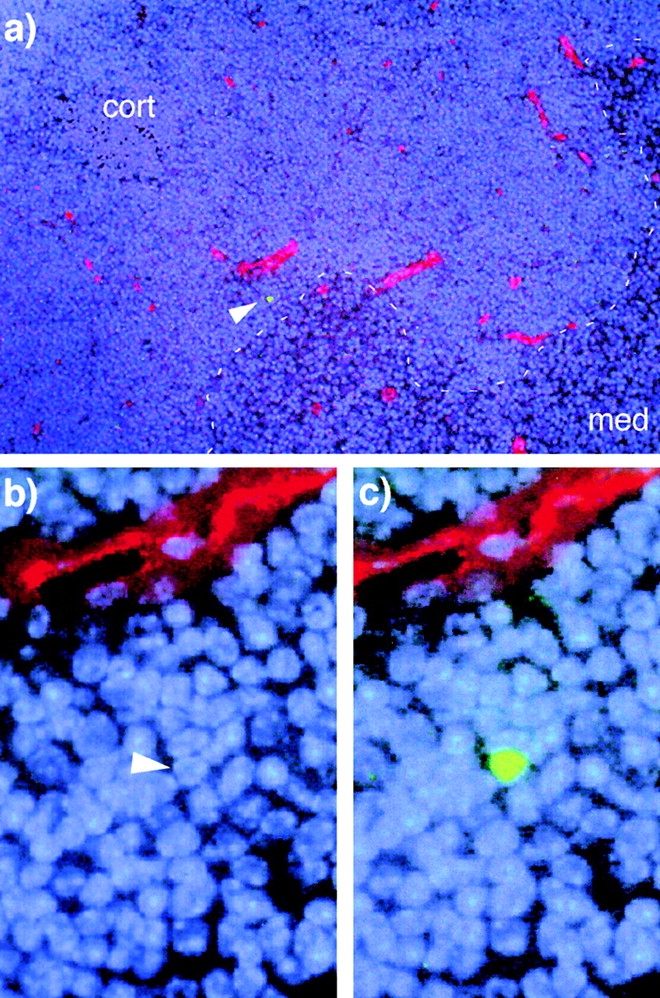
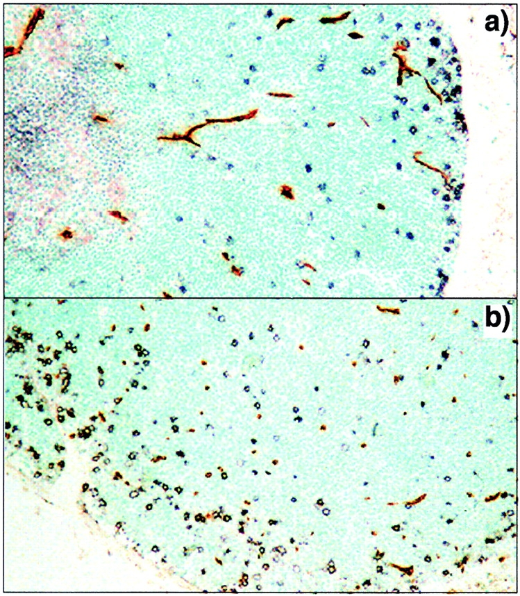
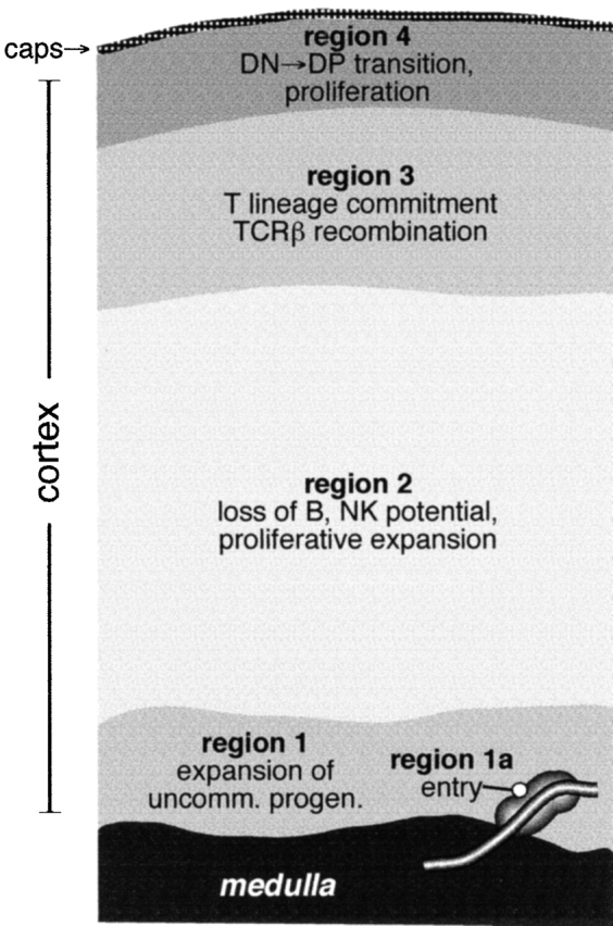
Cellular differentiation is a complex process involving integrated signals for lineage specification, proliferation, endowment of functional capacity, and survival or cell death. During embryogenesis, spatially discrete environments regulating these processes are established during the growth of tissue mass, a process that also results in temporal separation of developmental events. In tissues that undergo steady-state postnatal differentiation, another means for inducing spatial and temporal separation of developmental cues must be established. Here we show that in the postnatal thymus, this is achieved by inducing blood-borne precursors to enter the organ in a narrow region of the perimedullary cortex, followed by outward migration across the cortex before accumulation in the subcapsular zone. Notably, blood precursors do not transmigrate the cortex in an undifferentiated state, but rather undergo progressive developmental changes during this process, such that defined precursor stages appear in distinct cortical regions. Identification of these cortical regions, together with existing knowledge regarding the genetic potential of the corresponding lymphoid precursors, sets operational boundaries for stromal environments that are likely to induce these differentiative events. We conclude that active cell migration between morphologically similar but functionally distinct stromal regions is an integral component regulating differentiation and homeostasis in the steady-state thymus.


Figures

Figure 1
Figure 1
Anatomic localization of early precursors in the adult thymus. Low magnification (a and c, original magnifications: 40×) and high magnification (b and d, original magnifications: 100×) views of serial sections of adult thymus, stained with anti-CD117 (brown, a and b) or anti-CD25 (c and d) antibodies. The counterstain (blue) is hematoxylin. CD117+ cells are most frequent near the CMJ and are also scattered at lower density throughout the cortex, but are relatively infrequent in the SCZ. In contrast, CD25+ cells are rare in the vicinity of the CMJ, but increase in frequency with proximity to the SCZ. The total anatomic range for CD48 precursors is seen in e (original magnification: 40×) and f (original magnification: 200×), using a congenic marker for RAG-2–deficient cells (Thy-1.2) in thymuses from RAG-2/wild-type (Thy-1.1+) bone marrow chimeras. The relative distribution of CD117+ or CD25+ cells in the cortex, divided into quartiles, is illustrated in g; data are derived from counting multiple CD25/117 serial sections. h shows two-color immunofluorescent staining of CD117 (red) and CD25 (green) in a single cortical field (original magnification: 200×; left, CMJ). DN2 cells (yellow, CD117+CD25+) are generally found in the midcortical region. i and j illustrate the location of proliferating cells (brown, anti-BrdU) in the SCZ/outer cortex or inner cortex/CMJ, respectively (original magnification: 400×) after a single 4-h pulse of BrdU. As reported previously, the vast majority of cell proliferation is found to occur in the SCZ, although infrequent BrdU+ cells are found throughout the cortex. However, two-color staining (k) with CD25 (red) and BrdU (green) reveals that most of the proliferating cells in the SCZ are not DN cells (CD25+), and therefore represent amplification of the early DP compartment (preDP).
Figure 2
Figure 2
Marrow-derived precursors extravasate deep in the cortex adjacent to the medulla. CFSE-labeled, lineage-depleted bone marrow cells were administered intravenously to nonablated recipients. 1–2 d later, thymuses were recovered, cryogenically sectioned, and examined for the presence of CFSE+ cells. In all cases where CFSE+ cells (green) were found, they were in the deep cortex, near the CMJ (dashed line, a) and in close proximity to large blood vessels (red) that appear to be postcapillary venules. The outer capsule of the section shown in a is just outside of the field of view, to the left and above the image. The nuclear counterstain is DAPI (blue), blood vessels are revealed using Texas Red, and the single CFSE+ cell is indicated by an arrowhead. b and c show magnified views of this region; in b, green fluorescence is not shown so that the nucleus of the CFSE+ cell can be visualized.
Figure 3
Figure 3
Parenchymal entry correlates with the site of extravasation in the thymic cortex. The relationship between early precursors and vascular/perivascular tissues was examined using CD25 staining (black) as a marker of DN precursors and tomato lectin (brown) as a marker of vascular endothelium. The transverse section shown in a includes cortical and medullary regions and is parallel to cortical capillary loops. In b, the plane of sectioning is through the deep cortex perpendicular to the orientation of capillary loops. No correlation between CD25+ precursors and vascular tissue is seen in either orientation, indicating that the perivascular sheaths are not conduits for the transcortical migration of early thymocytes and that parenchymal entry occurs deep within the tissue.
Figure 4
Figure 4
Functionally distinct zones for lymphopoietic precursor differentiation in the postnatal thymus. A map of cortical regions, as defined by the approximate location of each lymphopoietic stage, is shown together with some of the developmental events that occur during migration between them.

References

    1. Shortman K., Egerton M., Spangrude G.J., Scollay R. The generation and fate of thymocytes. Semin. Immunol. 1990;2:3–12. - PubMed
    1. Pearse M., Wu L., Egerton M., Wilson A., Shortman K., Scollay R. A murine early thymocyte developmental sequence is marked by transient expression of the interleukin 2 receptor. Proc. Natl. Acad. Sci. USA. 1989;86:1614–1618. - PMC - PubMed
    1. Shimonkevitz R.P., Husmann L.A., Bevan M.J., Crispe I.N. Transient expression of IL-2 receptor precedes the differentiation of immature thymocytes. Nature. 1987;329:157–159. - PubMed
    1. Lesley J., Trotter J., Schulte R., Hyman R. Phenotypic analysis of the early events during repopulation of the thymus by the bone marrow prothymocyte. Cell. Immunol. 1990;128:63–78. - PubMed
    1. Ceredig R., Lowenthal J.W., Nabholz M., MacDonald H.R. Expression of interleukin-2 receptors as a differentiation marker on intrathymic stem cells. Nature. 1985;314:98–100. - PubMed

Publication types

MeSH terms