. 2001 Jul 25;123(29):7031-9.
doi: 10.1021/ja004306h.

Mn K-edge XANES and Kbeta XES studies of two Mn-oxo binuclear complexes: investigation of three different oxidation states relevant to the oxygen-evolving complex of photosystem II

Affiliations

Mn K-edge XANES and Kbeta XES studies of two Mn-oxo binuclear complexes: investigation of three different oxidation states relevant to the oxygen-evolving complex of photosystem II

H Visser et al. J Am Chem Soc. .

Abstract

Two structurally homologous Mn compounds in different oxidation states were studied to investigate the relative influence of oxidation state and ligand environment on Mn K-edge X-ray absorption near-edge structure (XANES) and Mn Kbeta X-ray emission spectroscopy (Kbeta XES). The two manganese compounds are the di-mu-oxo compound [L'2Mn(III)O2Mn(IV)L'2](ClO4)3, where L' is 1,10-phenanthroline (Cooper, S. R.; Calvin, M. J. Am. Chem. Soc. 1977, 99, 6623-6630) and the linear mono-mu-oxo compound [LMn(III)OMn(III)L](ClO4)2, where L- is the monoanionic N,N-bis(2-pyridylmethyl)-N'-salicylidene-1,2-diaminoethane ligand (Horner, O.; Anxolabéhère-Mallart, E.; Charlot, M. F.; Tchertanov, L.; Guilhem, J.; Mattioli, T. A.; Boussac, A.; Girerd, J.-J. Inorg. Chem. 1999, 38, 1222-1232). Preparative bulk electrolysis in acetonitrile was used to obtain higher oxidation states of the compounds: the Mn(IV)Mn(IV) species for the di-mu-oxo compound and the Mn(III)Mn(IV) and Mn(IV)Mn(IV) species for the mono-mu-oxo compound. IR, UV/vis, EPR, and EXAFS spectra were used to determine the purity and integrity of the various sample solutions. The Mn K-edge XANES spectra shift to higher energy upon oxidation when the ligand environment remains similar. However, shifts in energy are also observed when only the ligand environment is altered. This is achieved by comparing the di-mu-oxo and linear mono-mu-oxo Mn-Mn moieties in equivalent oxidation states, which represent major structural changes. The magnitude of an energy shift due to major changes in ligand environment can be as large as that of an oxidation-state change. Therefore, care must be exercised when correlating the Mn K-edge energies to manganese oxidation states without taking into account the nature of the ligand environment and the overall structure of the compound. In contrast to Mn K-edge XANES, Kbeta XES spectra show less dependence on ligand environment. The Kbeta1,3 peak energies are comparable for the di-mu-oxo and mono-mu-oxo compounds in equivalent oxidation states. The energy shifts observed due to oxidation are also similar for the two different compounds. The study of the different behavior of the XANES pre-edge and main-edge features in conjunction with Kbeta XES provides significant information about the oxidation state and character of the ligand environment of manganese atoms.


Figures

Figure 1
Figure 1
A schematic for the excitation and emission processes involved in XANES and Kβ XES spectroscopy. XANES spectra reflect the transition energy of 1s electrons excited to higher bound states, which depends on the overall charge and ligand environment of the metal. To enhance sensitivity, the absorption spectra are collected as excitation spectra using Mn Kα fluorescence detection. Kβ XES arise from the emission of a 3p electron to 1s hole, which is formed following X-ray absorption. In a simplified model, two final spin states exist with either a constructive (Kβ1,3) or destructive (Kβ′) spin exchange interaction between the unpaired 3p and 3d electrons. The magnitude of the interaction depends on the number of unpaired 3d electrons, which is related to the oxidation state of high-spin Mn. For a more accurate model, the ligand-field multiplet formalism needs to be applied, taking into account spin–spin and spin–orbit interactions, ligand-field splitting, and Jahn–Teller distortions.
Figure 2
Figure 2
(A) The di-μ-oxo manganese compound [L′2 MnIIIO2MnIVL′2]3+, with L′ as 1, 10-phenanthroline.,, (B) The linear mono-μ-oxo compound, [LMnIIIOMnIIIL]2+, with L as the monoanionic N,N-bis(2-pyridylmethyl)-N′-salicylidene-1,2-diaminoethane ligand.,
Figure 3
Figure 3
Scheme of the time course for the oxidation reaction of the di-μ-oxo compound. The arrows indicate sample extractions. The MnIIIMnIV compound (di(III,IV)) is an EPR-active di-μ-oxo species. The MnIVMnIV species is stable and EPR-silent. The EPR spectra of the di(IV,IV) samples are analyzed to determine the decline in the MnIIIMnIV concentration. The EPR spectrum of the starting sample (di(III,IV)) is used as the pure MnIIIMnIV quantitative standard. In the nomenclature of the electrochemical samples, “di” refers to the di-μ-oxo compound. The Roman numerals between brackets indicate the desired oxidation state of the compound. Different extraction time-points are indicated by subscripts.
Figure 4
Figure 4
Scheme of the time course for the oxidation reaction of the mono-μ-oxo compound. The arrows indicate sample extractions. UV/vis absorption coefficients of the MnIIIMnIII and MnIIIMnIV compounds are used to determine the starting concentration (mono(III,III)), and the degree of completion of the first oxidation step (mono(III,IV)). The MnIIIMnIV compound is the only EPR-active mono-μ-oxo species; it has a distinctive 18-line spectrum. When water is present in the electrochemical solution, the MnIVMnIV species decomposes into MnII, which has a distinctive six-line EPR spectrum. Therefore, the EPR spectra of the mono(IV,IV) samples are analyzed to determine the decline in the MnIIIMnIV concentration and the appearance of the decomposition product MnII. The EPR spectrum of sample mono(III,IV) is used as the MnIIIMnIV quantitative standard, and a MnCl2 acetonitrile solution as the MnII quantitative EPR standard. In the nomenclature of the electrochemical samples, “mono” refers to the mono-μ-oxo compound. The Roman numerals between brackets indicate the desired oxidation state of the compound. Different extraction time-points are indicated by subscripts.
Figure 5
Figure 5
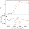
The deconvoluted Mn K-edge XANES (A) and second-derivative (B) spectra of the di-μ-oxo compound in its two oxidation states: MnIIIMnIV (black) and MnIVMnIV (red). The arrows in the second-derivative spectra indicate the first-inflection point energy for the two oxidation states: MnIIIMnIV = 6549.6 eV and MnIVMnIV = 6551.8 eV.
Figure 6
Figure 6
The deconvoluted Mn K-edge XANES (A) and second-derivative (B) spectra of the mono-μ-oxo compound in its three oxidation states: MnIIIMnIII (blue), MnIIIMnIV (black), and MnIVMnIV (red). The arrows in the second-derivative spectra indicate the first inflection-point energy for each of the three oxidation states: MnIIIMnIII = 6550.4 eV, MnIIIMnIV = 6551.6 eV, and MnIVMnIV = 6552.3 eV.
Figure 7
Figure 7
The deconvoluted Mn K-edge difference spectra for the different oxidation state transitions of the di-μ-oxo compound; MnIVMnIV – MnIIIMnIV (green), and of the mono-μ-oxo compound: MnIIIMnIV – MnIIIMnIII (black) and MnIVMnIV – MnIIIMnIV (purple).
Figure 8
Figure 8
The deconvoluted Kβ XES of the di-μ-oxo compound (A) and the mono-μ-oxo compound (B) in their respective oxidation states: MnIIIMnIII (blue), MnIIIMnIV (black), and MnIVMnIV (red). The spectra show the entire spectral region with the two emission bands: Kβ′ (weak, left) and Kβ1,3 (strong, right). The insets show the Mn Kβ1,3 XES spectra on an expanded scale.
Figure 9
Figure 9
The deconvoluted Kβ XES difference spectra for the different oxidation-state transitions of the di-μ-oxo compound: MnIVMnIV – MnIIIMnIV (green), and of the mono-μ-oxo compound: MnIIIMnIV – MnIIIMnIII (black) and MnIVMnIV – MnIIIMnIV (purple).

Similar articles

Cited by

References

    1. Cooper SR, Calvin M. J Am Chem Soc. 1977;99:6623–6630.
    1. Horner O, Anxolabéhère-Mallart E, Charlot MF, Tchertanov L, Guilhem J, Mattioli TA, Boussac A, Girerd J-J. Inorg Chem. 1999;38:1222–1232. - PubMed
    1. Kirby JA, Goodin DB, Wydrzynski T, Robertson AS, Klein MP. J Am Chem Soc. 1981;103:5537–5542.
    1. Yachandra VK, Sauer K, Klein MP. Chem Rev. 1996;96:2927–2950. - PubMed
    1. Liang W, Roelofs TA, Cinco RM, Rompel A, Latimer MJ, Yu WO, Sauer K, Klein MP, Yachandra VK. J Am Chem Soc. 2000;122:3399–3412. - PMC - PubMed

Publication types