

doi: 10.1101/gad.897901.
Inhibition of Cdh1-APC by the MAD2-related protein MAD2L2: a novel mechanism for regulating Cdh1
Affiliations
- PMID: 11459825
- PMCID: PMC312740
- DOI: 10.1101/gad.897901
Item in Clipboard
Inhibition of Cdh1-APC by the MAD2-related protein MAD2L2: a novel mechanism for regulating Cdh1
Genes Dev.
.
Abstract
Exit from mitosis requires the degradation of regulatory proteins including the mitotic cyclins and securin through ubiquitination by the anaphase promoting complex (APC) bound to Cdc20 or Cdh1. Cdc20-APC is regulated through inhibition by the spindle assembly checkpoint protein MAD2. Knowledge of Cdh1-APC regulation is limited to the phosphorylation-dependent dissociation of Cdh1 from APC. We report a novel means of regulating Cdh1 by the MAD2-related gene, MAD2L2. MAD2L2 specifically binds and inhibits Cdh1-APC, paralleling the effect of MAD2 on Cdc20. We suggest that MAD2L2 and MAD2 inhibit the release of substrates from APC and propose a mechanism of inhibition.
Figures
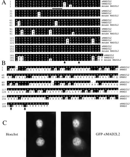
MAD2L2 sequence conservation. (A) Comparison of xMAD2L2 with MAD2L2 sequences from human and mouse. Shading indicates residues in common with xMAD2L2; a bold line indicates a putative nuclear localization signal. (B) Sequence comparison of xMAD2L2 with XMAD2. Arrows indicate a conserved C-terminal lysine and valine (A,B). (C) Hoechst staining of eGFP-xMAD2L2 transfected BHK cells (left); eGFP-xMAD2L2 signal (right).

MAD2L2 binds to Cdh1 and not Cdc20. (A) 35S-labeled in vitro-translated Cdc20 and Cdh1 prebound to APC on αCdc27 beads. (B) Binding of in vitro-translated 35S-labeled xMAD2L2 to Cdh1–APC on αCdc27 beads but not to Cdc20–APC on beads, iAPC on beads, or αCdc27 beads alone.

xMAD2L2 inhibits Cdh1 but not Cdc20 activity. (A) Degradation assays in interphase extracts, Cdh1-supplemented interphase extracts, and Cdh1 extracts in the presence of recombinant His–MAD2L2 for mB99, Xkid, securin, and cyc B. Phosphorylation of mB99 in interphase extracts causes a shift; mB99 samples were CIP treated to clarify the difference between phosphorylation and degradation. (B) Gel filtration profiles of recombinant MBP-MAD2L2 on a superdex 200 column using the SMART system (upper panel) and tetramer, trimer, and monomer peaks rerun over the same column (lower panel). (C) t1/2 of Xkid in Cdh1 extracts (upper panel) or Δ90 extracts (lower panel) with increasing concentrations of MAD2M, MAD2O, MBP-xMAD2L2M or MBP-xMAD2L2O. Graph reflects typical results from one extract; t1/2 varies between extracts, but the effect on t1/2 is consistent. (D) Ubiquitination assays of cyc B using Cdh1-activated APC in the presence of 5 μM MAD2O or 5 μM MBP–xMAD2L2.

xMAD2L2-mediated inhibition of gastrulation is rescued by coinjection with Cdh1. Vegetal views of stage 11 embryos injected (left half of each embryo) at the two-cell stage with (A) 2.5 ng MBP–MAD2L2, (B) 2.5 ng His6–Cdh1, (C) 2.5 ng MBP–MAD2L2 plus 2.5 ng His6–Cdh1, or (D) buffer. Injections of MBD–MAD2L2 result in cells that have enlarged and enhanced nuclear staining properties. One cell of two-cell stage embryos injected with 2.5 ng of MBP–MAD2L2, allowed to develop to stages 8–9, fixed, and stained with propidium iodide (E,F) or Hoechst (G). (E) Uninjected side and (F) injected side of the same embryo. (G) A view showing an injected region (left) next to an uninjected region. (H) Injections of Cdh1 RNA result in enlarged cells that give diffuse nuclear staining. One cell of two-cell embryos were injected with 500 pg of Cdh1 (left side), allowed to develop to stages 10.5, fixed, and stained with Hoechst. (I) Summary of the gastrulation arrest and rescue of the embryos shown in A–D.

MAD2 and MAD2L2 prevent release of substrate bound to Cdc20 or Cdh1, respectively. Graph of substrate binding assay showing prebound Xkid retained by Cdc20 or Cdh1 beads over time after addition of excess cold Xkid in the presence of 5 μM MAD2O or MAD2L2, respectively, compared to buffer alone.
References
-
- Baumer M, Braus GH, Irniger S. Two different modes of cyclin Clb2 proteolysis during mitosis in Saccharomyces cerevisiae. FEBS Lett. 2000;468:142–148. - PubMed
-
- Cahill DP, da Costa LT, Carson-Walter EB, Kinzler KW, Vogelstein B, Lengauer C. Characterization of MAD2B and other mitotic spindle checkpoint genes. Genomics. 1999;58:181–187. - PubMed
-
- Fang G, Yu H, Kirschner MW. Direct binding of CDC20 protein family members activates the anaphase-promoting complex in Mitosis and G1. Mol Cell. 1998a;2:163–171. - PubMed
-
- Funabiki H, Murray AW. The Xenopus Chromokinesin Xkid is essential for metaphase chromosome alignment and must be degraded to allow anaphase chromosome movement. Cell. 2000;102:411–424. - PubMed
Publication types
MeSH terms
Substances
Grants and funding
LinkOut - more resources
Full Text Sources
Molecular Biology Databases
Miscellaneous