. 2001 Jul 15;15(14):1771-82.
doi: 10.1101/gad.892401.

Messenger RNAs are recruited for nuclear export during transcription

Affiliations

Messenger RNAs are recruited for nuclear export during transcription

E P Lei et al. Genes Dev. .

Abstract

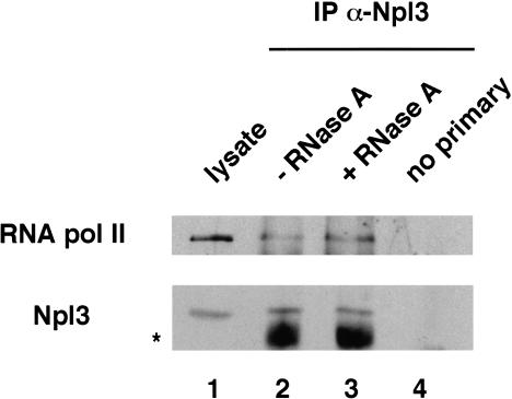
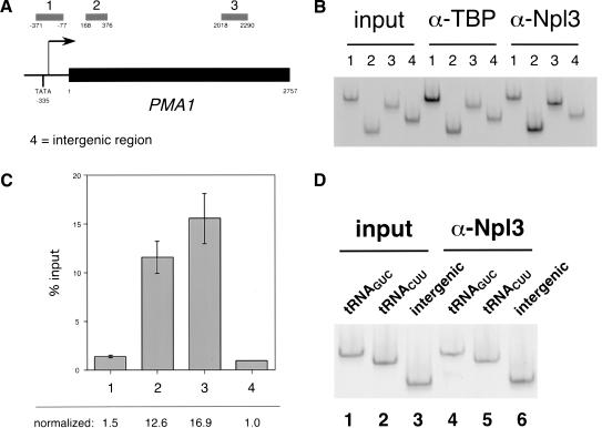
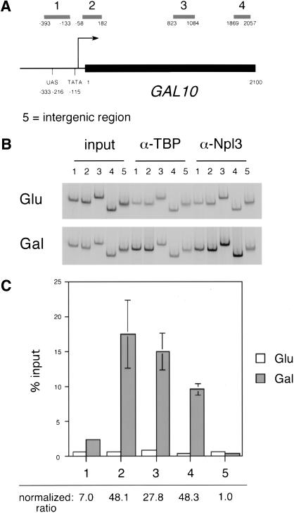
Following transcription and processing, eukaryotic mRNAs are exported from the nucleus to the cytoplasm for translation. Here we present evidence that mRNAs are targeted for nuclear export cotranscriptionally. Combined mutations in the Saccharomyces cerevisiae hnRNP Npl3 and TATA-binding protein (TBP) block mRNA export, implying that cotranscriptional recruitment of Npl3 is required for efficient export of mRNA. Furthermore, Npl3 can be found in a complex with RNA Pol II, indicating that Npl3 associates with the transcription machinery. Finally, Npl3 is recruited to genes in a transcription dependent manner as determined by chromatin immunoprecipitation. Another mRNA export factor, Yra1, also associates with chromatin cotranscriptionally but appears to be recruited at a later step. Taken together, our results suggest that export factors are recruited to the sites of transcription to promote efficient mRNA export.


Figures

Figure 1
Figure 1
spt15-ts1 npl3-27 cells are slowed for Npl3-27 export and display a synthetic mRNA export defect. (A) Localization of Npl3 or Npl3-27 in wild type (PSY580, a–c), npl3-27 (PSY1031, d–f), spt15-ts1 npl3-27 (PSY1698, g–i) and spt15-ts1 cells (PSY1702, j–l). Cells were shifted to 37°C for 1 h. Indirect immunofluorescence with polyclonal antibodies to Npl3 (left), DAPI (center), and Nomarski images (right) are shown. (B) Localization of poly(A)+ RNA in wild type (a–c), npl3-27 (d–f), spt15-ts1 npl3-27 (g–i), and spt15-ts1 cells (j–l). Cells were shifted to 37°C for 1 h. in situ hybridization with an oligo (dT)50 probe (left), DAPI (center), and Nomarski images (right) are shown.
Figure 1
Figure 1
spt15-ts1 npl3-27 cells are slowed for Npl3-27 export and display a synthetic mRNA export defect. (A) Localization of Npl3 or Npl3-27 in wild type (PSY580, a–c), npl3-27 (PSY1031, d–f), spt15-ts1 npl3-27 (PSY1698, g–i) and spt15-ts1 cells (PSY1702, j–l). Cells were shifted to 37°C for 1 h. Indirect immunofluorescence with polyclonal antibodies to Npl3 (left), DAPI (center), and Nomarski images (right) are shown. (B) Localization of poly(A)+ RNA in wild type (a–c), npl3-27 (d–f), spt15-ts1 npl3-27 (g–i), and spt15-ts1 cells (j–l). Cells were shifted to 37°C for 1 h. in situ hybridization with an oligo (dT)50 probe (left), DAPI (center), and Nomarski images (right) are shown.
Figure 2
Figure 2
npl3-27 has no effect on total mRNA levels. (A) Slot blot hybridization of total poly(A)+ RNA of wild type (lanes 1–2, PSY580 and 603), spt15-ts1 npl3-27 (lanes 3–4, PSY1698 and 1699), spt15-ts1 (lanes 5–6, PSY1702 and 1703), and npl3-27 cells (lanes 7–8, PSY1031 and 1032). Cells were grown to log phase at 25°C, cultures were split, one half was shifted to 37°C for 1 h, and total RNA was isolated. Two μg of total RNA was probed with a 32P-labeled poly dT probe. (B) ACT1 Northern of wild type (lanes 1–2), spt15-ts1 npl3-27 (lanes 3–4), spt15-ts1 (lanes 5–6), and npl3-27 cells (lanes 7–8). Cells were grown to log phase at 25°C, cultures were split, one half was shifted to 37°C for 1 h, and total RNA was isolated. Fifteen μg of total RNA separated by agarose gel electrophoresis was probed with a 32P-labeled ACT1 probe.
Figure 3
Figure 3
Npl3 and RNA Pol II form a complex that is not dependent on RNA. Npl3 from cell lysates is immunoprecipitated with α-Npl3 antibodies (bottom). Heavy chain of the primary antibody is denoted with an asterisk. RNA Pol II is coimmunoprecipitated with Npl3 (top). Total cell lysate is loaded (lane 1). After binding and washing, samples were treated with 50 μg/mL RNase A in wash buffer (lane 3) or wash buffer alone (lane 2) and incubated at RT for 20 min. Samples were washed once and raised in sample loading buffer, run on a 7% SDS-PAGE gel and Western blotted with α-Npl3 antibodies or 8WG16 against Pol II CTD. No primary samples were mock IPs lacking primary antibody (lane 4). Blots shown are from two different gels from the same experiment. Approximately 20% of Npl3 and 0.02% of Pol II were immunoprecipitated from the lysate.
Figure 4
Figure 4
Npl3 crosslinks to the promoter and coding sequence of the constitutively expressed Pol II transcribed PMA1 gene. (A) Diagram of PMA1. Primer set 1 spans the promoter including the TATA box. Primer set 2 spans the 5′ region of the coding sequence. Primer set 3 spans the 3′ region of the coding sequence. (ATG = +1). Primer set 4 spans a nontranscribed intergenic region. (B) Npl3 immunoprecipitates promoter and coding sequence of PMA1. Quantitative PCR of input (left), α-TBP immunoprecipitate (center) and α-Npl3 immunoprecipitate (right) using primers sets 1–4 spanning regions as indicated in A. PCR products were separated on an 8% TBE polyacrylamide gel. Single dilutions of each template are shown and are in the linear range of PCR (data not shown). (C) Quantitation of α-Npl3 immunoprecipitated material. Raw values are expressed in graphical form as percentage of input. Error bars >0.1 are shown for a single experiment. Normalized values were obtained by dividing the percentage obtained for each primer set by the percentage obtained of primer 4 (bottom). (D) Npl3 does not associate with Pol III transcribed genes. Quantitative PCR of input (lanes 1–3) and Npl3 immunoprecipitate (lanes 4–6) of DNA spanning the tRNAGUC gene (lanes 1,4), tRNACUU gene (lanes 2,5), and intergenic region (lanes 3,6).
Figure 5
Figure 5
Npl3 crosslinks to the promoter and coding sequence of GAL10 in a transcription dependent manner. (A) Diagram of GAL10. Primer set 1 spans the upstream activating sequence. Primer set 2 spans the promoter and 5′ coding sequence. Primer set 3 spans the middle of the coding sequence. Primer set 4 spans the 3′ coding sequence. Primer set 5 spans a nontranscribed intergenic region. (B) Npl3 immunoprecipitates promoter and coding sequence of GAL10 under inducing conditions. Quantitative PCR of input (left), α-TBP immunoprecipitate (center), and α-Npl3 immunoprecipitate (right) using primers sets 1–5 spanning regions as indicated in A. Cells were grown in glucose (top) or galactose (bottom). (C) Quantitation of α-Npl3 immunoprecipitated material. Raw values are expressed for cells grown in glucose (white) and galactose (gray) in graphical form as a percentage of input. Error bars >0.3 are shown for one experiment. Normalized ratio values were obtained by dividing galactose values by glucose values for each primer set and normalizing to primer set 5.
Figure 6
Figure 6
Yra1 is associated with the 3′ end of genes in a transcription dependent manner. (A) Quantitation of Yra1-myc association with the PMA1 promoter (bar 1), 5′ coding sequence (bar 2), and 3′ coding sequence (bar 3) and intergenic region (bar 4). Values normalized to intergenic region are shown (bottom). (B) Quantitation of Yra1-myc association with the GAL10 UAS (bars 1), 5′ coding sequence (bars 2), middle coding sequence (bars 3), 3′ coding sequence (bars 4), and intergenic region (bars 5) in cells grown in raffinose (white) and galactose (gray). Normalized ratio values are shown (bottom).
Figure 7
Figure 7
Model for cotranscriptional recruitment of mRNA export factors. Npl3 is recruited to the transcription machinery perhaps by Pol II in the preinitiation complex and/or elongating Pol II and then transferred to the nascent transcript. Further recruitment of Npl3 continues as elongation proceeds. CBC is also recruited to the nascent transcript possibly dependent on Npl3. Yra1 associates with the nascent RNA at a late step of transcription. Other RNA-binding proteins are indicated (black). Cotranscriptional recruitment of these factors promotes efficient export of the RNP.

Similar articles

Cited by

References

    1. Alzhanova-Ericsson AT, Sun X, Visa N, Kiseleva E, Wurtz T, Daneholt B. A protein of the SR family of splicing factors binds extensively to exonic Balbiani ring pre-mRNA and accompanies the RNA from the gene to the nuclear pore. Genes & Dev. 1996;10:2881–2893. - PubMed
    1. Amberg DC, Goldstein AL, Cole CN. Isolation and characterization of RAT1: An essential gene of Saccharomyces cerevisiae required for the efficient nucleocytoplasmic trafficking of mRNA. Genes & Dev. 1992;6:1173–1189. - PubMed
    1. Arndt KM, Ricupero-Hovasse S, Winston F. TBP mutants defective in activated transcription in vivo. EMBO J. 1995;14:1490–1497. - PMC - PubMed
    1. Bachi A, Braun IC, Rodrigues JP, Pante N, Ribbeck K, von Kobbe C, Kutay U, Wilm M, Gorlich D, Carmo-Fonseca M, et al. The C-terminal domain of TAP interacts with the nuclear pore complex and promotes export of specific CTE-bearing RNA substrates. RNA. 2000;6:136–158. - PMC - PubMed
    1. Bossie MA, DeHoratius C, Barcelo G, Silver P. A mutant nuclear protein with similarity to RNA binding proteins interferes with nuclear import in yeast. Mol Biol Cell. 1992;3:875–893. - PMC - PubMed

Publication types

MeSH terms

LinkOut - more resources