. 2001 Jul 15;15(14):1845-58.
doi: 10.1101/gad.193901.

The set1Delta mutation unveils a novel signaling pathway relayed by the Rad53-dependent hyperphosphorylation of replication protein A that leads to transcriptional activation of repair genes

Affiliations

The set1Delta mutation unveils a novel signaling pathway relayed by the Rad53-dependent hyperphosphorylation of replication protein A that leads to transcriptional activation of repair genes

V Schramke et al. Genes Dev. .

Abstract

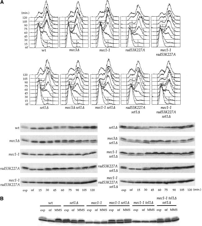
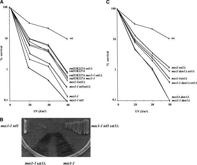
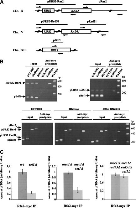
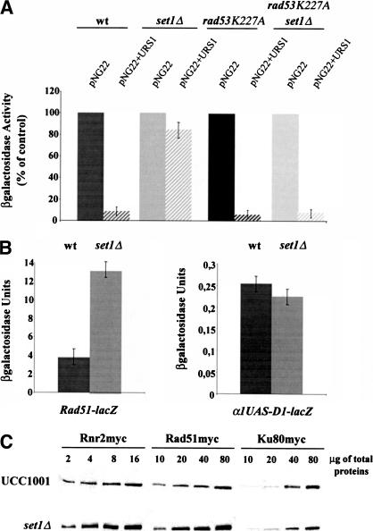
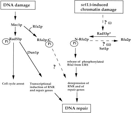
SET domain proteins are present in chromosomal proteins involved in epigenetic control of transcription. The yeast SET domain protein Set1p regulates chromatin structure, DNA repair, and telomeric functions. We investigated the mechanism by which the absence of Set1p increases DNA repair capacities of checkpoint mutants. We show that deletion of SET1 induces a response relayed by the signaling kinase Rad53p that leads to the MEC1/TEL1-independent hyperphosphorylation of replication protein A middle subunit (Rfa2p). Consequently, the binding of Rfa2p to upstream repressing sequences (URS) of repair genes is decreased, thereby leading to their derepression. Our results correlate the set1Delta-dependent phosphorylation of Rfa2p with the transcriptional induction of repair genes. Moreover, we show that the deletion of the amino-terminal region of Rfa2p suppresses the sensitivity to ultraviolet radiation of a mec3Delta checkpoint mutant, abolishes the URS-mediated repression, and increases the expression of repair genes. This work provides an additional link for the role of Rfa2p in the regulation of the repair capacity of the cell and reveals a role for the phosphorylation of Rfa2p and unveils unsuspected connections between chromatin, signaling pathways, telomeres, and DNA repair.


Figures

Figure 1
Figure 1
set1Δ-induced hyperphosphorylation of Rfa2p. (A, top) Exponentially growing cells (exp) of K699 derivative strains with the indicated relevant genotypes were synchronized with α-factor and released at time zero. FACS analysis of the synchronized cultures at the indicated times after α-factor release. (Bottom) Protein extracts prepared at the indicated times after release from α-factor were analyzed by SDS-PAGE and immunoblotting with anti-pRfa2p polyclonal antibodies. The upper band corresponds to the phosphorylated form of Rfa2p. (B) set1Δ-dependent phosphorylation of Rfa2p after MMS treatment in sml1 mec1-1 and sml1 mec1-1 tel1 mutants. Protein extracts from exponentially growing cultures (exp), from G1-arrested cultures (α-F), and MMS treated G1-arrested cultures (MMS), were analyzed by Western blot with anti-pRfa2p polyclonal antibodies.
Figure 2
Figure 2
Phosphorylation of Rfa2p Δ40. (A) Exponentially growing cells of K699 derivative strains with the indicated relevant genotypes were synchronized with α-factor and released at time zero. Protein extracts prepared at the indicated times after release from α-factor were analyzed by Western blot with anti-pRfa2p polyclonal antibodies. The upper band corresponds to the phosphorylated form of Rfa2p Δ40. (B) Native extracts of exponentially growing rfa2 Δ40 and rfa2 Δ40 set1Δ. Cells were divided in two samples and treated (+) or not (−) with λ-phosphatase.
Figure 3
Figure 3
Viability after UV irradiation. Approximately 1000 cells of K699 derivative strains with the indicated relevant genotypes were spread on YPD plates and exposed to ultraviolet irradiation. Cells were grown at 30°C for 5 d before we counted the number of colonies. (A) The set1Δ mutation partially rescues ultraviolet sensitivity of sml1 mec1-1 but not of rad53K227A and sml1 mec1-1 rad53K227A mutants; (B) the deletion of SET1 suppresses the growth defect of a sml1 mec1-1 tel1 double mutant; (C) the set1Δ mutation increases the viability after ultraviolet of mec3Δ dun1Δ and sml1 mec1-1 dun1Δ cells.
Figure 4
Figure 4
set1Δ-dependent phosphorylation of Rfa2p decreases its binding to URS elements and leads to transcriptional induction of repair genes. (A) Scheme of the DNA regions flanking the URS elements of RNR2 (pURS2–Rnr2) and RAD51 (pURS2–Rad51) that were used to analyze the binding of Rfa2p to the URS elements. The pairs of primers used to amplify pURS2–Rnr2, pURS2–Rad51, and the control fragments pRnr2, pRad51, and pBdf1 are indicated. (B, top) Rfa2p binds to URS elements in vivo. The ChIP assay was performed as described in Materials and Methods. Rpa2myc and Ku80myc indicate that the parental strain UCC1001 bears a 18-mer Myc epitope fused to the carboxyl terminus of the genomic copy of RFA2 and HDF2, respectively. Immunoprecipitation was performed with anti-myc antibodies (9 E10). After DNA purification, PCR was performed with primers for the sequences flanking the URS2 of RNR2 (pURS2–Rnr2) and RAD51 (pURS2–Rad51), and for the control sequence in BDF1 (pBdf1). PCR reactions were performed on immunoprecipitated DNA and on the corresponding imput DNA. PCR products (indicated by arrows) were resolved on 2% agarose gels. (Bottom) Rfa2p binds to URS2–Rnr2 and URS2–Rad51 but not to the adjacent sequences, pRnr2 and pRad51. DNA regions pRnr2 and pRad51 are localized at ∼1400 bp of the URS2 sequences of RNR2 and RAD51, respectively. (C) In vivo association of Rfa2p with the URS2 of RAD51 is diminished in set1Δ cells. Chromatin solutions from wild-type and set1Δ cells expressing the Rfa2-myc were subjected to ChIP. The amount of precipitated DNA was analyzed by quantitative PCR (see Materials and Methods). The signals obtained by PCR in the Rfa2-myc immunoprecipitates were quantitated by referring them to a standard curve. This standard curve was obtained by plotting the signals obtained from PCRs performed with the same primers on serial dilutions of the corresponding input DNA. The effect of the set1Δ mutation was analyzed in different parental strains. For each couple of strains (UCC1001 (wt), UCC1001 set1Δ), (UCC1001 mec1Δ sml1Δ, UCC1001 mec1Δ sml1Δ set1Δ), and (UCC1001 mec1Δ rad53Δ sml1Δ UCC1001, mec1Δ rad53Δ sml1Δ set1Δ), amplification of the input DNA samples with the specific and control primers produced similar amount of pBdf1 and pURS2–Rad51 PCR DNA fragments. For each graph, the amount of DNA corresponding to pURS2–Rad51 in the precipitated DNA from the parental strain is arbitrary plotted as 1. The experiments shown were done in triplicate
Figure 5
Figure 5
(A) The set1Δ mutation abolishes the repression mediated by the MAG URS1 element. We compared for each strain the β-galactosidase activity associated with the CYC1–lacZ vector (pNG22) and with pNG22 containing the MAG URS1 element (pNG22+URS1). The enzymatic activity associated with pNG22 was taken as control. (B, left) The set1Δ mutation increases the RAD51–lacZ expression; (right) the set1Δ mutation does not act via the D1 element of the RNR2 promoter. (C) protein levels of Rad51p and Rnr2p increase in set1Δ cells. A 13-mer Myc epitope was fused to the carboxyl terminus of the genomic copy of RAD51 and RNR2. Ku80myc was used as an internal control. Identical amounts of total protein extracts were analyzed by SDS-10% PAGE and immunoblotting. The presence of Rnr2p, Rad51p, and Ku80p was revealed with 9 E10 (anti-myc) antibodies. Relative amounts of proteins were determined by analyzing the chemiluminescence signals.
Figure 6
Figure 6
(A) The sensitivity to ultraviolet radiation of the mec3Δ mutant is suppressed by the deletion of the amino-terminal region of Rfa2p. Approximately 1000 cells of K699 derivative strains with the indicated relevant genotypes were spread on YPD plates and exposed to ultraviolet irradiation. Cells were grown at 30°C for 5 d before we counted the number of colonies. (B) Analysis of intraS checkpoint of the rfa2 Δ40 mutants. Exponentially growing (exp) wild-type, mec3Δ rfa2 Δ40 and mec3Δ rfa2 Δ40 cells were synchronized with α-factor and released in YPD medium with (+MMS) or without 0.02% MMS. (Left) Untreated or MMS-treated samples were taken at the indicated times after α-factor release and analyzed. (Right) Aliquots were removed from the MMS-treated cultures to determine cell number and to score for colony-forming units on YPD plates at 25°C. (C) The rfa2 Δ40 mutation abolishes the repression mediated by the MAG URS2 element. We measured the β-galactosidase activity associated with the CYC1–lacZ vector (pNG22) and with pNG22 containing the MAG URS1 element (pNG22+URS1). (D) The rfa2 Δ40 mutation increases the RAD51–lacZ expression.
Figure 7
Figure 7
A schematic representation of the set1Δ-induced cellular response that leads to the transcriptional activation of repair genes. We propose that phosphorylation of Rfa2p can result either from (1) the activation of Rad53p (Rad53*) in response to a set1Δ-induced chromatin damage, or (2) from the deregulation of a phosphorylation pathway controlled by Set1p. Both models are not exclusive. The set1Δ cellular response is shown in parallel with the DNA damage response. N-Pi and C-Pi indicated phosphorylation in the amino and carboxyl terminus of Rfa2p, respectively.

Similar articles

Cited by

References

    1. Aboussekhra A, Chanet R, Adjiri A, Fabre F. Semidominant suppressors of Srs2 helicase mutations of Saccharomyces cerevisiae map in the RAD51 gene, whose sequence predicts a protein with similarities to procaryotic RecA proteins. Mol Cell Biol. 1992;12:3224–3234. - PMC - PubMed
    1. Aboussekhra A, Vialard JE, Morrison DE, de la Torre-Ruiz MA, Cernakova L, Fabre F, Lowndes NF. A novel role for the budding yeast RAD9 checkpoint gene in DNA damage-dependent transcription. EMBO J. 1996;15:3912–3922. - PMC - PubMed
    1. Allen JB, Zhou Z, Siede W, Friedberg EC, Elledge SJ. The SAD1/RAD53 protein kinase controls multiple checkpoints and DNA damage-induced transcription in yeast. Genes & Dev. 1994;8:2416–2428. - PubMed
    1. Brill SJ, Stillman B. Replication factor-A from Saccharomyces cerevisiae is encoded by three essential genes coordinately expressed at S phase. Genes & Dev. 1991;5:1589–1600. - PubMed
    1. Brush GS, Kelly TJ. Phosphorylation of the replication protein A large subunit in the Saccharomyces cerevisiae checkpoint response. Nucleic Acids Res. 2000;19:3725–3732. - PMC - PubMed

Publication types

MeSH terms

Substances