. 2001 Jul 31;98(16):9265-70.
doi: 10.1073/pnas.151151198. Epub 2001 Jul 17.

Angiogenic role for glycodelin in tumorigenesis

Affiliations

Angiogenic role for glycodelin in tumorigenesis

M Song et al. Proc Natl Acad Sci U S A. .

Abstract

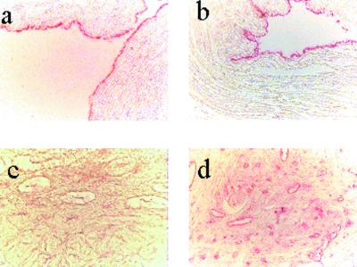
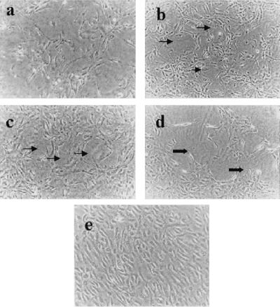
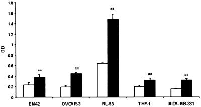
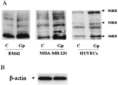
Angiogenesis plays an important role in neovascularization in tumors. Glycodelin, a hormone-responsive protein, has been detected in tumors of reproductive organs and is found in high levels in the plasma of subjects with gynecological malignancies. Glycodelin is also found in the endothelial cells of the umbilical cord and in the blood vessels of tumors. In this study, we tested whether glycodelin-rich amniotic fluid and a synthetic peptide derived from the sequence of glycodelin peptide (Gp) might promote angiogenic response by examining the migration and tube formation in human umbilical cord vein endothelial cells (HUVECs). Increased migration and tube formation of HUVECs were found in the presence of amniotic fluid and Gp, and this increase was blocked by antibody to Gp and by an anti-vascular endothelial growth factor (VEGF) antibody, suggesting that the angiogenic effects of glycodelin might be mediated by VEGF. The results also showed that Gp significantly increased the release of VEGF protein and mRNA expression in HUVECs, RL-95 (human endometrial carcinoma cells), OVCAR-3 (human ovarian adenocarcinoma cells), EM42 (human endometrial epithelial cells), THP-1 (human monocyte), and MCF-7 and MDA-MB-231 (human breast adenocarcinoma cells) cell lines. VEGF receptor Fit-1 mRNA expression in HUVECs was also increased in the presence of Gp. These findings, together with the suggestion from the literature that glycodelin may have immunosuppressive properties, suggest that glycodelin might play an important role in neovascularization during embryogenesis and tumor development.


Figures

Figure 1
Figure 1
(a) Immunohistochemistry with anti-Gp antibody in human umbilical cord vein, and (b) with anti-von Willebrand factor in human umbilical cord vein. (c and d) Immunohistochemistry with anti-Gp antibody in normal and adenocarcinoma endometrium tissue. In c, no obvious staining in endothelial cells of the blood vessels was seen, but in d, intense pink staining in endothelial cells of the blood vessels could be seen in endometrium adenocarcinoma tissue.
Figure 2
Figure 2
(A) Effect of Gp on migration of HUVECs. HUVECs were incubated with medium only (bar 1), or with Gp (bar 2), Gp plus anti-Gp antibody (bar 3), Gp plus anti-VEGF antibody (bar 4), Gp plus anti-gastrin I antibody (bar 5), Gp plus chicken IgG (bar 6), AF (bar 7), and AF plus anti-Gp antibody (bar 8). The number of migrating cells was counted under a phase-contrast light microscope at magnification = ×400. Results are presented as mean ± SD of each group. **, P < 0.01 vs. control; $, P < 0.01 vs. Gp; @, P > 0.05 vs. Gp; *, P < 0.05 vs. control; #, P < 0.01 vs. AF. (B) Staining of migrating cells with Diff-Quick staining solution. (i) HUVECs (control). (ii) HUVECs exposed to Gp. (iii) HUVECs exposed to AF. (iv) HUVECs exposed to Gp plus anti-Gp antibody. More cell migration was observed in cells treated with Gp and AF. (Magnification = ×200.)
Figure 3
Figure 3
Tube formation of HUVECs in collagen gels. (a) Control. (b) HUVECs exposed to Gp. (c) HUVECs exposed to AF. (d) HUVECs exposed to Gp plus anti-Gp antibody. (e) HUVECs exposed to Gp plus anti-VEGF antibody. Increased tube formation (thin arrows, b and c) was observed in HUVECs exposed to Gp and AF when compared with untreated cells. Compared with Gp alone, HUVECs exposed to Gp plus the antibodies to Gp or VEGF showed broken tube formation (thick arrows, d) or less tube formation (e), respectively. (Magnification = ×100.)
Figure 4
Figure 4
Effect of Gp on VEGF level in different cell culture media by ELISA analysis. C, control (white bars); GP, cultured with 50 ng/ml Gp (black bars). **, P < 0.01 vs. control.
Figure 5
Figure 5
(A) Western blot analysis of VEGF expression in EM42, MDA-MB-231, and HUVECs cells. C, control; Gp, cells cultured in presence of 50 ng/ml Gp. (B) β-Actin was used as internal control.
Figure 6
Figure 6
(A) RT-PCR analysis of VEGF expression in the cells cultured with or without Gp. The 440-, 572-, 644-, and 695-bp products correspond to VEGF121, VEGF165, VEGF189, and VEGF206 isoforms, respectively; the 500-bp product may correspond to the VEGF145 isoform. (B) Flt-1 expression in HUVECs cultured with or without Gp. M, 100-bp DNA marker; C, control; Gp, cells cultured in presence of 50 ng/ml Gp. Glyceraldehyde-3-phosphate dehydrogenase (GAPDH) mRNA was used as internal control in A and B.

References

    1. Ferrara N, Davis-Smyth T. Endocr Rev. 1997;18:4–25. - PubMed
    1. Folkman J. Cancer Res. 1986;46:467–473. - PubMed
    1. Weidner N, Semple J P, Welch W R, Folkman J. N Engl J Med. 1991;324:1–8. - PubMed
    1. Ferrara N. Breast Cancer Res Treat. 1995;36:127–137. - PubMed
    1. Mattern J, Koomagi R, Volm M. Br J Cancer. 1996;73:931–934. - PMC - PubMed

Publication types

LinkOut - more resources