. 2001 Jul 31;98(16):9348-52.
doi: 10.1073/pnas.151247598. Epub 2001 Jul 17.

Holins kill without warning

Affiliations

Holins kill without warning

A Gründling et al. Proc Natl Acad Sci U S A. .

Abstract

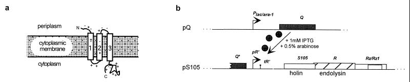
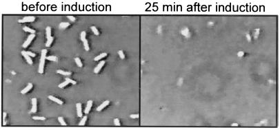
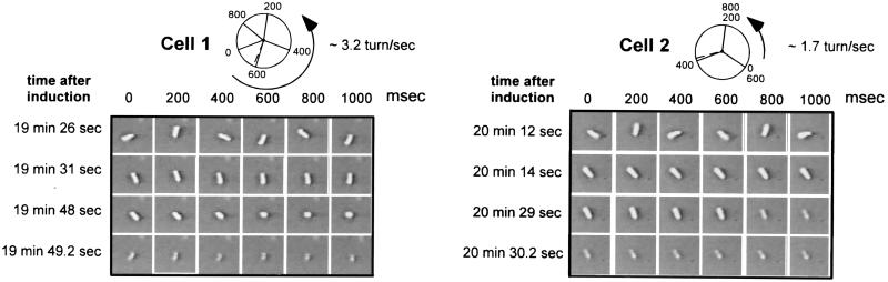
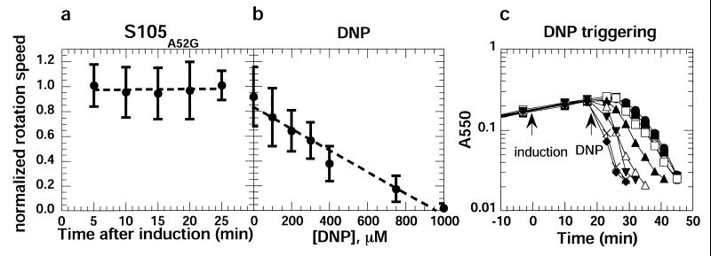
Holins comprise the most diverse functional group of proteins known. They are small bacteriophage-encoded proteins that accumulate during the period of late-protein synthesis after infection and cause lysis of the host cell at a precise genetically programmed time. It is unknown how holins achieve temporal precision, but a conserved feature of their function is that energy poisons subvert the normal scheduling mechanism and instantly trigger membrane disruption. On this basis, timing has been proposed to involve a progressive decrease in the energized state of the membrane until a critical triggering level is reached. Here, we report that membrane integrity is not compromised after the induction of holin synthesis until seconds before lysis. The proton motive force was monitored by the rotation of individual cells tethered by a single flagellum. The results suggest an alternative explanation for the lysis "clock," in which holin concentrations build to a critical level that leads to formation of an oligomeric complex that disrupts the membrane.


Figures

Figure 1
Figure 1
(a) Topology of the λ S holin. It is not known how the holin oligomerizes to form the permeabilizing lethal membrane lesion. (b) Scheme for inducible expression of the cloned lysis genes. The λ antiterminator gene Q is cloned in the plasmid pQ under control of a lac/ara hybrid promoter, which is inducible with IPTG and arabinose. The λ lysis genes lie in an overlapping cluster in the λ lysis cassette located downstream of the late gene promoter, pR′, on the compatible plasmid pS105. Expression of the lysis genes follows transactivation of pR′ by Q.
Figure 2
Figure 2
Typical microscope field as seen in a tethering assay before and after induction of the λ lysis genes. Bacterial cells of the strain RY8797pQpS105A52G were grown, tethered, and induced as described in Materials and Methods. A field of tethered cells is shown 1 min before induction (Left) and 25 min after induction (Right).
Figure 3
Figure 3
Lysis of individual cells. Cells were tethered to coverslips as described in Fig. 2. At t = 0, expression of the lysis genes from the plasmid pS105A52G was induced by IPTG and arabinose. Single frames were chosen from the recordings of two representative cells and are depicted here to illustrate the process of cell lysis. Starting from the time point indicated (Left), single frames were captured every 200 ms. After induction of the lysis genes, the tethered cells rotate at high and constant speed (first row). About 20 min after induction, rotation of the cell abruptly slows and stops completely within 1–3 sec (second row). Cell lysis, because of digestion of the cell wall by the λ R endolysin, occurs within several seconds after the sudden stop in rotation (third and fourth rows). A digitized recording (quicktime movie) can be found as Movie 1, which is published as supplemental data on the PNAS web site, www.pnas.org.
Figure 4
Figure 4
Rotation speeds after induction of the S gene or addition of DNP. (a) Cells of strain RY8797pQpS105A52G were tethered to a glass coverslip, induced at t = 0, recorded, and analyzed for rotation speed, as described in Materials and Methods. The mean of the normalized rotation speeds for 10 individual cells at each time is plotted, except for the 25-min time point, where the value is for seven surviving cells. The vertical bars indicate one standard deviation. (b) RY8797 cells were tethered to glass slides, equilibrated with prewarmed TB medium, and exposed to a 5-min flow of prewarmed TB medium containing DNP at the indicated concentrations. The rotation speed of at least 20 individual cells was determined 5 min after the addition of DNP and normalized to the rotation speed determined before addition. The vertical bars indicate one standard deviation. (c) DNP-induced triggering of S. Cultures of strain RY8797pQpS105 were grown in TB medium at 37°C to an A550 = 0.2 and induced at t = 0 with IPTG and arabinose. At t = 20 min, DNP was added to a final concentration of 0 μM (●), 5 μM (○), 50 μM (■), 100 μM (□), 200 μM (▴), 400 μM (Δ), 600 μM (▾), 1,000 μM (×), and 1,500 μM (⧫). After DNP addition, the turbidity of the cultures was monitored as A550 until lysis was complete.

References

    1. Wang I-N, Smith D L, Young R. Annu Rev Microbiol. 2000;54:799–825. - PubMed
    1. Young R, Wang I-N, Roof W D. Trends Microbiol. 2000;8:120–128. - PubMed
    1. Wang I-N, Dykhuizen D E, Slobodkin L B. Evol Ecol. 1996;10:545–558.
    1. Raab R, Neal G, Sohaskey C, Smith J, Young R. J Mol Biol. 1988;199:95–105. - PubMed
    1. Johnson-Boaz R, Chang C-Y, Young R. Mol Microbiol. 1994;13:495–504. - PubMed

Publication types

MeSH terms

LinkOut - more resources