

Review
doi: 10.1073/pnas.151265198.
Structural basis for bending tropomyosin around actin in muscle thin filaments
Affiliations
- PMID: 11459946
- PMCID: PMC37414
- DOI: 10.1073/pnas.151265198
Item in Clipboard
Review
Structural basis for bending tropomyosin around actin in muscle thin filaments
Proc Natl Acad Sci U S A.
.
No abstract available
Figures

Interaction between the two α-helices in the tropomyosin coiled-coil. Each α-helix is shown with seven residues (a–g) in two turns. (A) End-on view looking from N terminus. The interface between the α-helices derives primarily from hydrophobic residues in core positions a and d, although there are also some salt bridges formed between residues e and g. (B) The core interface viewed parallel to the coiled-coil axis shows how residues from one chain occupy the spaces between the corresponding residues from the second chain to give “knobs in holes” packing (18). In tropomyosin there are slightly more than 3.6 residues per turn, which produces a left-handed supercoil.
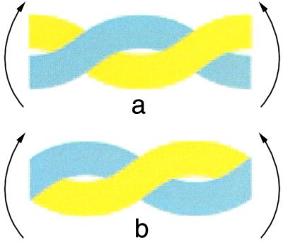
Bending coiled-coils about an axis perpendicular to the plane of the page. Bending about the broad face (a) involves a shearing motion so that one chain (yellow) follows a longer path and so the two chains need to slide relative to one another elsewhere in the coiled-coil to compensate. However, a bend about the narrow edge of the supercoil (b) simply bends both α-helices in the same way. Both types of bending are likely to be present when tropomyosin wraps around actin in muscle thin filaments.
Comment on
-
Deciphering the design of the tropomyosin molecule.Proc Natl Acad Sci U S A. 2001 Jul 17;98(15):8496-501. doi: 10.1073/pnas.131219198. Epub 2001 Jul 3. Proc Natl Acad Sci U S A. 2001. PMID: 11438684 Free PMC article.
References
-
- Stone D, Sodek J, Johnson P, Smillie L B. Proc IX FEBS Meeting. 1975;31:125–136.
-
- McLachlan A D, Stewart M. J Mol Biol. 1975;98:293–304. - PubMed
-
- Parry D A D. J Mol Biol. 1975;98:519–535. - PubMed
-
- McLachlan A D, Stewart M, Smillie L B. J Mol Biol. 1975;98:281–291. - PubMed
-
- McLachlan A D, Stewart M. J Mol Biol. 1976;103:271–298. - PubMed
Publication types
MeSH terms
Substances
LinkOut - more resources
Full Text Sources
