Review
. 2001 Jul 17;98(15):8181-8.
doi: 10.1073/pnas.111008798.

Rescue of arrested replication forks by homologous recombination

Affiliations
Review

Rescue of arrested replication forks by homologous recombination

B Michel et al. Proc Natl Acad Sci U S A. .

Abstract

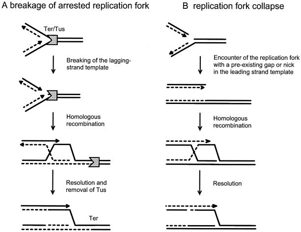
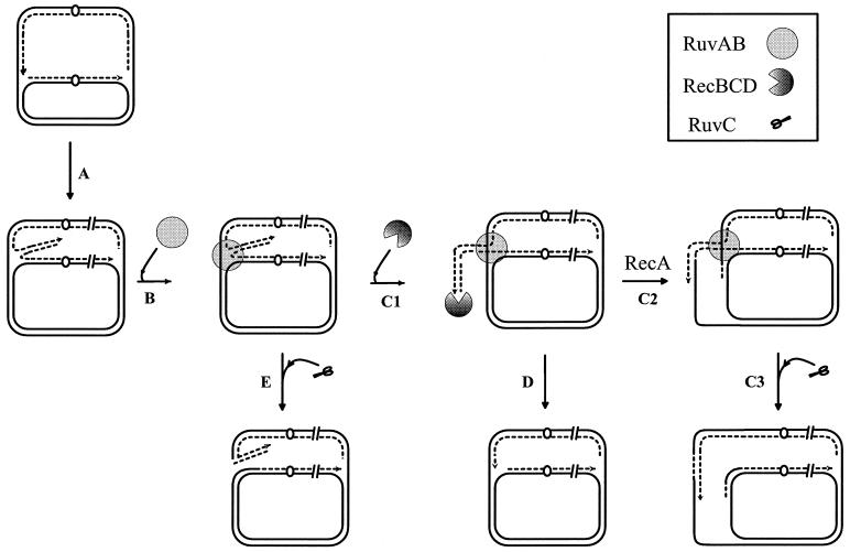
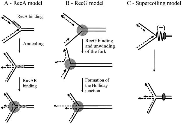
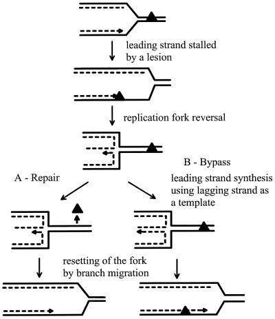
DNA synthesis is an accurate and very processive phenomenon; nevertheless, replication fork progression on chromosomes can be impeded by DNA lesions, DNA secondary structures, or DNA-bound proteins. Elements interfering with the progression of replication forks have been reported to induce rearrangements and/or render homologous recombination essential for viability, in all organisms from bacteria to human. Arrested replication forks may be the target of nucleases, thereby providing a substrate for double-strand break repair enzyme. For example in bacteria, direct fork breakage was proposed to occur at replication forks blocked by a bona fide replication terminator sequence, a specific site that arrests bacterial chromosome replication. Alternatively, an arrested replication fork may be transformed into a recombination substrate by reversal of the forked structures. In reversed forks, the last duplicated portions of the template strands reanneal, allowing the newly synthesized strands to pair. In bacteria, this reaction was proposed to occur in replication mutants, in which fork arrest is caused by a defect in a replication protein, and in UV irradiated cells. Recent studies suggest that it may also occur in eukaryote organisms. We will review here observations that link replication hindrance with DNA rearrangements and the possible underlying molecular processes.


Figures

Figure 1
Figure 1
Recombination repair of broken replication forks. (A) Rescue of blocked replication forks (adapted from ref. 17). The replication fork is blocked at the Ter site in the presence of Tus. A DSB occurs in the lagging-strand template. The RecBCD enzyme enters at the double-strand end and initiates homologous recombination catalyzed by RecA. Completion of the recombination reaction by resolution of the Holliday junction leads to restoration of a replication fork. Binding of the primosome allows loading of a new replisome to promote DNA replication restart. To account for the viability of a strain carrying an ectopic Ter site in a recombination-proficient background, one needs to assume that the newly reconstituted replication fork is not arrested again, and hence that Tus has been removed from Ter during the recombination reaction. DSB on the lagging strand is shown, but a similar model can apply to breakage and repair of the leading strand. (B) Replication fork collapse (adapted from ref. 31). The progressing fork encounters a single-strand interruption in the leading-strand template, because of a defect in closure of the lagging strand at the previous replication round. Reincorporation of the broken DNA strand by homologous recombination and replication restart are catalyzed by the same enzymes as on breakage of the fork. The full lines represent the two DNA strands, the dashed lines represents newly synthesized DNA strands. The arrowhead corresponds to DNA 3′ ends.
Figure 2
Figure 2
RuvAB/RecBCD-mediated rescue of blocked replication forks (adapted from ref. 44). In the first step (A) the replication fork is blocked and the two newly synthesized strands anneal, forming a Holliday junction (see Fig. 3 for the different pathways proposed to promote this step). In a second step (B) the junction is stabilized by RuvAB binding. (C) In recombination proficient strains, RecBCD binds to the double-strand tail (C1); degradation takes place until the first recognized CHI site (CHI is an octameric sequence that switches RecBCD from an exonuclease to a recombinase enzyme) and is followed by a genetic exchange mediated by RecA (C2); RuvC resolves the first Holliday junction bound by RuvAB (C3). In C2 and C3, the double-strand end is reincorporated into the circular chromosome by homologous recombination and the Holliday junction is resolved, which results in the reconstitution of a replication fork. This pathway is presumably used in recombination-proficient cells. (D) RecBCD-mediated degradation of the tail progresses up to the RuvAB-bound Holliday junction. Replication can restart when RecBCD has displaced the RuvAB complex. D can take place in recombination-proficient strains if RecBCD reaches RuvAB before encountering a CHI site; it is the only pathway that leads to a viable chromosome in recA and ruvC mutants. (E) RuvC resolves the RuvAB-bound Holliday junction. This pathway is used in the absence of RecBCD and leads to the RuvABC-dependent DSBs observed in recBC mutants. Continuous and discontinuous lines represent the template and the newly synthesized strand of the chromosome respectively; the arrowheads indicate the 3′end of the growing strands.
Figure 3
Figure 3
Models for formation of Holliday junctions at arrested replication forks by RFR. (A) RecA binds to the single-stranded region of the lagging-strand template, polymerizing in the 5′ to 3′ direction. Pairing of the lagging-strand template with the leading-strand template renders the leading strand free to anneal with the 5′ end of the lagging strand. This results in the formation of a Holliday junction that can be bound directly by RuvAB. (Adapted from ref. .) (B) RecG binds to the replication fork and migrates toward the chromosomal replication origins, displacing the 5′ end of the lagging strand. RecG activity ultimately creates a four-stranded junction. (Adapted with modifications from ref. .) (C) (+) Topological stress that accumulates downstream of the fork on arrest is relaxed by unwinding of the two newly synthesized strands from the template strands and their annealing. (Schematic representation based on results in ref. .) Full and dashed lines represent the template and the newly synthesized DNA, respectively; the arrowheads indicate the 3′ end of the growing strands.
Figure 4
Figure 4
RFR in UV-irradiated cells (adapted from refs. , , and 92). The replication fork is blocked by a UV photo-product (black triangle) in the leading-strand template. RFR, proposed to be catalyzed by RecG in E. coli (76), or by Rad51 (the yeast RecA homologue) in S. pombe (80), renders the damaged DNA double stranded and thereby allows direct repair by nucleotide excision repair enzymes (A). If the lagging-strand polymerase has continued synthesis past the lesion, leading-strand DNA synthesis using the lagging strand as template followed by reverse branch migration [proposed to be catalyzed by RecG in E. coli (76) and by Rqh in S. pombe (80)] reconstitutes a fork on which the lesion has been bypassed (B; ref. 94). Full and dashed lines represent the template and the newly synthesized DNA, respectively; the arrowhead indicates the 3′ end of the growing strand.

References

    1. Seitz E M, Kowalczykowski S C. Mol Microbiol. 2000;37:555–560. - PubMed
    1. Egelman E. Trends Biochem Sci. 2000;25:180–184. - PubMed
    1. Michel B. In: Organization of the Prokaryotic Genome. Charlebois R L, editor. Washington, DC: Am. Soc. Microbiol.; 1999. pp. 129–150.
    1. Lewis L K, Resnick M A. Mutat Res. 2000;451:71–89. - PubMed
    1. Lieber M R. Genes Cells. 1999;4:77–85. - PubMed

Publication types

LinkOut - more resources