. 2001 Jul 17;98(15):8241-6.
doi: 10.1073/pnas.131009198.

Single-strand interruptions in replicating chromosomes cause double-strand breaks

Affiliations

Single-strand interruptions in replicating chromosomes cause double-strand breaks

A Kuzminov. Proc Natl Acad Sci U S A. .

Abstract

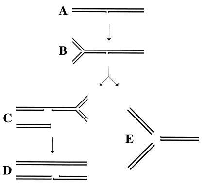
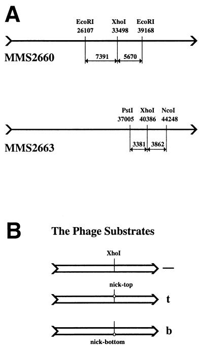
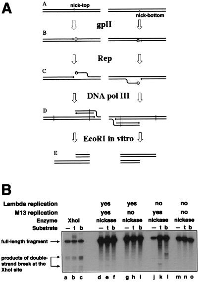
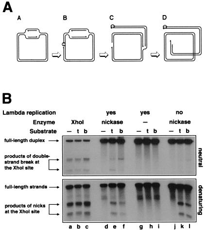
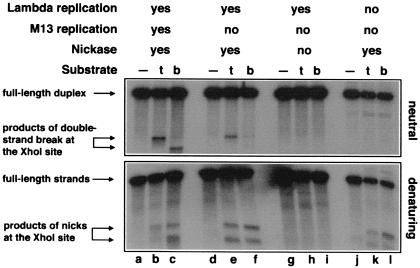
Replication-dependent chromosomal breakage suggests that replication forks occasionally run into nicks in template DNA and collapse, generating double-strand ends. To model replication fork collapse in vivo, I constructed phage lambda chromosomes carrying the nicking site of M13 bacteriophage and infected with these substrates Escherichia coli cells, producing M13 nicking enzyme. I detected double-strand breaks at the nicking sites in lambda DNA purified from these cells. The double-strand breakage depends on (i) the presence of the nicking site; (ii) the production of the nicking enzyme; and (iii) replication of the nick-containing chromosome. Replication fork collapse at nicks in template DNA explains diverse phenomena, including eukaryotic cell killing by DNA topoisomerase inhibitors and inviability of recombination-deficient vertebrate cell lines.


Figures

Figure 1
Figure 1
The idea of replication fork collapse at a single-strand interruption in template DNA. (A) A DNA segment with a long-lived nick. (B) A replication fork is approaching the nick. (C) The replication fork has reached the nick and collapsed; another replication fork is approaching the remaining single-strand interruption from the opposite direction. (D) The second replication fork has reached the interruption and collapsed; as a result, one of the daughter chromosomes has a double-strand break. (E) An alternative scenario of double-strand breakage at replication forks: replication fork “explosion” at the nick.
Figure 2
Figure 2
Experimental substrates. (A) The two λ phages used to construct the nicking site-containing phages. The XhoI-sites at which the nicking sites were inserted and the restriction sites used in Southern analysis are shown, together with their coordinates on wild-type λ chromosome and the sizes of the generated fragments. (Upper) MMS2660, the progenitor of λAK2 (nick in the top strand) and λAK3 (nick in the bottom strand). These three phages are used in Figs. 3 and 4. (Lower) MMS2663, the progenitor of λAK4 (nick in the top strand) and λAK5 (nick in the bottom strand). These three phages are used in Fig. 5. (B) A set of three phage substrates (either MMS2660, λAK2, and λAK3 or MMS2663, λAK4, and λAK5) run in parallel in one experiment. All three phages within a set have a unique XhoI site at approximately the same location. The control phage (marked “—”) has nothing else; the middle phage carries the nicking site in the top strand and is therefore marked “t;” the bottom phage carries the nicking site in the bottom strand and is therefore marked “b.”
Figure 3
Figure 3
The interplay between λ and M13 DNA replication. (A) Sigma replication in Rep+ cells is predicted to generate the “half-break” pattern at the nicking sites. 3′-ends are indicated by arrows. (A) The substrate chromosomes carrying nicking sites at the same location, either in the top or in the bottom strand. (B) The nicking sites are nicked by gpII (open circles attached to the 5′-sides of the nicks). (C) The Rep helicase unwinds the 5′-ends of the nicks, attracting replisomes to the replication fork structures. (D) Sigma replication from the nicks. (E) Restriction cutting in vitro (at sites indicated by vertical lines in D) reveals the “half-break” pattern. (B) Interference between λ DNA replication and nicking by the wild-type nicking enzyme. Phages MMS2660, λAK2, or λAK3, indicated in the entry “substrate” as “—”, “t,” or “b,” respectively, were infected at moi = 6 into the strains described below; the cells were incubated as indicated, and the phage DNA was extracted and analyzed by restriction digestion with EcoRI and blot hybridization with probe 1. Molecular weight markers for double-strand break at the natural XhoI site were generated in vivo (lanes a–c). The strains and conditions are as follows: lanes a–c, recB268 pK107, 10 min at 28°C; lanes d–f, recB270 recC271 pCL475, 30 min at 42°C; lanes g–i, Δrepcam recB270 recC271 pCL475, 30 min at 42°C; lanes j–l, recB270 recC271 pCL475, incubated for 30 min at 42°C before phage injection and 60 min at 28°C after phage injection; lanes m–o, Δrepcam recB270 recC271 pCL475, incubated for 30 min at 42°C before phage injection and 60 min at 28°C after phage injection.
Figure 4
Figure 4
Replication-induced double-strand breaks at persistent nicks at the natural XhoI-site. (A) Replication of a circular λ chromosome containing a nick is predicted to generate one circular chromosome and one linear chromosome with a double-strand break at the nick. Nicking enzyme is shown as an open circle attached to the 5′-end of the nick; 3′-ends are indicated by half-arrows, and RNA primers are marked by wavy lines. (A) Initiation of theta replication in the circular λ chromosome. (B) The nicking site is nicked while the theta replication continues. (C) The first replication fork runs into the nick and collapses, switching the chromosome to sigma replication. (D) The second replication fork reaches the single-strand interruption, resulting in a double-strand break in one of the replicated chromosomes. (B) Phages MMS2660, λAK2, or λAK3, indicated in the entry “substrate” as “—”, “t,” or “b,” respectively, were infected at moi = 10 into the strains described below; the cells were incubated at 37°C for 40 min (unless indicated otherwise), and the phage DNA was extracted and analyzed by restriction digestion with EcoRI and blot hybridization with probe 1 under both neutral (Upper gel) and denaturing (Lower gel) conditions. Molecular weight markers for double-strand break at the XhoI site were generated in vivo (lanes a–c). The strains and conditions are as follows: lanes a–c, recB268 pK107, incubated for 10 min at 37°C; lanes d–f, Δrepkan recD1011 pK125 + IPTG; lanes g–i, Δrepkan recD1011 pK125, no induction; lanes j–l, Su Δrepcam recC1010 pK125 + IPTG. To compensate for the smaller amount of λ DNA because of the inhibition of λ replication, 25 times more total DNA has been loaded in the case of Su samples (lanes j–l).
Figure 5
Figure 5
Replication-induced double-strand breaks at persistent nicks at the XhoI-site on the other side of the λ replication origin. Phages MMS2663, λAK4, or λAK5, indicated in the entry “substrate” as “—”, “t,” or “b,” respectively, were infected at moi = 6 into the strains described below; the cells were incubated at 28°C for 80 min, and the phage DNA was extracted and analyzed by restriction digestion with PstI + NcoI and blot hybridization with probe 2 under both neutral (Upper gel) and denaturing (Bottom gel) conditions. Molecular weight markers for “half-breaks” at the XhoI site were generated in vivo (lanes a–c). The strains and conditions are as follows: lanes a–c, recB268 pK133 + IPTG; lanes d–f, Δrepcam recD1011 pK125 + IPTG; lanes g–i, Δrepcam recD1011 pK125, no induction; lanes j–l, Su Δrepcam recC1010 pK125 + IPTG. To compensate for the smaller amount of λ DNA because of the inhibition of λ replication, 25 times more total DNA has been loaded in the case of Su samples (lanes j–l).

References

    1. Ginsberg D M, Jagger J. J Gen Microbiol. 1965;40:171–184. - PubMed
    1. Hanawalt P C. Photochem Photobiol. 1966;5:1–12. - PubMed
    1. Li L H, Fraser T J, Olin E J, Bhuyan B K. Cancer Res. 1972;32:2643–2650. - PubMed
    1. Tyrrell R M, Moss S H, Davies D J G. Mutat Res. 1972;16:1–12. - PubMed
    1. Friedberg E C, Walker G C, Siede W. DNA Repair and Mutagenesis. Washington, DC: Am. Soc. Microbiol.; 1995.

Publication types