

Review
doi: 10.1073/pnas.111006398.
Homologous DNA recombination in vertebrate cells
Affiliations
- PMID: 11459980
- PMCID: PMC37448
- DOI: 10.1073/pnas.111006398
Item in Clipboard
Review
Homologous DNA recombination in vertebrate cells
Proc Natl Acad Sci U S A.
.
Abstract
The RAD52 epistasis group genes are involved in homologous DNA recombination, and their primary structures are conserved from yeast to humans. Although biochemical studies have suggested that the fundamental mechanism of homologous DNA recombination is conserved from yeast to mammals, recent studies of vertebrate cells deficient in genes of the RAD52 epistasis group reveal that the role of each protein is not necessarily the same as that of the corresponding yeast gene product. This review addresses the roles and mechanisms of homologous recombination-mediated repair with a special emphasis on differences between yeast and vertebrate cells.
Figures
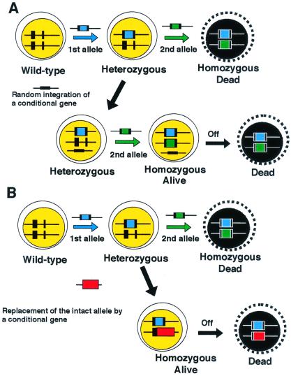
Strategy for generating conditionally gene targeted clones. Ts mutant cDNAs of a gene of interest can be designed based on information from yeast ts mutants, if available. Following the standard protocol to generate ts mutant cells, each mutated cDNA is introduced by targeted integration into the intact endogenous locus of the gene in heterozygous mutant (+/−) DT40 cells. The resulting cells should be homozygous mutant (−/−) cells and express only the mutated protein.
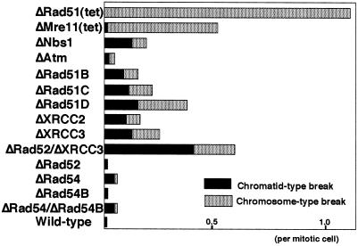
The level of spontaneous chromosomal aberrations in various DT40 mutant clones. Data are presented as macrochromosomal (1–5 and Z) aberrations per mitotic cell.
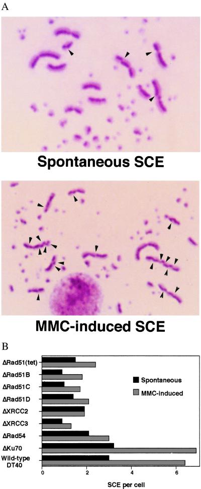
(A) SCE in wild-type DT40 cells. (A) Spontaneous SCE and (B) mitomycin C (MMC)-induced SCE are shown. Arrowheads indicate the sites of SCE. [Reproduced with permission from ref. (Copyright 1999, American Society for Microbiology)]. (B) Reduced levels of SCE in cells deficient in homologous recombination. Cells were labeled with BrdUrd during two cell cycle periods with or without MMC treatment (50 ng/ml) for the last 8 h. Spontaneous and MMC-induced SCEs were scored in the macrochromosomes of metaphase cells. Histograms show the mean value of SCE per cell. The SCE levels of all mutants except ΔKu70 differ significantly (P < 0.002) from wild-type control SCE levels; statistical significance was calculated by the Mann–Whitney nonparametric u test.
References
-
- Reynaud C A, Bertocci B, Dahan A, Weill J C. Adv Immunol. 1994;57:353–378. - PubMed
-
- Arakawa H, Kuma K, Yasuda M, Furusawa S, Ekino S, Yamagishi H. J Immunol. 1998;160:4232–4241. - PubMed
-
- Baba T W, Giroir B P, Humphries E H. Virology. 1985;144:139–151. - PubMed
-
- Buerstedde J M, Takeda S. Cell. 1991;67:179–188. - PubMed
Publication types
MeSH terms
Substances
LinkOut - more resources
Full Text Sources
Other Literature Sources
Molecular Biology Databases
Research Materials
