. 2001 Jul 17;98(15):8419-24.
doi: 10.1073/pnas.111005398.

Domain structure and dynamics in the helical filaments formed by RecA and Rad51 on DNA

Affiliations

Domain structure and dynamics in the helical filaments formed by RecA and Rad51 on DNA

X Yu et al. Proc Natl Acad Sci U S A. .

Abstract

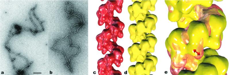
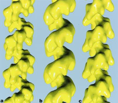
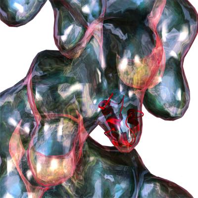
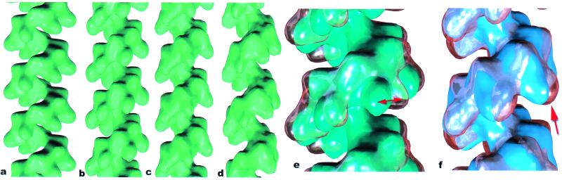
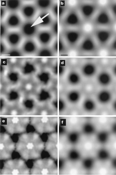
Both the bacterial RecA protein and the eukaryotic Rad51 protein form helical nucleoprotein filaments on DNA that catalyze strand transfer between two homologous DNA molecules. However, only the ATP-binding cores of these proteins have been conserved, and this same core is also found within helicases and the F1-ATPase. The C-terminal domain of the RecA protein forms lobes within the helical RecA filament. However, the Rad51 proteins do not have the C-terminal domain found in RecA, but have an N-terminal extension that is absent in the RecA protein. Both the RecA C-terminal domain and the Rad51 N-terminal domain bind DNA. We have used electron microscopy to show that the lobes of the yeast and human Rad51 filaments appear to be formed by N-terminal domains. These lobes are conformationally flexible in both RecA and Rad51. Within RecA filaments, the change between the "active" and "inactive" states appears to mainly involve a large movement of the C-terminal lobe. The N-terminal domain of Rad51 and the C-terminal domain of RecA may have arisen from convergent evolution to play similar roles in the filaments.


Figures

Figure 1
Figure 1
Electron micrograph of hRad51 after incubation with ssDNA in the presence of ADP and AlFformula image (used as a non-hydrolyzable analog of ATP; a) and after incubation with ssDNA and ATP-γ-S (b). Average from filaments of hRad51 formed on ssDNA in the presence of ADP and AlFformula image (c) is compared with average of hRad51 filaments on ssDNA in the presence of ATP-γ-S (d). The average in d was generated from 4,199 segments and has a pitch of 76 Å. The symmetry of this filament, 6.43 subunits per turn, is quite close to the symmetry of the 99-Å pitch filament in c, 6.39 subunits per turn. When the two structures are superimposed (e), it can be seen that the main difference is a rotation of the subunit lobes (red double arrow). Because of the large difference in pitch between the ADP-AlFformula image filament (shown as a glass surface) and the ATP-γ-S filament (in yellow), the comparison shown in e is meaningful only for the subunit labeled with the arrow. The scale bar in a is 1,000 Å.
Figure 2
Figure 2
Surfaces of reconstructions of filaments formed by hRad51 (a), ScRad51 (b), and E. coli RecA (c) on DNA. The hRad51 filaments were formed on ssDNA in the presence of ADP and AlFformula image, and 7,620 filament segments were used to generate the reconstruction. The ScRad51 filaments were formed on dsDNA in the presence of ATP-γ-S, and 10,757 segments were used. The RecA filaments were formed on dsDNA with ATP-γ-S, and 8,635 segments were used. The helical parameters that were determined from the real-space refinement (27) for the hRad51 were a pitch of 99 Å and 6.39 subunits/turn. The parameters for the ScRad51 were a pitch of 94 Å with 6.28 subunits per turn, and the parameters for RecA were a pitch of 91 Å with 6.16 subunits/turn.
Figure 3
Figure 3
An atomic structure for the N-terminal domain of hRad51 (18) can be easily fit into the lobe of the hRad51 reconstruction (glass surface). Although an ambiguity exists about the rotational orientation of this domain within the filament, the residues (nos. 61–69) involved in binding DNA are located at the bottom of the lobe.
Figure 4
Figure 4
A comparison of a low-resolution rendering of the RecA crystal structure (ref. ; a) with reconstructions of the “inactive” RecA filament (b) and two states of the “active” filament (c and d). Whereas the inactive filament, in general, has a shorter pitch than the active filament, the pitch of the two states can overlap substantially. The filament in b (averaged from 3,014 segments) has a pitch of 82 Å with 6.09 subunits/turn. The reconstruction of the active RecA filament in Fig. 1c (from 8,635 segments) had a pitch of 91 Å with 6.16 subunits/turn. However, smaller averages can be obtained of filaments in this state with very different values for the pitch. The filament in c (averaged from 1,294 segments) has a pitch of 84 Å with 5.97 subunits/turn, whereas the filament in d (averaged from 1,551 segments) has a pitch of 97 Å with 6.20 subunits/turn. When the inactive filament in b is displayed as a glass surface and superimposed on the RecA crystal structure (e), it can be seen that there is a large rotation (≈10°-15°) of the C-terminal lobe between the two (red double arrow). A comparison (f) of the inactive 82 Å pitch filament (b), shown as a glass surface, and the active 84 Å pitch filament (c), displayed in blue, shows that the main difference is due to a large shift in the C-terminal domain (arrow).
Figure 5
Figure 5
Projections down the filament axis from the crystal of RecA alone (ref. ; a) and from an in situ crystal of RecA that has been shown to contain DNA (ref. ; b). The arrow in a indicates the lack of any density (black) near the filament axis in the RecA crystal. The two different states of the RecA filament described in this paper have very different axial projections. Pseudocrystalline lattices of these filaments are shown in c for the ADP-state and e for the ATP-state. The lattices are not exactly crystalline, because the filaments do not have 6.0 subunits/turn, but have 6.09 subunits/turn (c) and 6.20 subunits/turn (e). Low-pass filtered images of c and e are shown in d and f, respectively. It can be seen that the lattice generated from the ADP-dsDNA-RecA filaments provides a very good match to the in situ crystal (b), but the ATP-dsDNA-filament lattice (f) would be easily distinguishable, even at very low resolution. Further, the density because of the DNA and the putative DNA-binding loops is most likely responsible for the large difference in axial density between the RecA crystal (a) and the RecA-dsDNA filaments (c and d). [a and b are reproduced with permission from ref. (Copyright 2000, PNAS).]

References

    1. Kowalczykowski S C, Dixon D A, Eggleston A K, Lauder S D, Rehrauer W M. Microbiol Rev. 1994;58:401–465. - PMC - PubMed
    1. Roca A I, Cox M M. Prog Nucleic Acid Res Mol Biol. 1997;56:129–223. - PubMed
    1. Tashiro S, Walter J, Shinohara A, Kamada N, Cremer T. J Cell Biol. 2000;150:283–291. - PMC - PubMed
    1. Tashiro S, Kotomura N, Shinohara A, Tanaka K, Ueda K, Kamada N. Oncogene. 1996;12:2165–2170. - PubMed
    1. Kato M, Yano K, Matsuo F, Saito H, Katagiri T, Kurumizaka H, Yoshimoto M, Kasumi F, Akiyama F, Sakamoto G, et al. J Hum Genet. 2000;45:133–137. - PubMed

Publication types