. 2001 Jul 17;98(15):8447-53.
doi: 10.1073/pnas.121009898.

Rad54 protein stimulates the postsynaptic phase of Rad51 protein-mediated DNA strand exchange

Affiliations

Rad54 protein stimulates the postsynaptic phase of Rad51 protein-mediated DNA strand exchange

J A Solinger et al. Proc Natl Acad Sci U S A. .

Abstract

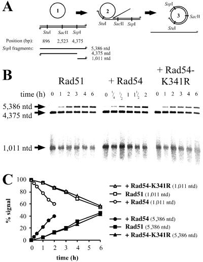
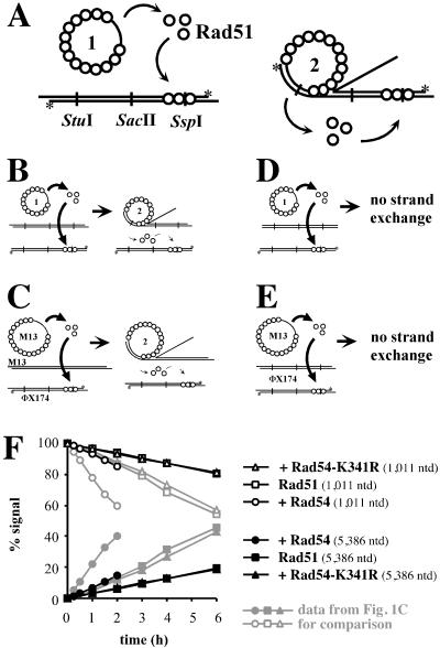
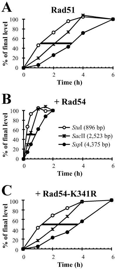
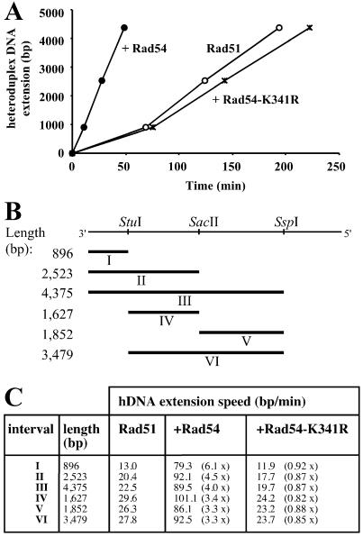
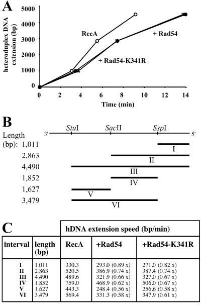
Rad54 and Rad51 are important proteins for the repair of double-stranded DNA breaks by homologous recombination in eukaryotes. As previously shown, Rad51 protein forms nucleoprotein filaments on single-stranded DNA, and Rad54 protein directly interacts with such filaments to enhance synapsis, the homologous pairing with a double-stranded DNA partner. Here we demonstrate that Saccharomyces cerevisiae Rad54 protein has an additional role in the postsynaptic phase of DNA strand exchange by stimulating heteroduplex DNA extension of established joint molecules in Rad51/Rpa-mediated DNA strand exchange. This function depended on the ATPase activity of Rad54 protein and on specific protein:protein interactions between the yeast Rad54 and Rad51 proteins.


Figures

Figure 1
Figure 1
DNA strand exchange and assay for hDNA extension. (A) Schematic representation of the hDNA extension assay (modified from ref. 39). DNA strand exchange with circular ssDNA and linear dsDNA substrates (1). The joint molecules (2) are resolved by hDNA extension to nicked circular and displaced ssDNA end products (3). Digestion with different restriction endonucleases followed by denaturing gel electrophoresis is used to observe hDNA extension. Note that the displaced strand cannot be digested once hDNA extends over the restriction site. The restriction endonucleases used and the positions of the corresponding restriction sites along the dsDNA are indicated. Radioactively labeled 5′ ends are marked with an asterisk. The expected fragments after digestion with SspI are indicated and data for this restriction enzyme are shown in B and C. (B) Denaturing agarose gels analyzing DNAs from DNA strand exchange reactions (A) containing Rad51 protein and Rpa. Storage buffer (Rad51 control), Rad54, or Rad54-K341R protein was added as indicated. All reactions shown were digested with SspI restriction endonuclease. The band corresponding to the full-length displaced strand (5,386 nt) appears in a time-dependent manner. At the same time, the amount of the corresponding shorter fragment (1,011 nt) declines. The sizes of the fragments are indicated on the left. The incubation times of the DNA strand exchange assays are shown for each lane. Note the shorter incubation times for the reactions containing Rad54 protein. A minor loss of label that was not dependent on Rad54 protein was observed at late time points. This loss of label did not affect the quantitation, as the relative amount of the 5,386-nt product was determined in comparison to the digested 1,011-nt fragment (see C). (C) Graphical representation of data shown in B. The 5,386-nt (●, ■, ▴) and 1,011-nt (○, □, ▵) bands were quantified by phosphorimaging, and the relative amounts of both fragments are displayed. At 0 h the signal for the 1,001-nt fragment was defined as 100% and the signal for the 5,386-nt fragment as 0%. Reactions contained S. cerevisiae Rad51 protein and Rpa, as well as storage buffer (Rad51 control; □, ■), Rad54 (○, ●), or Rad54-K341R (▵, ▴) protein. The relative decrease and increase of both fragments after quantitation by a phosphorimager is more evident than by visual inspection of the scanned image.
Figure 2
Figure 2
Control experiments for hDNA extension assay. Control reactions to measure the amount of inhibition of the restriction endonucleases due to the relocalization of Rad51 protein to the dsDNA. (A) Relocalization of Rad51 protein from ssDNA to dsDNA might occur either spontaneously (Right) or may be enhanced during DNA strand exchange (Left). (BE) To determine the amount of signal in the hDNA extension assay, which corresponds to uncleaved dsDNA analyzed on a denaturing gel because of interference by Rad51 protein relocating on dsDNA, four types of control reactions were performed: in the presence of ongoing DNA strand exchange (B + C) and in the absence of DNA strand exchange (D + E). (B + D) Reactions contained blunt-ended dsDNA (linearized with FspI and labeled at the 5′ ends by polynucleotide kinase as indicated with asterisks), which could not participate in DNA strand exchange (ref. and data not shown). The unlabeled dsDNA was either digested with PstI (B) to enable DNA strand exchange or with FspI (D) to preclude DNA strand exchange. (C + E) Control experiments with heterologous M13mp19 ssDNA. (C) Reactions with ongoing DNA strand exchange contained circular M13mp19 ssDNA and PstI linearized M13mp19 dsDNA. (E) Reactions without DNA strand exchange contained circular M13mp19 ssDNA and PstI linearized ΦX174 dsDNA. The relocalization of Rad51 protein was monitored with a radioactively end-labeled (*) ΦX174 dsDNA linearized with PstI. (F) Graphical representation of data from control reactions as in E by using SspI restriction endonuclease. To reactions with Rad51 protein and Rpa, storage buffer (Rad51 control) (□, ■), Rad54 (○, ●), or Rad54-K341R (▵, ▴) protein was added. All results were obtained in the same manner as in Fig. 1, and the data from Fig. 1C are indicated in gray for comparison.
Figure 3
Figure 3
Rad54 protein stimulates joint molecule formation and subsequent hDNA extension. DNA from DNA strand exchange reactions as described in Fig. 1A was digested with StuI (○), SacII (*), or SspI (●) and subjected to denaturing gel electrophoresis. All bands were quantified on a phosphorimager and the relative amount corresponding to the uncut fragment arising from hDNA extension past the restriction site was calculated (see Fig. 1 B and C). All data were corrected for the contribution to the accumulation of uncut fragment by the relocalization of Rad51 protein as described in Fig. 2. Reactions contained S. cerevisiae Rad51 protein and Rpa, to which storage buffer (Rad51 control; A), Rad54 (B), or Rad54-K341R protein (C) was added. The data were normalized by setting the endpoints of each reaction to 100%. This was possible because all reactions were at or very near to their endpoints and achieved very similar final yields (25–30% products). The lines at 50% represent the time needed for half of the population of joint molecules to migrate from the StuI site to the SspI site.
Figure 4
Figure 4
Rad54 protein stimulates the rate of hDNA extension in Rad51 protein-mediated DNA strand exchange. (A) Graphic representation of the effect of Rad54 protein on hDNA extension derived from the data shown in Fig. 3 AC. Plotted are the time points when 50% of the joint molecules have passed the physical distance defined by the three restriction sites. Reactions contained S. cerevisiae Rad51 protein and Rpa as well as storage buffer (Rad51 control; ○), Rad54 (●), or Rad54-K341R (*). (B) Intervals in DNA strand exchange. ΦX174 dsDNA linearized with PstI is represented on top and has a total length of 5,386 bp. The direction refers to the virion DNA (plus strand). The restriction sites used for this analysis are indicated. Rates for hDNA extension could be calculated for intervals I–VI. (C) Rad54 protein stimulates the rate of hDNA extension. The intervals are illustrated in B. The numbers in parentheses represent the fold stimulation in comparison to control reactions containing Rad51 protein to which protein storage buffer was added.
Figure 5
Figure 5
Rad54 protein does not stimulate the rate of hDNA extension in RecA-mediated DNA strand exchange. (A) Graphic representation of the effect of Rad54 protein on hDNA extension derived from data similar to those shown in Fig. 3 for Rad51 protein. Plotted are the time points when 50% of the joint molecules have passed the physical distance defined by the three restriction sites. Reactions contained E. coli RecA protein and Rpa as well as storage buffer (RecA control; ○), Rad54 (●), or Rad54-K341R (*). (B) Intervals in RecA-mediated DNA strand exchange. The restriction sites used for this analysis are indicated. Rates for hDNA extension speeds could be calculated for intervals I–VI. Note that the directionality of the reaction is opposite to that described in Fig. 4B. (C) Rad54 protein is unable to stimulate hDNA extension of joint molecules formed by RecA. The intervals are illustrated in B. The numbers in parentheses represent the fold stimulation in comparison to control reactions containing RecA protein to which protein storage buffer was added.
Figure 6
Figure 6
Comparison of proteins with homologies to RuvB and Rad54/Snf2. The protein sequences of E. coli RuvB and S. cerevisiae Rad54 were used in BLAST searches to define RuvB- and Rad54-like protein families. The GenBank accession numbers are given for each protein. The groups comprise the following proteins: (A) Bacterial RuvB proteins from Borrelia burgdorferi AAC66410, Bacillus halodurans BAB04944, Bacillus subtilis CAB75331, Campylobacter jejuni CAB73789, Chlamydia muridarum AAF39175, Chlamydophila pneumoniae BAA98598, Chlamydia trachomatis AAC67630, Deinococcus radiodurans AAF10176, E. coli BAA15671, Hemophilus influenzae AAC21975, Heliobacter pylori AAD08100, Mycoplasma pneumoniae AAB95954, Mycobacterium leprae AAA17098, Mycoplasma genitalium AAC71584, Mycobacterium tuberculosis CAB01285, Neisseria meningitidis AAF41624, Pseudomonas aeruginosa AAG04356, Rhizobium etli AAF36814, Rickettsia prowazekii CAA14843, Streptomyces coelicolor CAB70920, Synechocystis sp. BAA10350, Thermus thermophilus BAA76480, Thermotoga maritima AAB03727, Treponema pallidum AAC65150, Vibrio cholerae AAF94993, Xylella fastidiosa AAF84708; eukaryotic RuvB-like proteins from Arabidopsis thaliana CAB66921 and BAB08471, Drosophila melanogaster AAF43412 (reptin) and AAF43411 (pontin), Homo sapiens BAA28169 RuvBL1) and BAA76708 (RuvBL2), Mus musculus BAA76297, Rattus norvegicus BAA76313, Saccharomyces cerevisiae CAA88704 (Rvb1) and CAA97952 (Rvb2); archaeal Tip49 proteins from Archaeoglobus fulgidus AAB89434, Aeropyrum pernix BAA79281, Pyrococcus abyssi CAB49285, Pyrococcus horikoshii BAA30923, Sulfolobus solfataricus (see http://niji.imb.nrc.ca/sulfolobus). (B) Eukaryotic Rad54 proteins from Arabidopsis thaliana BAB02963, Caenorhabditis elegans CAA22254 (W06D4.6), Drosophila melanogaster AAC24577 (OKR), Gallus gallus AAB54115 and AAG09308 (Rad54B), Homo sapiens CAA66379 and AAD34331 (Rad54B), Mus musculus CAA66380, Neurospora crassa BAA93079, S. cerevisiae AAA34949 and CAA85017 (Rdh54/Tid1), Schizosaccharomyces pombe CAA82750 (Rhp54); S. cerevisiae Swi2/Snf2 AAA35059; bacterial Rad54-like proteins from Bacillus cereus CAA67095, Bacillus halodurans BAB06632, Bacillus subtilis CAB15645, Chlamydia muridarum AAF73609 and AAF73530, Chlamydophila pneumoniae AAF38809 and AAF73724, Chlamydia trachomatis AAC68157 and AAC68303, Deinococcus radiodurans AAF10831, Mycoplasma genitalium AAC71234, Mycoplasma pneumoniae AAB95782, Mycobacterium tuberculosis CAA17284, Pseudomonas aeruginosa AAG04188, Streptomyces coelicolor CAB60181 and CAB82825, Synechocystis sp. BAA18659; archaeal Rad54-like protein from Sulfolobus solfataricus (see http://niji.imb.nrc.ca/sulfolobus); bacterial HepA-like proteins form Bacillus halodurans BAB06536, Bacillus subtilis BAA12545, Dich elobacter nodosus AAC33384, Deinococcus radiodurans AAF12565, E. coli AAC73170, Hemophilus influenzae AAC22275, Thermotoga maritima AAD36069, Vibrio cholerae AAF95648; archaeal HepA-like proteins from Archaeoglobus fulgidus AAB91314, Aeropyrum pernix BAA79369, Halobacterium sp. AAG20812, Pyrococcus abyssi CAB49794, Pyrococcus horikoshii BAA29994. All possible pairwise alignments with RuvB and Rad54 homologs were carried out by using BLAST. The numbers indicate the average identities and similarities (in brackets) inside the different groups as well as between the groups. Comparison of an unrelated protein Xrn1 from S. cerevisiae (AAA35219) to all bacterial RuvB proteins resulted in 23% identity and 41% similarity. Comparison of a random shuffled E. coli RuvB sequence to all bacterial RuvB proteins (including E. coli RuvB protein) resulted in 23% identity and 39% similarity.

References

    1. Paques F, Haber J E. Microbiol Molec Biol Rev. 1999;63:349–404. - PMC - PubMed
    1. Bianco P R, Tracy R B, Kowalczykowski S C. Front Biosci. 1998;3:570–603. - PubMed
    1. Sung P. Science. 1994;265:1241–1243. - PubMed
    1. Sugiyama T, Zaitseva E M, Kowalczykowski S C. J Biol Chem. 1997;272:7940–7945. - PubMed
    1. Kowalczykowski S C, Dixon D A, Eggleston A K, Lauder S D, Rehrauer W M. Microbiol Rev. 1994;58:401–465. - PMC - PubMed

Publication types

MeSH terms