. 2001 Aug;75(16):7305-14.
doi: 10.1128/JVI.75.16.7305-7314.2001.

A human rotavirus with rearranged genes 7 and 11 encodes a modified NSP3 protein and suggests an additional mechanism for gene rearrangement

Affiliations

A human rotavirus with rearranged genes 7 and 11 encodes a modified NSP3 protein and suggests an additional mechanism for gene rearrangement

E Gault et al. J Virol. 2001 Aug.

Abstract

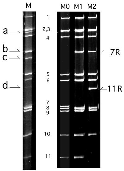
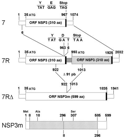
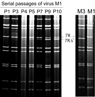
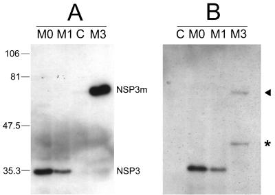
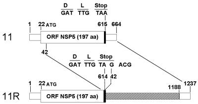
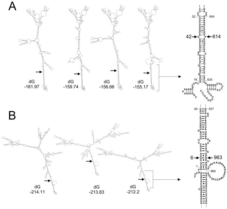
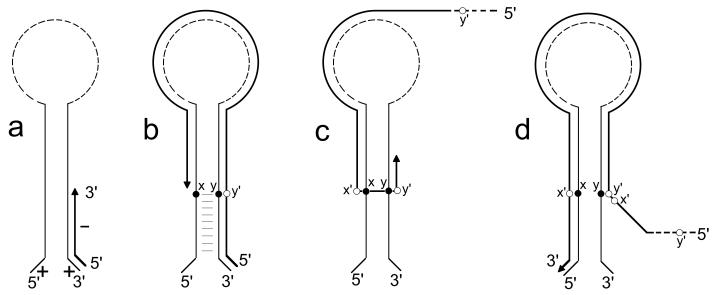
A human rotavirus (isolate M) with an atypical electropherotype with 14 apparent bands of double-stranded RNA was isolated from a chronically infected immunodeficient child. MA-104 cell culture adaptation showed that the M isolate was a mixture of viruses containing standard genes (M0) or rearranged genes: M1 (containing a rearranged gene 7) and M2 (containing rearranged genes 7 and 11). The rearranged gene 7 of virus M1 (gene 7R) was very unusual because it contained two complete open reading frames (ORF). Moreover, serial propagation of virus M1 in cell culture indicated that gene 7R rapidly evolved, leading to a virus with a deleted gene 7R (gene 7RDelta). Gene 7RDelta coded for a modified NSP3 protein (NSP3m) of 599 amino acids (aa) containing a repetition of aa 8 to 296. The virus M3 (containing gene 7RDelta) was not defective in cell culture and actually produced NSP3m. The rearranged gene 11 (gene 11R) had a more usual pattern, with a partial duplication leading to a normal ORF followed by a long 3' untranslated region. The rearrangement in gene 11R was almost identical to some of those previously described, suggesting that there is a hot spot for gene rearrangements at a specific location on the sequence. It has been suggested that in some cases the existence of short direct repeats could favor the occurrence of rearrangement at a specific site. The computer modeling of gene 7 and 11 mRNAs led us to propose a new mechanism for gene rearrangements in which secondary structures, besides short direct repeats, might facilitate and direct the transfer of the RNA polymerase from the 5' to the 3' end of the plus-strand RNA template during the replication step.


Figures

FIG. 1
FIG. 1
RNA profiles of the M isolate and of viruses M0, M1, and M2. Lanes: M, RNA profile of the M isolate obtained from the original stool sample; M0, M1, and M2, RNA profiles of viruses M0, M1, and M2 recovered in MA-104 cell culture performed under limiting-dilution conditions. Numbers indicate the rotavirus gene segments of standard size. a, b, c, and d represent the extra segments in isolate M. 7R and 11R indicate rearranged genes 7 and 11, respectively.
FIG. 2
FIG. 2
Schematic diagram of the standard and rearranged genes 7 and of the NSP3m protein. The ORFs are indicated by large boxes, and the UTRs are indicated by small boxes. Thick lines indicate the stop codons. The nucleotide positions corresponding to the gene rearrangement (gene 7R) and to the 91-bp deletion of gene 7RΔ are indicated. The nucleotide sequence of the junction region of the rearrangement is detailed. In gene 7R, the duplicated part of the gene is shaded. In NSP3m, residues are numbered referring to the first methionine codon (at nt 35) and the repeated residues (aa 8 to 296) are indicated by a cross-hatched box.
FIG. 3
FIG. 3
Serial propagation of virus M1 in cell culture. (Left) Evolution of the virus M1 RNA profile during serial passages in MA-104 cell culture. Pn indicates the number of passages (n) performed prior to the analysis of the electropherotype. At P4, segment 7RΔ appeared below segment 7R (arrows), increased in relative concentration along with further passages, and became a majority at P9. (Right) RNA profiles of the viruses obtained after subculture of the P10 viral mixture, allowing us to recover virus M3 containing segment 7RΔ and virus M1 containing segment 7R.
FIG. 4
FIG. 4
Western blot analyses of NSP3 and NSP3m. MA-104 cells infected by viruses M0, M1, and M3 were recovered 24 h postinfection, and whole-cell lysates were analyzed by Western blotting with the monoclonal antibody ID3 specific for NSP3. C indicates mock-infected cells as a control. Numbers indicate molecular mass markers in kilodaltons. The position of viral proteins NSP3 and NSP3m are indicated. (A) Western blotting was performed after cell lysis with a 2% SDS-containing buffer. The apparent molecular mass of NSP3m (virus M3) was approximately double the molecular mass of NSP3 (viruses M0 and M1). (B) Western blot analysis was performed after cell lysis without SDS. Unlike NSP3, NSP3m was only faintly detected (arrow) and an additional band of 40 kDa was observed (asterisk).
FIG. 5
FIG. 5
Schematic diagram of the standard and rearranged genes 11. The ORFs are indicated by large open boxes, and the UTRs are indicated by small open boxes. Thick lines indicate the stop codons. The nucleotide positions corresponding to the gene rearrangement (gene 11R) are indicated. The nucleotide sequence of the junction region of the rearrangement is detailed. The duplicated sequence of the gene 11R is shaded.
FIG. 6
FIG. 6
Predicted optimal secondary structures for gene 7 and 11 plus-strand RNAs. Examples of some optimal folding configurations predicted by the mfold program for genes 7 (A) and 11 (B) of virus M0 are shown. dG indicates the minimum free energy values (in kilocalories per mole). Arrows indicate the positions of the nucleotides involved in the rearrangements leading to genes 7R and 11R. On the right, the base pairing between the 3′ and 5′ ends of the RNA predicted for the most stable secondary structures is enlarged. The nucleotides involved in the gene rearrangements are indicated.
FIG. 7
FIG. 7
Possible mechanism for rearrangements of rotavirus genes. (a) During the replication step, the viral RNA polymerase initiates the minus-strand synthesis at the 3′ end of the plus-strand RNA. (b) The RNA polymerase reaches nucleotide position x, in the panhandle formed by the 5′ and 3′ ends of the RNA, and interrupts RNA synthesis. (c) Without releasing the nascent minus strand, the RNA polymerase falls back on its template at nucleotide y in the panhandle and reinitiates replication. (d) The RNA polymerase completes the replication up to the 5′ end, thus duplicating the sequence x′-y′ (complementary to x-y). Since minus-strand RNAs were predicted to fold quite differently from plus-strand RNAs (data not shown), a similar model could not be proposed to explain the occurrence of rearrangement at the transcription step. In addition, at the transcription stage, the minus-strand RNA is associated with the plus strand and is probably not subject to folding.

Similar articles

Cited by

References

    1. Allen A M, Desselberger U. Reassortment of human rotaviruses carrying rearranged genomes with bovine rotavirus. J Gen Virol. 1985;66:2703–2714. - PubMed
    1. Aponte C, Mattion N M, Estes M K, Charpilienne A, Cohen J. Expression of two bovine rotavirus non-structural proteins (NSP2, NSP3) in the baculovirus system and production of monoclonal antibodies directed against the expressed proteins. Arch Virol. 1993;133:85–95. - PubMed
    1. Ballard A, McCrae M A, Desselberger U. Nucleotide sequences of normal and rearranged RNA segments 10 of human rotaviruses. J Gen Virol. 1992;73:633–638. - PubMed
    1. Bellinzoni R C, Mattion N M, Burrone O, Gonzalez A, La Torre J L, Scodeller E A. Isolation of group A swine rotaviruses displaying atypical electropherotypes. J Clin Microbiol. 1987;25:952–954. - PMC - PubMed
    1. Besselaar T G, Rosenblatt A, Kidd A H. Atypical rotavirus from South African neonates. Arch Virol. 1986;87:327–330. - PubMed

Publication types

MeSH terms

LinkOut - more resources