. 2001 Aug;75(16):7339-50.
doi: 10.1128/JVI.75.16.7339-7350.2001.

Proteolysis of monomeric recombinant rotavirus VP4 yields an oligomeric VP5* core

Affiliations

Proteolysis of monomeric recombinant rotavirus VP4 yields an oligomeric VP5* core

P R Dormitzer et al. J Virol. 2001 Aug.

Abstract

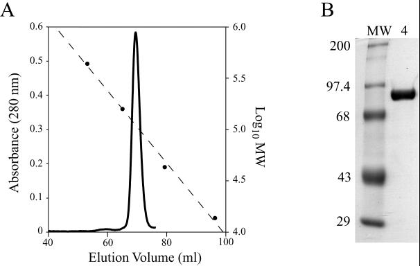
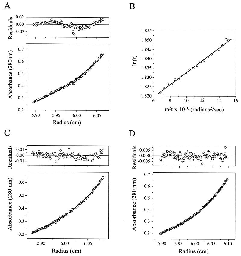
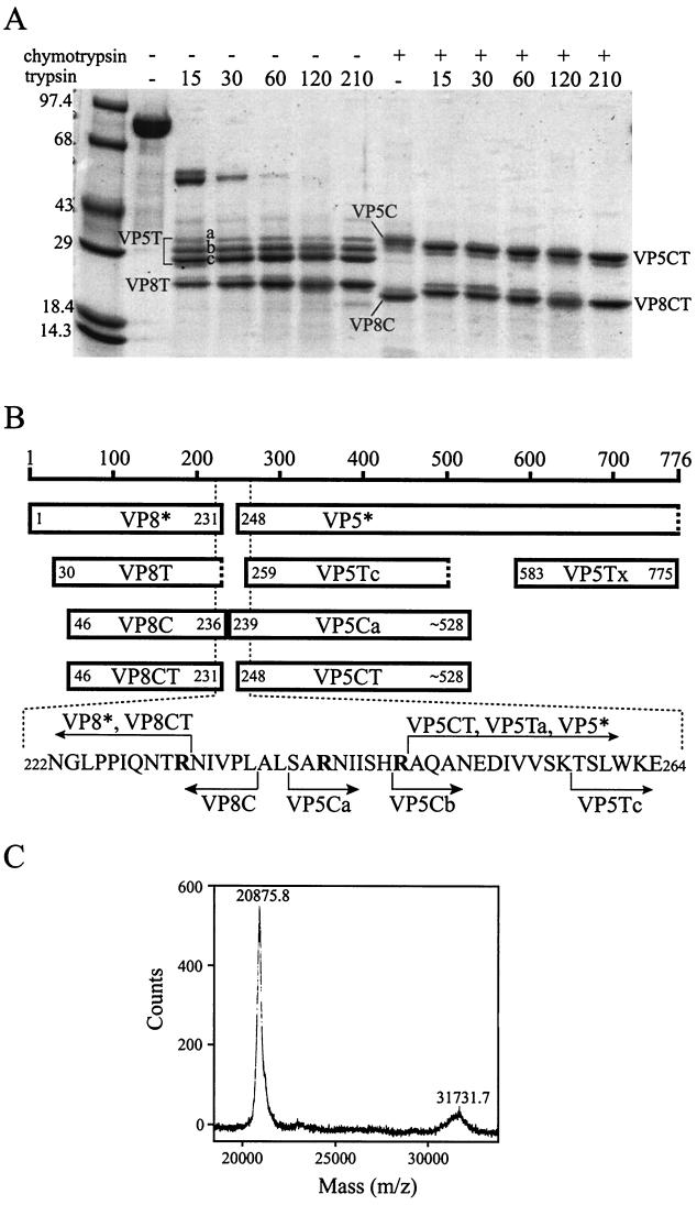
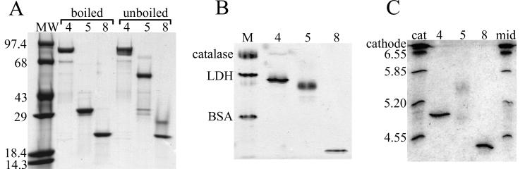
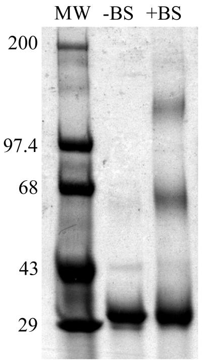
Rotavirus particles are activated for cell entry by trypsin cleavage of the outer capsid spike protein, VP4, into a hemagglutinin, VP8*, and a membrane penetration protein, VP5*. We have purified rhesus rotavirus VP4, expressed in baculovirus-infected insect cells. Purified VP4 is a soluble, elongated monomer, as determined by analytical ultracentrifugation. Trypsin cleaves purified VP4 at a number of sites that are protected on the virion and yields a heterogeneous group of protease-resistant cores of VP5*. The most abundant tryptic VP5* core is trimmed past the N terminus associated with activation for virus entry into cells. Sequential digestion of purified VP4 with chymotrypsin and trypsin generates homogeneous VP8* and VP5* cores (VP8CT and VP5CT, respectively), which have the authentic trypsin cleavages in the activation region. VP8CT is a soluble monomer composed primarily of beta-sheets. VP5CT forms sodium dodecyl sulfate-resistant dimers. These results suggest that trypsinization of rotavirus particles triggers a rearrangement in the VP5* region of VP4 to yield the dimeric spikes observed in icosahedral image reconstructions from electron cryomicroscopy of trypsinized rotavirus virions. The solubility of VP5CT and of trypsinized rotavirus particles suggests that the trypsin-triggered conformational change primes VP4 for a subsequent rearrangement that accomplishes membrane penetration. The domains of VP4 defined by protease analysis contain all mapped neutralizing epitopes, sialic acid binding residues, the heptad repeat region, and the membrane permeabilization region. This biochemical analysis of VP4 provides sequence-specific structural information that complements electron cryomicroscopy data and defines targets and strategies for atomic-resolution structural studies.


Figures

FIG. 1
FIG. 1
Purification of VP4. (A) Gel filtration chromatography. Chromatogram of VP4 separated on a Hi-Load 16/60 Superdex 200 gel filtration column as a final purification step. The Vo is 44.1 ml. Solid line, A280; dashed line, calibration curve; circles, elution volumes of MW markers. (B) SDS-PAGE. Coomassie blue-stained SDS-polyacrylamide gel of purified recombinant VP4. Lane MW, molecular mass standards labeled in kilodaltons; lane 4, VP4.
FIG. 2
FIG. 2
Analytical ultracentrifugation. Curves were fit and residuals were determined as described in Materials and Methods. (A) Equilibrium sedimentation of VP4. Shown is the A280-versus-radius plot of 453 μg of VP4 per ml in TNE following centrifugation at 9,000 rpm in an AN-60 Ti rotor (Beckman Coulter) at 4°C for 89 h. The curve, fit to data sets of VP4 centrifuged at starting concentrations of 112, 226, and 453 μg/ml, is the theoretical distribution of a 86.0-kDa particle with a partial specific volume of 0.7222 cm3/g in a buffer with a density of 1.005 g/ml using a baseline offset of 0.008 A280 unit. (B) Velocity sedimentation of VP4. Shown are results of a second-moment analysis of the sedimentation of 865 μg of VP4 per ml in TNE at 4°C during centrifugation at 25,000 rpm. The fitted line has a slope of 3.3 s. (C) Equilibrium sedimentation of VP8CT. Shown is an A280-versus-radius plot of 317 μg of VP8CT per ml in TNE following centrifugation at 20,000 rpm at 4°C for 45 h. The curve, fit to data sets of VP8CT centrifuged at starting concentrations of 79, 158, and 317 μg/ml, is the theoretical distribution of a 22.4-kDa particle with a partial specific volume of 0.7158 cm3/g in a buffer with a density of 1.005 g/ml using a baseline offset of −0.003 A280 unit. (D) Equilibrium sedimentation of VP5CT. Shown is a A280-versus-radius plot of 392 μg of VP5CT per ml in TNE following centrifugation at 9,000 rpm at 4°C for 90 h. The curve, fit to data sets of VP5CT centrifuged at starting concentrations of 98, 196, and 392 μg/ml, is the theoretical distribution of a 63.4-kDa particle with a partial specific volume of 0.7215 cm3/g that dimerizes with an association constant of 5.1 in a buffer with a density of 1.005 g/ml using a baseline offset of 0.002 A280 unit.
FIG. 3
FIG. 3
Sequential digestion of VP4 with chymotrypsin and trypsin. (A) Coomassie blue stained SDS-polyacrylamide gel. A 1.1-mg/ml solution of VP4 was either digested with 4.2 μg of chymotrypsin per ml for 30 min at 37°C (lanes marked with +) or incubated on ice for 30 min (lanes marked with −). Trypsin was then added to a concentration of 3.6 μg/ml (except in those samples marked − immediately above the lane), and the samples were incubated at room temperature for an additional 15 to 210 min (indicated by numbers immediately above the lanes). Digestions were stopped by the addition of PMSF to 0.625 mM prior to denaturation by boiling in reducing SDS-PAGE sample buffer and separation on a 4-to-15% polyacrylamide gradient gel. Molecular mass standards are identified in kilodaltons adjacent to marker bands. The gel has a slight distortion that causes bands on the right-hand side to appear to migrate further. (B) Schematic. The scale indicates residue numbers of intact VP4. “VP8*” and “VP5*” designate fragments generated by trypsinization of virions (3). “VP5Ca” and “VP5Cb” designate alternate N termini found on fragments with the electrophoretic mobility of band VP5C in panel A. “VP5Tx” designates the fragment first cleaved from intact VP4 upon exposure to trypsin. Other fragment designations refer to the digestion products labeled in panel A. Amino acids specified by number at the N termini of bars were determined by Edman degradation. Amino acids specified at the C termini of bars are preceded by “∼” if an alternate C terminus is also compatible with the data. Dashed ends of boxes designate C termini estimated from SDS-PAGE data. Bold letters in the amino acid sequence designate residues after which cleavages occur upon trypsinization of virion-associated VP4 (3). (C) Mass spectrometry of VP8CT and VP5CT. A dual digestion of VP4 was performed as described in Materials and Methods, and the products were analyzed by MALDI-time of flight mass spectrometry. The amount of VP5CT in the sample was diminished by its relatively poor elution from a benzamidine-Sepharose column. The range of the mass spectrum containing the single ionization peaks for VP8CT and VP5CT is displayed.
FIG. 4
FIG. 4
Purification of VP5CT and VP8CT. (A) Gel filtration chromatography. Shown is a chromatogram of VP5CT and VP8CT, produced by sequential digestion of VP4 with chymotrypsin and trypsin (protocol is described in Materials and Methods), separated on a Superdex 200 HR 10/30 column. The Vo is 8.43 ml. Solid line, A280; dashed line, calibration curve; circles, elution volumes of MW markers; peak 5, main VP5CT peak; peak 8, VP8CT peak. Fractions are numbered above the abscissa. (B) Coomassie blue-stained SDS-polyacrylamide gel. The fractions labeled in panel A were analyzed on a reducing 4-to-15% polyacrylamide gradient gel. Fraction numbers are indicated immediately above the lanes. Fractions pooled for further analysis of VP5CT and VP8CT are indicated above the brackets. Molecular mass standards are identified in kilodaltons adjacent to the marker bands.
FIG. 5
FIG. 5
Electrophoresis of VP4, VP5CT, and VP8CT. (A) Coomassie blue-stained SDS-polyacrylamide gel. Purified proteins were denatured with SDS-PAGE sample buffer containing 1% β-mercaptoethanol and either boiled or not boiled (indicated above brackets) prior to separation on a 4-to-15% polyacrylamide gradient gel. Lane MW, molecular mass standards (in kilodaltons); lanes 4, VP4; lanes 5, VP5CT; lanes 8, VP8CT. (B) Coomassie blue-stained native polyacrylamide gel. Shown are purified proteins in TNE electrophoresed on a 4-to-25% polyacrylamide gradient gel with a discontinuous native buffer system (described in Materials and Methods). Lane M, native PAGE markers; LDH, lactate dehydrogenase; BSA, bovine serum albumin. Samples are labeled as in panel A. (C) Coomassie blue stained IEF gel. Purified proteins in TNE were applied to the cathode end of a polyacrylamide gel containing carrier ampholytes in the pH range 4 to 6.5. Markers were applied either at the cathode end (cat) or in the middle of the gel (mid), as indicated above the lanes. IEF was performed until the markers applied at either location had focused to the same position. The pI of each marker is indicated adjacent to the corresponding band, and the position of the cathode is indicated. Samples are labeled as in panel A.
FIG. 6
FIG. 6
Coomassie blue-stained SDS-polyacrylamide gel of chemically cross-linked VP5CT. VP5CT, untreated or cross-linked with BS3 (indicated above lanes), was boiled in reducing SDS-PAGE sample buffer and separated on a 4-to-15% polyacrylamide gradient gel. Molecular mass standards are identified in kilodaltons adjacent to the marker bands.
FIG. 7
FIG. 7
CD spectroscopy. In each plot, the experimentally measured mean residue molar ellipticity is depicted by open circles (measured); the ellipticity of a model protein with the composition indi- cated in Table 1 is depicted by filled circles (fit). The fit composition was determined using CONTIN (30). (A) VP4; (B) VP5CT; (C) VP8CT. deg, degree.

Similar articles

Cited by

References

    1. Allison S L, Schalich J, Stiasny K, Mandl C W, Kunz C, Heinz F X. Oligomeric rearrangement of tick-borne encephalitis virus envelope proteins induced by an acidic pH. J Virol. 1995;69:695–700. - PMC - PubMed
    1. Anonymous. Rotavirus vaccines. Wkly Epidemiol Rec. 1999;74:33–38. - PubMed
    1. Arias C F, Romero P, Alvarez V, Lopez S. Trypsin activation pathway of rotavirus infectivity. J Virol. 1996;70:5832–5839. - PMC - PubMed
    1. Barnett B B, Spendlove R S, Clark M L. Effect of enzymes on rotavirus infectivity. J Clin Microbiol. 1979;10:111–113. - PMC - PubMed
    1. Bullough P A, Hughson F M, Skehel J J, Wiley D C. Structure of influenza haemagglutinin at the pH of membrane fusion. Nature. 1994;371:37–43. - PubMed

Publication types

LinkOut - more resources