. 2001 Aug;75(16):7517-27.
doi: 10.1128/JVI.75.16.7517-7527.2001.

Human herpesvirus 8 envelope glycoprotein K8.1A interaction with the target cells involves heparan sulfate

Affiliations

Human herpesvirus 8 envelope glycoprotein K8.1A interaction with the target cells involves heparan sulfate

F Z Wang et al. J Virol. 2001 Aug.

Abstract

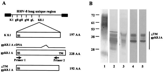
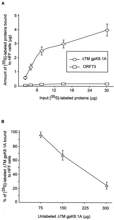
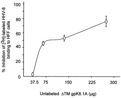
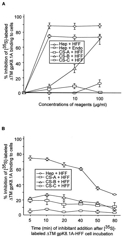
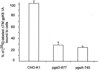
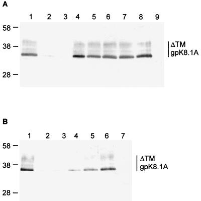
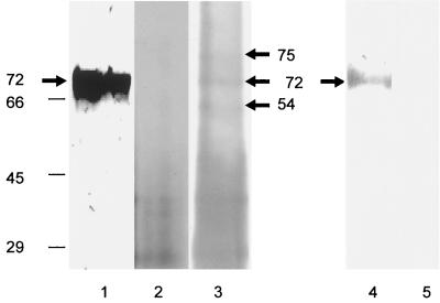
Human herpesvirus-8 (HHV-8) or Kaposi's sarcoma-associated herpesvirus K8.1 gene encodes for two immunogenic glycoproteins, gpK8.1A and gpK8.1B, originating from spliced messages. The 228-amino-acid (aa) gpK8.1A is the predominant form associated with the virion envelope, consisting of a 167-aa region identical to gpK8.1B and a 61-aa unique region (L. Zhu, V. Puri, and B. Chandran, Virology 262:237-249, 1999). HHV-8 has a broad in vivo and in vitro cellular tropism, and our studies showed that this may be in part due to HHV-8's interaction with the ubiquitous host cell surface molecule, heparan sulfate (HS). Since HHV-8 K8.1 gene is positionally colinear to the Epstein-Barr virus (EBV) gene encoding the gp350/gp220 protein involved in EBV binding to the target cells, gpK8.1A's ability to interact with the target cells was examined. The gpK8.1A without the transmembrane and carboxyl domains (DeltaTMgpK8.1A) was expressed in a baculovirus system and purified. Radiolabeled purified DeltaTMgpK8.1A protein bound to the target cells, which was blocked by unlabeled DeltaTMgpK8.1A. Unlabeled DeltaTMgpK8.1A blocked the binding of [(3)H]thymidine-labeled purified HHV-8 to the target cells. Binding of radiolabeled DeltaTMgpK8.1A to the target cells was inhibited in a dose-dependent manner by soluble heparin, a glycosaminoglycan (GAG) closely related to HS, but not by other GAGs such as chondroitin sulfate A and C, N-acetyl heparin and de-N-sulfated heparin. Cell surface absorbed DeltaTMgpK8.1A was displaced by soluble heparin. Radiolabeled DeltaTMgpK8.1A also bound to HS expressing Chinese hamster ovary (CHO-K1) cells, and binding to mutant CHO cell lines deficient in HS was significantly reduced. The DeltaTMgpK8.1A specifically bound to heparin-agarose beads, which was inhibited by HS and heparin, but not by other GAGs. Virion envelope-associated gpK8.1A was specifically precipitated by heparin-agarose beads. These findings suggest that gpK8.1A interaction with target cells involves cell surface HS-like moieties, and HHV-8 interaction with HS could be in part mediated by virion envelope-associated gpK8.1A.


Figures

FIG. 1
FIG. 1
(A) Construction of ΔTMgpK8.1A without the transmembrane and carboxyl domains. The top line shows the schematic diagram of HHV-8 genome and the location of encoded glycoprotein ORFs. The genomic K8.1 ORF is 197 aa long with a signal sequence (SS) and without the transmembrane (TM) sequence. The 228-aa gpK8.1A ORF with signal and transmembrane sequences is derived from a spliced mRNA (10). The ΔTMgpK8.1A was constructed by using primers amplifying aa 1 to 192 with the signal sequence but lacking the transmembrane and the carboxyl domains. (B) Expression and purification of ΔTMgpK8.1A in the baculovirus expression system. Sf9 insect cells were infected with ΔTMgpK8.1A-baculovirus for 2 days and labeled with [35S]methionine for 20 h. His-tagged ΔTMgpK8.1A protein from the cell pellet was purified by use of a nickel column. Protein purity was analyzed by SDS–12% PAGE gels, Western blots with anti-gpK8.1 MAb, and autoradiography. Lane 1, full-length gpK8.1A affinity purified from HHV-8-infected BCBL-1 cells detected by Western blot reaction with anti-gpK8.1A MAb; lane 2, ΔTMgpK8.1A-expressing Sf9 insect cell lysate in Western blot reactions with anti-gpK8.1A MAb; lane 3, ΔTMgpK8.1A-expressing Sf9 cells culture supernatant in Western blot reactions with anti-gpK8.1A MAb; lane 4, [35S]methionine-labeled purified ΔTMgpK8.1A in Western blot reactions with anti-gpK8.1A MAb; lane 5, autoradiography of [35S]methionine-labeled purified ΔTMgpK8.1A. The numbers on the left indicate the molecular masses (in kilodaltons) of the standard protein markers run in parallel lanes. The glycosylated forms of ΔTMgpK8.1A are marked on the right.
FIG. 2
FIG. 2
HHV-8 gpK8.1A binds to the target cells. Binding of purified full-length gpK8.1A and ΔTMgpK8.1A to the target cells was detected by surface immunofluorescence assay. Paraformaldehyde-treated BJAB, 293, HFF, or HMVEC-d cells were incubated with medium alone (controls) or medium with purified proteins for 30 min at 37°C. After cells were washed, anti-gpK8.1A-specific MAb or anti-HHV-8 ORF 59 MAb (11) or rabbit anti-HHV-8 ORF 73 antibodies (34) were added, incubated for 30 min at 37°C, washed, and incubated for an additional 30 min at 37°C with FITC-conjugated goat anti-mouse or anti-rabbit IgG antibodies. Cells were washed, mounted, and examined under a fluorescence microscope. (A and B) BJAB and 293 cells, respectively, incubated with the full-length affinity-purified gpK8.1A and anti-gpK8.1A MAb. (C) BJAB cells incubated with anti-gpK8.1A MAb alone. (D) BJAB cells incubated with the purified His-tagged ΔTMgpK8.1A and anti-gpK8.1A MAb. Fluorescence signals detected on the surface of cells indicate the cell-bound gpK8.1A and ΔTMgpK8.1A.
FIG. 3
FIG. 3
(A) Binding of radiolabeled ΔTMgpK8.1A to HFF cells. Different concentrations of [35S]methionine-labeled purified ΔTMgpK8.1A (7,666 cpm/μg of protein) or ORF 73 (14,672 cpm/μg of protein) proteins were incubated for 90 min at 4°C with HFF cells in 96- or 24-well plates. After incubation, cells were washed five times and lysed with 1% SDS and 1% Triton X-100, and the cell-bound ΔTMgpK8.1A radioactivity was counted. Each reaction was done in triplicate and each point represents the average ± the standard deviation (SD) of three experiments. Similar results were seen with cells in 96- and 24-well plates, and the results with the 96-well plates are shown here. (B) Inhibition of labeled ΔTMK8.1A binding to cells by unlabeled ΔTMK8.1A protein. HFF cells were preincubated with the indicated concentrations of nonlabeled ΔTMgpK8.1A for 15 min and then incubated with 3.5 μg (for cells in the 96-well plate) or 15 μg (for cells in the 24-well plate) of 35S-labeled ΔTMgpK8.1A (7,666 cpm/μg of protein) for 90 min at 4°C. Cells were washed five times and lysed with 1% SDS and 1% Trition X-100, and the cell-bound ΔTMgpK8.1A radioactivity was counted. The cell-associated radiolabeled ΔTMgpK8.1A cpm in the presence or absence of unlabeled protein was calculated. In the absence of unlabeled ΔTMgpK8.1A protein, about 30% of the input labeled ΔTMgpK8.1A (1.1 and 4.5 μg for cells in the 96-well and 24-well plates, respectively) became associated with the cells. Each reaction was done in triplicate, and each point represents the average ± the SD of three experiments.
FIG. 4
FIG. 4
Nonradiolabeled ΔTMgpK8.1A blocks HHV-8 attachment. HFF cells were incubated with increasing concentrations of purified unlabeled ΔTMgpK8.1A for 90 min at 4°C, followed by the addition of a fixed quantity of [3H]thymidine-labeled purified HHV-8 (2,684 cpm) (2). For a control, a fixed quantity of [3H]thymidine-labeled purified HHV-8 (2,684 cpm) was mixed with 10 μg of heparin per ml for 90 min at 4°C and then added to HFF cells. After incubation for 90 min at 4°C with the virus, cells were washed five times and lysed with 1% SDS and 1% Triton X-100, and the radioactivity was precipitated with TCA and counted. The cell-associated virus cpm in the absence or presence of unlabeled ΔTMgpK8.1A and heparin and the percentage of inhibition of virus binding were calculated. In the absence of heparin or ΔTMgpK8.1A, approximately 21% of the input HHV-8 radioactivity (552 cpm) became associated with the cells. Approximately 90% of HHV-8 attachment to the cells was blocked by heparin. Each reaction was done in triplicate, and each point represents the average ± the SD of three experiments.
FIG. 5
FIG. 5
Inhibition of [35S]methionine-labeled purified ΔTMgpK8.1A binding to target cells by heparin. (A) A constant quantity of purified labeled ΔTMgpK8.1A (7,666 cpm/μg of protein) within the linear range of the dose-response curve (3.5 μg for cells in the 96-well plate or 15 μg for cells in the 24-well plate) (Fig. 3A) was mixed with medium alone or with different concentrations of heparin or CS-A, CS-B, or CS-C and then incubated for 90 min at 4°C. These mixtures were then incubated with HFF or adult HMVEC-d (Endo) for 90 min at 4°C and washed five times. Cells were lysed with 1% SDS–1% Triton X-100 and counted. The cell-associated ΔTMgpK8.1A cpm in the presence or absence of heparin and the percentage of inhibition of ΔTMgpK8.1A binding were calculated. In the absence of heparin, approximately 30% of the input ΔTMgpK8.1A radioactivity (1.1 and 4.5 μg for cells in the 96-well and 24-well plates, respectively) became associated with the cells. Each reaction was done in triplicate and each point represents the average ± the SD of three experiments. (B) Displacement of adsorbed ΔTMgpK8.1A from the HFF cell surface by heparin. HFF cell. monolayers in 96-well plates were incubated with a constant quantity (3.5 μg) of purified labeled ΔTMgpK8.1A (7,666 cpm/μg of protein). At the indicated time points, cells were incubated with medium (controls) or with medium containing 10 μg of heparin or CS-A, CS-B, or CS-C per ml. Cells were further incubated for a total of 90 min at 4°C, washed five times, and then counted. The cell-associated ΔTMgpK8.1A cpm in the presence or absence of heparin and the percentage of inhibition of ΔTMgpK8.1A binding were calculated. In the absence of heparin, approximately 30% of the input ΔTMgpK8.1A radioactivity (1.1 μg) became associated with the cells. Each reaction was done in triplicate, and each point represents the average ± the SD of three experiments.
FIG. 6
FIG. 6
Binding of radiolabeled ΔTMgpK8.1A to CHO-K1 cells. Confluent monolayers of wild-type CHO-K1 cells and of two CHO mutants, pgsD-677 (lacking HS but not chondroitin sulfate) and pgsA-745 (lacking both HS and chondroitin sulfate), in 24-well plates were incubated with 15 μg of [35S]methionine-labeled purified ΔTMgpK8.1A (3,310 cpm/μg of protein) for 90 min at 4°C. The cells were washed five times and lysed in 1% SDS–1% Triton X-100, and the cell-associated radioactivity was counted. About 4 μg or 26% of the input ΔTMgpK8.1A radioactivity bound to CHO-K1 cells. The results are expressed as the percentage of radioactivity bound to the wild-type CHO-K1 cells. Each reaction was done in triplicate, and each point represents the average ± the SD of three experiments.
FIG. 7
FIG. 7
(A) HHV-8 ΔTMgpK8.1A binding to heparin-agarose beads. Purified ΔTMgpK8.1A (2.5 μg) was incubated with or without 350 μg of various GAGs for 1 h at 4°C and then with heparin-agarose beads for 2 h at 4°C. These mixtures were washed five times, and bound material was eluted by boiling in sample buffer, analyzed by SDS–12% PAGE gels, and tested with anti-gpK8.1A MAb in Western blot reactions. Lane 1, purified ΔTMgpK8.1A with heparin-agarose beads; lanes 2 to 8, purified ΔTMgpK8.1A preincubated with heparin (lane 2), HS (lane 3), CS-A (lane 4), CS-B (lane 5), CS-C (lane 6), N-acetyl heparin (lane 7), and de-N-sulfated heparin (lane 8) before the addition of heparin-agarose beads; lane 9, purified ΔTMgpK8.1A with agarose beads. The numbers on the left indicate the molecular masses (in kilodaltons) of the standard protein markers run in parallel lanes. The glycosylated forms of ΔTMgpK8.1A are marked on the right. (B) Dose-response results of heparin blocking HHV-8 ΔTMgpK8.1A binding to heparin-agarose beads. Purified ΔTMgpK8.1A (2.5 μg) was preincubated with different concentrations of heparin for 1 h at 4°C and then incubated with heparin-agarose beads for 2 h at 4°C. These mixtures were washed five times, and bound material was eluted by boiling the beads in sample buffer and then analyzed by SDS–12% PAGE gels and in Western blot reactions with anti-gpK8.1A MAb. Lane 1, purified ΔTMgpK8.1A with heparin-agarose beads; lanes 2 to 7, purified ΔTMgpK8.1A preincubated with 300 μg (lane 2), 150 μg (lane 3), 75 μg (lane 4), 38 μg (lane 5), or 19 μg (lane 6) of heparin before the addition of heparin-agarose beads; lane 7, purified ΔTMgpK8.1A with agarose beads. The numbers on the left indicate the molecular masses (in kilodaltons) of the standard protein markers run in parallel lanes. The glycosylated forms of ΔTMgpK8.1A are marked on the right.
FIG. 8
FIG. 8
Virion envelope-associated gpK8.1A binding with heparin-agarose. Biotin-labeled purified HHV-8 was lysed with RIPA buffer, sonicated, and centrifuged at 100,000 × g for 1 h at 4°C. The resulting supernatant containing soluble biotinylated envelope proteins was mixed with heparin-agarose or agarose beads, mixed for 2 h at 4°C, and washed five times in RIPA buffer. The bound material was eluted by boiling the beads in sample buffer with 2-ME, resolved by SDS–12% PAGE, Western blotted, and analyzed. Lane 1, purified virus solubilized by sample buffer in Western blot reactions with anti-gpK8.1A MAb; lane 2, biotinylated proteins eluted from the agarose beads reacted with AP-labeled streptavidin and substrate; lane 3, biotinylated proteins eluted from the heparin-agarose beads reacted with AP-labeled streptavidin and substrate; lane 4, biotinylated proteins eluted from the heparin-agarose beads in Western blot reactions with anti-gpK8.1A MAb; lane 5, biotinylated proteins eluted from the heparin-agarose beads in Western blot reactions with rabbit anti-HHV-8 gL IgG antibodies. The numbers on the left indicate the molecular masses (in kilodaltons) of the standard protein markers run in parallel lanes.

Similar articles

Cited by

References

    1. Albrecht J C, Nicholas J, Biller D, Cameron K R, Biesinger B, Newman C, Wittmann S, Craxton M A, Coleman H, Fleckenstein B, Honness R W. Primary structure of the herpesvirus saimiri genome. J Virol. 1992;66:5047–5058. - PMC - PubMed
    1. Akula S M, Wang F-Z, Vierira J, Chandran B. Human herpesvirus 8 (HHV-8/KSHV) infection of target cells involves interaction with heparan sulfate. Virology. 2001;282:245–255. - PubMed
    1. Ballestas M E, Chatis P A, Kaye K M. Efficient persistence of extrachromosomal KSHV DNA mediated by latency-associated nuclear antigen. Science. 1999;284:641–644. - PubMed
    1. Blasig C, Zietz C, Haar B, Neipel F, Esser S, Brockmeyer N H, Tschachler E, Colombini S, Ensoli B, Sturzl M. Monocytes in Kaposi's sarcoma lesions are productively infected by human herpesvirus 8. J Virol. 1997;71:7963–7968. - PMC - PubMed
    1. Boshoff C, Schulz T F, Kennedy M M, Graham A K, Fisher C, Thomas A, McGee J O, Weiss R A, O'Leary J J. Kaposi's sarcoma-associated herpesvirus infects endothelial and spindle cells. Nat Med. 1995;1:1274–1278. - PubMed

Publication types