. 2001 Aug;75(16):7572-82.
doi: 10.1128/JVI.75.16.7572-7582.2001.

Inhibition of p53 tumor suppressor by viral interferon regulatory factor

Affiliations

Inhibition of p53 tumor suppressor by viral interferon regulatory factor

H Nakamura et al. J Virol. 2001 Aug.

Abstract

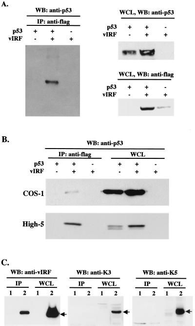
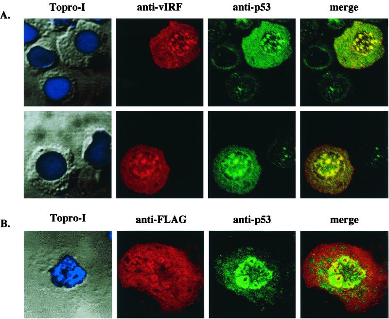
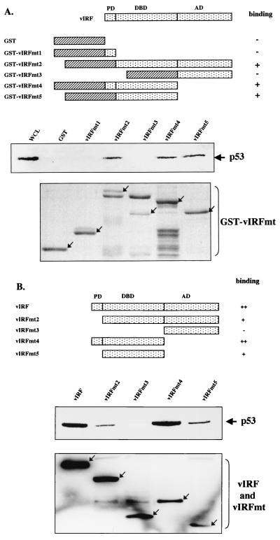
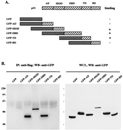
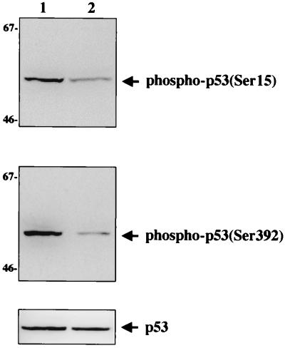
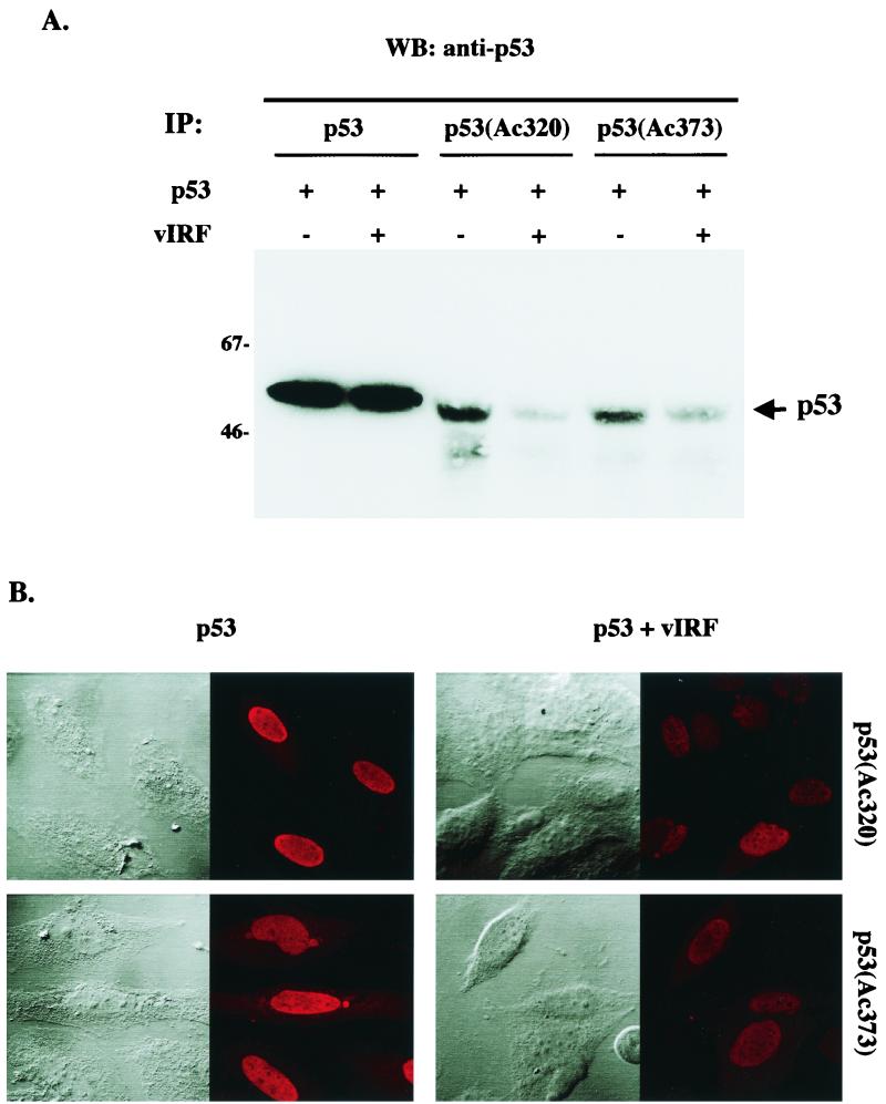
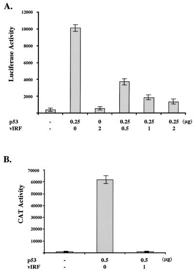
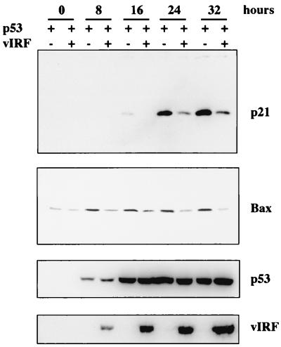
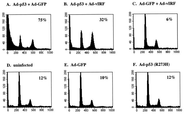
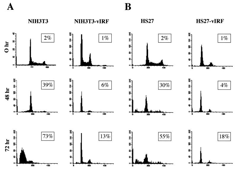
The irreversible cell cycle arrest and apoptosis induced by p53 are part of the host surveillance mechanisms for viral infection and tumor induction. Kaposi's sarcoma-associated herpesvirus (KSHV), the most recently discovered human tumor virus, is associated with the pathogenesis of Kaposi's sarcoma, primary effusion lymphoma, and multicentric Castleman's disease. The K9 open reading frame of KSHV encodes a viral interferon (IFN) regulatory factor (vIRF) which functions as a repressor for cellular IFN-mediated signal transduction and as an oncoprotein to induce cell growth transformation. Here, we demonstrate that KSHV vIRF interacts with the cellular p53 tumor suppressor through the putative DNA binding region of vIRF and the central region of p53. This interaction suppresses the level of phosphorylation and acetylation of p53 and inhibits transcriptional activation of p53. As a consequence, vIRF efficiently prevents p53-mediated apoptosis. These results suggest that KSHV vIRF interacts with and inhibits the p53 tumor suppressor to circumvent host growth surveillance and to facilitate uncontrolled cell proliferation.


Figures

FIG. 1
FIG. 1
Interaction of vIRF with p53. (A) Interaction of vIRF with p53 in recombinant adenovirus-infected Saos-2 cells. p53-null Saos-2 cells were infected with Ad-p53 and/or Ad-vIRF as indicated at the top. After 48 h, cell extracts were used for immunoprecipitations (IP) with an anti-Flag antibody, followed by Western blot assay with an anti-p53 antibody. p53 and vIRF expression in whole-cell lysates (WCL) of infected Saos-2 cells were determined by Western blotting with anti-p53 and anti-Flag antibodies. (B) Interaction of vIRF with p53 in COS-1 cells transfected with p53 and vIRF expression vectors and in High-5 cells infected with recombinant p53 and vIRF baculoviruses. As indicated at the top, COS-1 cells were transfected with p53 and/or Flag-tagged vIRF expression vector, and High-5 insect cells were infected with recombinant p53 and/or vIRF baculoviruses. After 48 h, cell extracts were used for immunoprecipitations with an anti-Flag antibody, followed by Western blot assay (WB) with an anti-p53 antibody (left). Whole-cell lysates (WCL) of transfected COS-1 cells and infected High-5 cells were used to show p53 expression (right) and vIRF expression (data not shown). In addition, vIRF expression did not alter the level of SV40 large T antigen interaction of p53 in COS-1 cells (data not shown). (C) Interaction of vIRF with p53 in KSHV-infected BCBL-1 cells. Lysates of KSHV-negative BJAB (lane 1) and KSHV-infected BCBL-1 (lane 2) cells were used for immunoprecipitation (IP) with an anti-p53 antibody, followed by Western blot assay (WB) with anti-vIRF, anti-K3, and anti-K5 antibodies. A whole-cell lysate (WCL) was used to show vIRF, K3, and K5 expression.
FIG. 2
FIG. 2
Colocalization of vIRF with p53. (A) Colocalization of vIRF with p53 in KSHV-infected BCBL-1 cells. KSHV-infected BCBL-1 cells were fixed and reacted with rabbit polyclonal anti-vIRF and mouse monoclonal anti-p53 antibodies. vIRF protein was detected with an anti-rabbit secondary antibody conjugated with Alexa 568 (red), and p53 protein was detected with an anti-mouse secondary antibody conjugated with Alexa 488 (green). Cells were visualized with Nomarski optics after Topro-I nuclear staining (blue). These two panels are representatives of 10 different fields. (B) Colocalization of vIRF with p53 in COS-1 cells. COS-1 cells were transfected with expression vector containing the Flag-tagged vIRF gene. At 48 h posttransfection, cells were fixed and reacted with mouse monoclonal anti-Flag and rabbit polyclonal anti-p53 antibodies. p53 protein was detected with an anti-rabbit secondary antibody conjugated with Alexa 488 (green), and the Flag-tagged vIRF protein was detected with an anti-mouse secondary antibody conjugated with Alexa 568 (red). Cells were visualized with Nomarski optics after Topro-I nuclear staining (blue). The immunofluorescence test was performed with a Leica confocal immunofluorescence microscope. The yellow color in merged panels indicates colocalization of the red and green labels. The data were reproduced in three independent experiments.
FIG. 3
FIG. 3
Mutational analysis of vIRF for p53 interaction. (A) Summary of GST-vIRF fusion constructs and in vitro GST pull-down assay. Individual domains of vIRF were cloned into the pGEX4T-1 vector to generate GST-vIRF fusion proteins. Lysates of Saos-2 cells infected with Ad-p53 were precleared with 5 μg of GST, followed by incubation with 5 μg of GST or GST-vIRF fusion proteins. Polypeptides associated with the GST-vIRF fusion proteins were subject to Western blotting with an anti-p53 antibody. A whole-cell lysate (WCL) which represents 5% of cellular p53 was used for a positive control. Arrows in the bottom indicate GST and GST-vIRF mutant fusion proteins (GST-vIRFmt) stained with Coomassie blue solution. Boxes with slashed lines indicate GST, and boxes with dots indicate a domain of vIRF, PD, proline-rich domain; DBD, DNA binding domain; AD, activation domain. + and − indicate positive and negative binding of GST-vIRF fusion proteins to p53. (B) Summary of vIRF mutants and in vivo interaction of vIRF mutants with p53. COS-1 cells were transfected with the Flag-tagged wt vIRF and mutants vIRFmt2 to -5. Cell lysates were used for immunoprecipitation with an anti-Flag antibody, followed by Western blotting with an anti-p53 antibody to detect p53. PD, proline-rich domain; DBD, DNA binding domain; AD, activation domain. Western blotting of whole-cell lysates with an anti-Flag antibody showed equivalent expression of wt and mutant vIRF: arrows indicate the Flag-tagged wt and mutant vIRF (bottom). +, ++, and − indicate weak, strong, and no binding of vIRF mutants to p53.
FIG. 4
FIG. 4
Mutational analysis of p53 for vIRF interaction. (A) Summary of p53 mutants. Individual domains of p53 were cloned in frame into the GFP vector to generate GFP-p53 fusion proteins. Boxes with large cross lines indicate individual domains of p53; boxes with small cross lines indicate GFP. AD, activation domain; SH3D, SH3B domain; DBD, DNA binding domain; TD, tetramerization domain; BD, basic domain. + and − indicate positive and negative binding of GFP-p53 fusion proteins to vIRF. (B) Identification of the vIRF binding domains of p53. 293T cells were cotransfected with Flag-tagged wt vIRF and GFP-p53 fusion constructs. Cell lysates were used for immunoprecipitation (IP) with an anti-Flag antibody, followed by Western blotting (WB) with an anti-GFP antibody to detect GFP-p53 fusion proteins (left). Western blotting of whole-cell lysates with an anti-GFP antibody showed GFP-p53 expression in transfected 293T cells (right). Sizes are indicated in kilodaltons.
FIG. 5
FIG. 5
Suppression of in vivo p53 phosphorylation by vIRF expression. Identical amounts of proteins from Saos-2 cells (lane 1) and Saos-2/vIRF (lane 2) cells were used for Western blot analysis with antibodies specific for p53 phosphorylated at serine residue 15 (top) and at serine residue 392 (middle). Western blot assay of whole-cell lysate with horseradish peroxidase-conjugated anti-pan-p53 antibody showed equivalent expression of p53 in both cells (bottom). Sizes are indicated in kilodaltons.
FIG. 6
FIG. 6
Suppression of in vivo p53 acetylation by vIRF expression. (A) Western blot assay of in vivo p53 acetylation. Saos-2 cells were infected with recombinant Ad-p53 and/or Ad-vIRF as indicated at the top. At 48 h postinfection, cell lysates were used for immunoprecipitation (IP) with anti-p53, anti-p53(Ac320), and anti-p53(Ac373) antibodies, followed by Western blot (WB) analysis with the horseradish peroxidase-conjugated anti-pan-p53 antibody. The data were reproduced in two independent experiments. (B) Immunofluorescence test of in vivo p53 acetylation. The Saos-2 cells described above were stained with anti-p53(Ac320) and anti-p53(Ac373) antibodies. Cells were visualized with Nomarski optics. Equivalent levels of p53 were detected in both cells with an anti-p53 antibody (see above). The immunofluorescence test was performed with a Leica confocal immunofluorescence microscope.
FIG. 7
FIG. 7
Inhibition of p53 transcriptional activation by vIRF. (A) Inhibition of p53-mediated activation of PG13 promoter activity by vIRF. PG13-luciferase (0.25 μg) and pGKβgal (0.25 μg) reporter plasmids were transfected into p53-null Saos-2 cells together with 0.25 μg of p53 expression vector and different amounts of vIRF expression vector as indicated at the bottom. Luciferase activity was measured 48 h posttransfection, and luciferase values were normalized by β-galactosidase activity. Luciferase activity is represented as the average of three independent experiments. (B) Inhibition of p53-mediated activation of p21 promoter activity by vIRF. p21-CAT (0.25 μg) and pGKβgal (0.25 μg) reporter plasmids were transfected into p53-null Saos-2 cells together with 0.5 μg of p53 expression vector and 1 μg of vIRF expression vector as indicated at the bottom. CAT activity was measured 48 h posttransfection by a Fuji phosphoimager, and values were normalized by β-galactosidase activity. CAT activity is represented as the average of two independent experiments.
FIG. 8
FIG. 8
Inhibition of p53-mediated upregulation of p21 and Bax protein by vIRF. Lysates of p53-null Saos-2 cells were collected at different time points after infection with a mixture of Ad-p53 plus Ad-GFP or Ad-p53 plus Ad-vIRF as indicated at the top and subject to Western blotting with anti-p21, anti-Bax, anti-p53, and anti-Flag (vIRF) antibodies, as indicated at the right. Equivalent titers of recombinant adenoviruses were used for infection.
FIG. 9
FIG. 9
Inhibition of p53-mediated apoptosis by vIRF. Exponentially growing Saos-2 cells were infected with equivalent titers of recombinant adenoviruses as indicated. At 48 h postinfection, cells were stained for the chromosomal DNA with PI and analyzed on a FACScan flow cytometer. Numbers inside boxes indicate percentages of cells in the subdiploid phase of cell cycle, representing apoptotic cells. Results are representative of three individual experiments.
FIG. 10
FIG. 10
vIRF expression confers resistance to apoptosis induced by growth factor depletion. A total of 5 × 10⁶ NIH 3T3 and NIH 3T3-vIRF mouse fibroblasts and HS27 and HS27-vIRF human fibroblasts were collected at 0, 48, and 72 h after incubation with medium containing 0.5% FBS, stained for chromosomal DNA with PI, and analyzed on a FACScan flow cytometer. Numbers inside boxes indicate percentages of subdiploid cells of the cell cycle, representing apoptotic cells. Results are representative of three individual experiments.

Similar articles

Cited by

References

    1. Banin S, Moyal L, Shieh S, Taya Y, Anderson C W, Chessa L, Smorodinsky N I, Prives C, Reiss Y, Shiloh Y, Ziv Y. Enhanced phosphorylation of p53 by ATM in response to DNA damage. Science. 1998;281:1674–1677. - PubMed
    1. Bates S, Hickman E S, Vousden K H. Reversal of p53-induced cell-cycle arrest. Mol Carcinog. 1999;24:7–14. - PubMed
    1. Bates S, Vousden K H. Mechanisms of p53-mediated apoptosis. Cell Mol Life Sci. 1999;55:28–37. - PMC - PubMed
    1. Burysek L, Yeow W S, Lubyova B, Kellum M, Schafer S L, Huang Y Q, Pitha P M. Functional analysis of human herpesvirus 8-encoded viral interferon regulatory factor 1 and its association with cellular interferon regulatory factors and p300. J Virol. 1999;73:7334–7342. - PMC - PubMed
    1. Cesarman E, Chang Y, Moore P S, Said J W, Knowles D M. Kaposi's sarcoma-associated herpesvirus-like DNA sequences in AIDS-related body-cavity-based lymphomas. N Engl J Med. 1995;332:1186–1191. - PubMed

Publication types