. 2001 Aug;75(16):7583-91.
doi: 10.1128/JVI.75.16.7583-7591.2001.

Degradation of the retinoblastoma tumor suppressor by the human papillomavirus type 16 E7 oncoprotein is important for functional inactivation and is separable from proteasomal degradation of E7

Affiliations

Degradation of the retinoblastoma tumor suppressor by the human papillomavirus type 16 E7 oncoprotein is important for functional inactivation and is separable from proteasomal degradation of E7

S L Gonzalez et al. J Virol. 2001 Aug.

Abstract

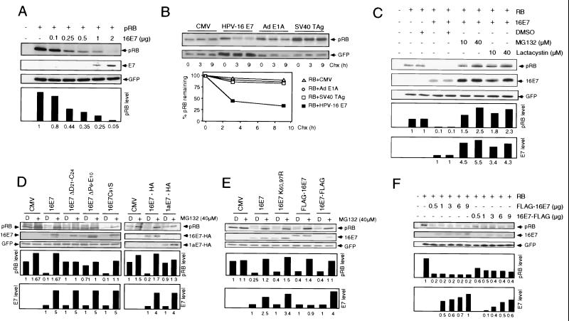
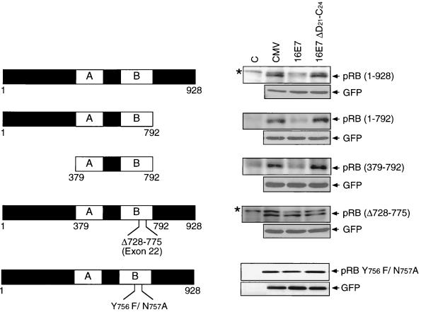
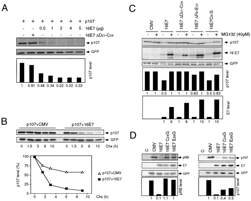
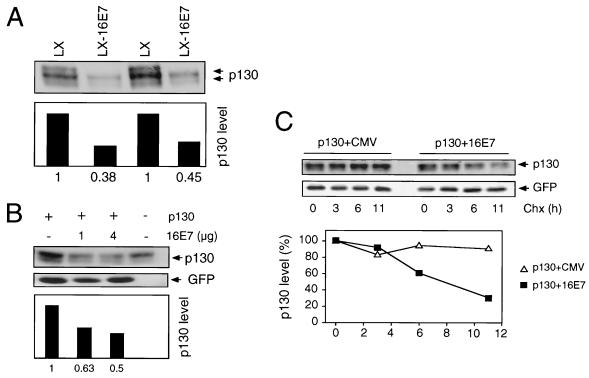
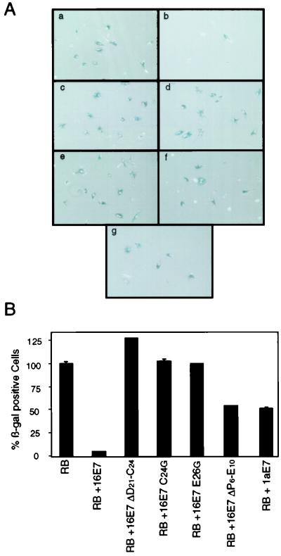
The steady-state level and metabolic half-life of retinoblastoma tumor suppressor protein pRB are decreased in cells that express high-risk human papillomavirus (HPV) E7 proteins. Here we show that pRB degradation is a direct activity of E7 and does not reflect a property of cell lines acquired during the selection process for E7 expression. An amino-terminal domain of E7 that does not directly contribute to pRB binding but is required for transformation is also necessary for E7-mediated pRB degradation. Treatment with inhibitors of the 26S proteasome not only blocks E7-mediated pRB degradation but also causes the stabilization of E7. Mutagenic analyses, however, reveal that the processes of proteasomal degradation of E7 and pRB are not linked processes. HPV type 16 E7 also targets the pRB-related proteins p107 and p130 for destabilization by a proteasome-dependent mechanism. Using the SAOS2 flat-cell assay as a biological indicator for pRB function, we demonstrate that pRB degradation, not solely binding, is important for the E7-induced inactivation of pRB.


Figures

FIG. 1
FIG. 1
HPV-16 E7 degradation and E7-mediated pRB degradation are 26S proteasome-dependent but separable processes. (A) The human osteosarcoma cell line SAOS2 was transiently transfected with the indicated combinations of plasmids encoding wild-type pRB and wild-type HPV-16 E7 (16E7). Samples (100 μg) were subjected to SDS-PAGE and immunoblot analysis for pRB (upper panel), HPV-16 E7 (middle panel), and cotransfected GFP control (lower panel). Quantitation of pRB levels, normalized for GFP expression, is shown underneath those panels (in arbitrary units based on densitometry). (B) HPV-16 E7, adenovirus E1A, and SV40 T antigen oncoproteins differ in their ability to destabilize pRB. SAOS2 cells were transfected with the indicated combinations of plasmids encoding pRB, HPV-16 E7, adenovirus (Ad) E1A, and SV40 T antigen (TAg) or control plasmid (CMV). At 24 h posttransfection, protein synthesis was blocked by treatment with cycloheximide (Chx). At the indicated times, samples (100 μg) were processed for immunoblot analysis for pRB (upper panel) and cotransfected GFP control (lower panel). Quantitation of pRB (RB) levels, normalized for GFP expression, is shown underneath these panels. (C) Effect of MG132 and Lactacystin on HPV-16 E7 and pRB steady-state levels. SAOS2 cells were transfected with expression plasmids encoding pRB alone or in combination with 16E7. At 40 h posttransfection, cells were treated with 10 or 40 μM MG132 or Lactacystin or mock treated with dimethyl sulfoxide (DMSO) for 4 h. Samples (100 μg) were subjected to SDS-PAGE and immunoblot analysis for pRB (upper panel), HPV-16 E7 (middle panel), and cotransfected GFP control (lower panel). Quantitation of pRB and HPV-16 E7 levels, normalized for GFP expression, is shown underneath these panels. (D) Effect of proteasome inhibition on various HPV E7 proteins. SAOS2 cells were transfected with the indicated combinations of plasmids encoding pRB, HPV-16 E7 (16E7 and 16E7-HA), a pRB-binding- and pRB degradation-deficient 16E7 mutant (16E7 ΔD21-C24), pRB-binding-competent and pRB degradation-deficient versions of E7 (16E7 ΔP6-E10 and 1aE7-HA), and a pRB-binding- and pRB degradation-competent 16E7 mutant (16E7 C91S) or control vector (CMV). Cells were treated with 40 μM MG132 (+) or mock treated with DMSO (D) for 4 h. Samples (100 μg) were subjected to SDS-PAGE and immunoblot analysis for pRB (upper panel), HPV-16 E7 or HPV-16 E7 tagged with HA (middle panel), and cotransfected GFP control or HPV-1a E7 tagged with HA (lower panel). Quantitation of pRB and HPV-16 E7 levels, normalized for GFP expression, is shown underneath these panels. (E) E7 degradation and E7-mediated pRB degradation are not linked. SAOS2 cells were transfected with the indicated combinations of plasmids encoding pRB, HPV-16 E7 (16E7), a lysineless mutant of 16E7 (16E7 K60,97R), an amino-terminal FLAG-tagged version of 16E7 (FLAG-16E7), and a carboxyl-terminal FLAG-tagged version of 16E7 (16E7-FLAG) or control vector (CMV). Cells were treated with 40 μM MG132 (+) or mock treated with DMSO (D) for 4 h. Samples (100 μg) were subjected to SDS-PAGE and immunoblot analysis for pRB (upper panel), HPV-16 E7 (middle panel), and cotransfected GFP control (lower panel). Quantitation of pRB and HPV-16 E7 levels, normalized for GFP expression, is shown underneath these panels. (F) HPV-16 E7 stability correlates with E7-mediated pRB degradation. SAOS2 cells were transiently transfected with the indicated combinations of plasmids encoding pRB and amino- and carboxyl-terminal FLAG-tagged HPV-16 E7. Samples (100 μg) were subjected to SDS-PAGE and immunoblot analysis for pRB (upper panel), HPV-16 E7 (middle panel), and cotransfected GFP control (lower panel). Quantitation of pRB and E7 levels, normalized for GFP expression, is shown underneath these panels.
FIG. 2
FIG. 2
The small pocket of pRB is sufficient for E7-mediated degradation. SAOS2 cells were transiently transfected with the indicated combinations of plasmids encoding HA-tagged wild-type or mutant forms of pRB, the E7-binding-defective pRB double point mutant (pRB Y756F/N757A), wild-type HPV-16 E7 (16E7), and a pRB-binding-deficient HPV-16 E7 mutant (16E7 ΔD21-C24) or control vector (CMV). Samples (100 μg) were subjected to SDS-PAGE and immunoblot analysis for the pRB mutants using an HA antibody or a pRB antibody (upper panels) and cotransfected GFP control (lower panels). Untransfected cells were used as controls (C). Background bands are denoted by asterisks.
FIG. 3
FIG. 3
HPV-16 E7 destabilizes p107 by a proteasome-dependent mechanism. (A) HPV-16 E7 decreases steady-state levels of p107. SAOS2 cells were transiently transfected with the indicated combinations of plasmids encoding HA-tagged p107, wild-type HPV-16 E7 (16E7), or the pRB-binding-deficient mutant 16E7 ΔD21-C24. Samples (100 μg) were subjected to SDS-PAGE and immunoblot analysis for p107 using an HA antibody (upper panel) and cotransfected GFP control (lower panel). Quantitation of p107 levels, normalized for GFP expression, is shown underneath these panels. (B) HPV-16 E7 decreases the half-life of p107. SAOS2 cells were transiently transfected with the indicated combinations of plasmids encoding HA-tagged p107 and HPV-16 E7 (16E7) or control plasmid (CMV). At 24 h posttransfection, protein synthesis was blocked by treatment with cycloheximide (Chx). At the indicated times, samples (100 μg) were harvested and then processed for immunoblot analysis for p107 using an HA antibody (upper panel) or cotransfected GFP control (lower panel). Quantitation of p107 levels, normalized for GFP expression, is shown underneath these panels. (C) Inhibition of the 26S proteasome blocks HPV-16 E7-mediated p107 degradation. SAOS2 cells were transfected with the indicated combinations of plasmids encoding HA-tagged p107, HPV-16 E7 (16E7), and HPV-16 E7 mutants 16E7 ΔD21-C24 (pRB binding and degradation deficient), 16E7 ΔP6-E10 (pRB binding competent and pRB degradation deficient), and 16E7 C91S (pRB binding and degradation competent) or control vector (CMV). Cells were treated with 40 μM MG132 (+) or mock treated with DMSO (−) for 4 h. Samples (100 μg) were subjected to SDS-PAGE and immunoblot analysis for p107 using an HA antibody (upper panel), HPV-16 E7 (middle panel), and cotransfected GFP control (lower panel). Quantitation of p107 and HPV-16 E7 levels, normalized for GFP expression, is shown underneath these panels. (D) Differential destabilization of pRB and p107 by pRB-binding-site E7 point mutants. SAOS2 cells were transfected with the indicated combinations of plasmids encoding pRB or HA-tagged p107, HPV-16 E7 (16E7), and HPV-16 E7 mutants 16E7 C24G and 16E7 E26G (pRB binding and degradation deficient, partially p107 binding competent) or control vector (CMV). C, untransfected controls. Samples (100 μg) were subjected to SDS-PAGE and immunoblot analysis for pRB (left upper panel), p107 using an HA antibody (right upper panel), HPV-16 E7 (middle panels), and cotransfected GFP control (lower panels). Quantitation of pRB and p107 levels, normalized for GFP expression, is shown underneath these panels.
FIG. 4
FIG. 4
HPV-16 E7 destabilizes p130. (A) Decreased p130 steady-state levels in HPV-16 E7-expressing pools of HFKs. Two independent, matched populations of HFKs infected with control LXSN (LX) and HPV-16 E7-expressing LXSN (LX-16E7) vectors are shown. Pools of cells were obtained after G418 selection. Extracts (100 μg) were prepared and subjected to SDS-PAGE and p130 immunoblot analysis. Quantitation of p130 levels is shown underneath the blot. (B) HPV-16 E7 decreases p130 steady-state levels in the SAOS2 cell transient assay. SAOS2 cells were transiently transfected with the indicated combinations of plasmids encoding p130 and HPV-16 E7 (16E7). Samples (100 μg) were subjected to SDS-PAGE and immunoblot analysis for p130 (upper panel) and cotransfected GFP control (lower panel). Quantitation of p130 levels, normalized for GFP expression, is shown underneath these panels. Untransfected cells are shown as controls. (C) HPV-16 E7 decreases the half-life of p130. SAOS2 cells were transiently transfected with the indicated combinations of plasmids encoding p130 and HPV-16 E7 (16E7) or control plasmid (CMV). At 24 h posttransfection, protein synthesis was blocked by treatment with cycloheximide (Chx). At the indicated times, samples (100 μg) were harvested and then processed for immunoblot analysis for p130 (upper panel) or cotransfected GFP control (lower panel). Quantitation of p130 levels, normalized for GFP expression, is shown underneath these panels.
FIG. 5
FIG. 5
Abrogation of pRB functions by HPV-16 E7-mediated degradation. (A) HPV-16 E7 inactivates pRB in an SAOS2 flat-cell assay. SAOS2 cells were transfected with the indicated combinations of plasmids encoding pRB alone (panel a) or in combination with carboxyl-terminal HA-tagged HPV-16 E7 (16E7) (panel b), HPV-16 E7 mutants 16E7 ΔD21-C24, 16E7 C24G, 16E7 E26G, and 16E7 ΔP6-E10 (panels c, d, e, and f, respectively), and HPV-1a E7 (panel g). At 10 days posttransfection, cells were stained for senescence-associated β-galactosidase (β-gal) activity. (B) Quantitation of the results from panel A. The results represent averages and standard deviations from two independent experiments. RB, pRB.

Similar articles

Cited by

References

    1. Alunni-Fabbroni M, Littlewood T, Deleu L, Caldeira S, Giarre M, Dell' Orco M, Tommasino M. Induction of S phase and apoptosis by the human papillomavirus type 16 E7 protein are separable events in immortalized rodent fibroblasts. Oncogene. 2000;19:2277–2285. - PubMed
    1. An B, Dou Q P. Cleavage of retinoblastoma protein during apoptosis: an interleukin 1 beta-converting enzyme-like protease as candidate. Cancer Res. 1996;56:438–442. - PubMed
    1. Baker S J, Markowitz S, Fearon E R, Willson J K V, Vogelstein B. Suppression of human colorectal carcinoma cell growth by wild-type p53. Science. 1990;249:912–915. - PubMed
    1. Banks L, Edmonds C, Vousden K. Ability of the HPV16 E7 protein to bind RB and induce DNA synthesis is not sufficient for efficient transforming activity in NIH 3T3 cells. Oncogene. 1990;5:1383–1389. - PubMed
    1. Berezutskaya E, Bagchi S. The human papillomavirus E7 oncoprotein functionally interacts with the S4 subunit of the 26S proteasome. J Biol Chem. 1997;272:30135–30140. - PubMed

Publication types

MeSH terms

Substances

LinkOut - more resources