

Induction of potent human immunodeficiency virus type 1-specific T-cell-restricted immunity by genetically modified dendritic cells
- PMID: 11462034
- PMCID: PMC114997
- DOI: 10.1128/JVI.75.16.7621-7628.2001
Induction of potent human immunodeficiency virus type 1-specific T-cell-restricted immunity by genetically modified dendritic cells
Abstract
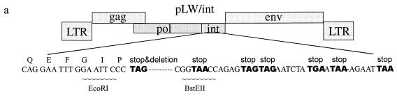
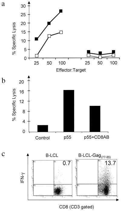
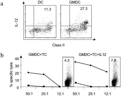
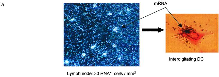
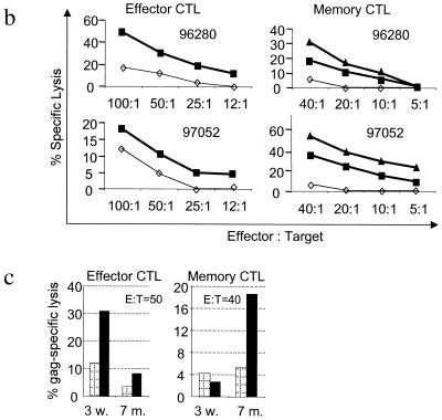
A novel technology combining replication- and integration-defective human immunodeficiency virus type 1 (HIV-1) vectors with genetically modified dendritic cells was developed in order to induce T-cell immunity. We introduced the vector into dendritic cells as a plasmid DNA using polyethylenimine as the gene delivery system, thereby circumventing the problem of obtaining viral vector expression in the absence of integration. Genetically modified dendritic cells (GMDC) presented viral epitopes efficiently, secreted interleukin 12, and primed both CD4(+) and CD8(+) HIV-specific T cells capable of producing gamma interferon and exerting potent HIV-1-specific cytotoxicity in vitro. In nonhuman primates, subcutaneously injected GMDC migrated into the draining lymph node at an unprecedentedly high rate and expressed the plasmid DNA. The animals presented a vigorous HIV-specific effector cytotoxic-T-lymphocyte (CTL) response as early as 3 weeks after a single immunization, which later developed into a memory CTL response. Interestingly, antibodies did not accompany these CTL responses, indicating that GMDC can induce a pure Th1 type of immune response. Successful induction of a broad and long-lasting HIV-specific cellular immunity is expected to control virus replication in infected individuals.
Figures
Similar articles
-
mRNA-based dendritic cell vaccination induces potent antiviral T-cell responses in HIV-1-infected patients.AIDS. 2012 Feb 20;26(4):F1-12. doi: 10.1097/QAD.0b013e32834f33e8. AIDS. 2012. PMID: 22156965 Clinical Trial.
-
Induction of novel CD8+ T-cell responses during chronic untreated HIV-1 infection by immunization with subdominant cytotoxic T-lymphocyte epitopes.AIDS. 2009 Jul 17;23(11):1329-40. doi: 10.1097/QAD.0b013e32832d9b00. AIDS. 2009. PMID: 19528789
-
Primary proliferative and cytotoxic T-cell responses to HIV induced in vitro by human dendritic cells.Immunology. 1991 Nov;74(3):399-406. Immunology. 1991. PMID: 1769689 Free PMC article.
-
Nanochemistry-based immunotherapy for HIV-1.Curr Med Chem. 2007;14(18):1911-9. doi: 10.2174/092986707781368513. Curr Med Chem. 2007. PMID: 17691933 Review.
-
HIV-1 and the hijacking of dendritic cells: a tug of war.Springer Semin Immunopathol. 2005 Jan;26(3):309-28. doi: 10.1007/s00281-004-0178-y. Springer Semin Immunopathol. 2005. PMID: 15609004 Review.
Cited by
-
Polymeric Materials for Gene Delivery and DNA Vaccination.Adv Mater. 2009 Feb 23;21(8):847-867. doi: 10.1002/adma.200801478. Epub 2008 Dec 4. Adv Mater. 2009. PMID: 28413262 Free PMC article.
-
Macrophage-specific nanotechnology-driven CD163 overexpression in human macrophages results in an M2 phenotype under inflammatory conditions.Immunobiology. 2017 Aug;222(8-9):900-912. doi: 10.1016/j.imbio.2017.05.011. Epub 2017 May 16. Immunobiology. 2017. PMID: 28545809 Free PMC article.
-
Single DermaVir immunization: dose-dependent expansion of precursor/memory T cells against all HIV antigens in HIV-1 infected individuals.PLoS One. 2012;7(5):e35416. doi: 10.1371/journal.pone.0035416. Epub 2012 May 9. PLoS One. 2012. PMID: 22590502 Free PMC article. Clinical Trial.
-
Cytomegalovirus, Macrophages and Breast Cancer.Open Virol J. 2017 Mar 31;11:15-27. doi: 10.2174/1874357901711010015. eCollection 2017. Open Virol J. 2017. PMID: 28567162 Free PMC article. Review.
-
Complementary antiviral efficacy of hydroxyurea and protease inhibitors in human immunodeficiency virus-infected dendritic cells and lymphocytes.J Virol. 2002 Mar;76(5):2274-8. doi: 10.1128/jvi.76.5.2274-2278.2002. J Virol. 2002. PMID: 11836405 Free PMC article.
References
-
- Agy M B, Schmidt A, Florey M J, Kennedy B J, Schaefer G, Katze M G, Corey L, Morton W R, Bosch M L. Serial in vivo passage of HIV-1 infection in Macaca nemestrina. Virology. 1997;238:336–343. - PubMed
-
- Banchereau J, Steinman R M. Dendritic cells and the control of immunity. Nature. 1998;392:245–252. - PubMed
-
- Bhardwaj N, Bender A, Gonzalez N, Bui L K, Garrett M C, Steinman R M. Stimulation of human anti-viral CD8+ cytolytic T lymphocytes by dendritic cells. Adv Exp Med Biol. 1995;378:375–379. - PubMed
-
- Cara A, Cereseto A, Lori F, Reitz M S., Jr HIV-1 protein expression from synthetic circles of DNA mimicking the extrachromosomal forms of viral DNA. J Biol Chem. 1996;271:5393–5397. - PubMed
MeSH terms
Substances
LinkOut - more resources
Full Text Sources
Other Literature Sources
Medical
Research Materials
