. 2001 Aug;2(8):736-42.
doi: 10.1093/embo-reports/kve155. Epub 2001 Jul 19.

CARD4/Nod1 mediates NF-kappaB and JNK activation by invasive Shigella flexneri

Affiliations

CARD4/Nod1 mediates NF-kappaB and JNK activation by invasive Shigella flexneri

S E Girardin et al. EMBO Rep. 2001 Aug.

Abstract

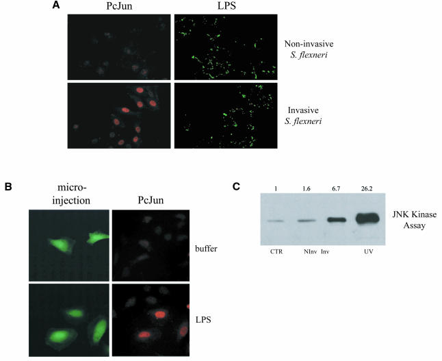
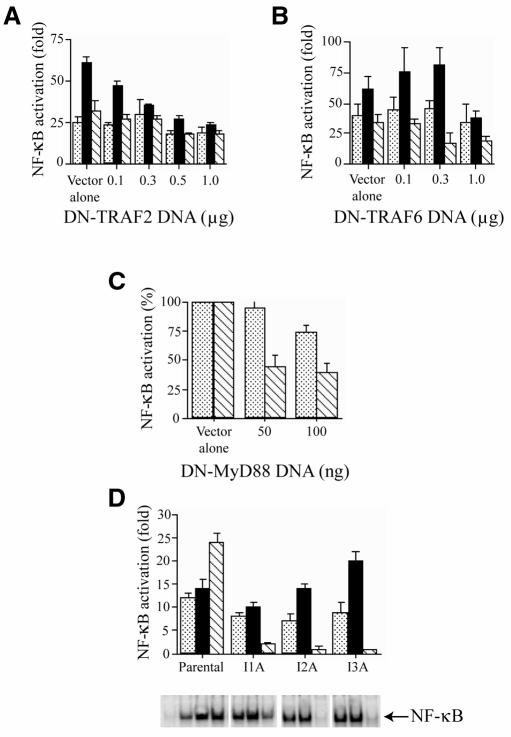
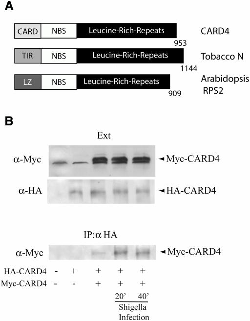
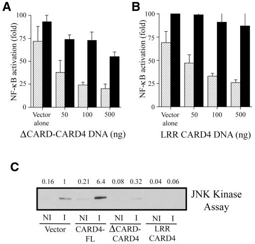
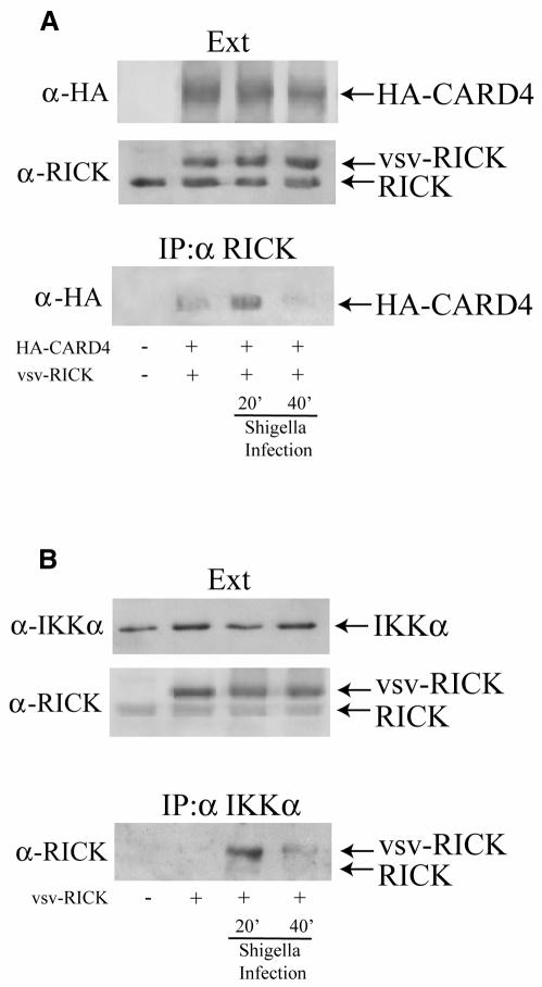
Epithelial cells are refractory to extracellular lipopolysaccharide (LPS), yet when presented inside the cell, it is capable of initiating an inflammatory response. Using invasive Shigella flexneri to deliver LPS into the cytosol, we examined how this factor, once intracellular, activates both NF-kappaB and c-Jun N-terminal kinase (JNK). Surprisingly, the mode of activation is distinct from that induced by toll-like receptors (TLRs), which mediate LPS responsiveness from the outside-in. Instead, our findings demonstrate that this response is mediated by a cytosolic, plant disease resistance-like protein called CARD4/Nod1. Biochemical studies reveal enhanced oligomerization of CARD4 upon S. flexneri infection, an event necessary for NF-kappaB induction. Dominant-negative versions of CARD4 block activation of NF-kappaB and JNK by S. flexneri as well as microinjected LPS. Finally, we showed that invasive S. flexneri triggers the formation of a transient complex involving CARD4, RICK and the IKK complex. This study demonstrates that in addition to the extracellular LPS sensing system mediated by TLRs, mammalian cells also possess a cytoplasmic means of LPS detection via a molecule that is related to plant disease-resistance proteins.


Figures

None
Fig. 1. (A) Shigella flexneri infection leads to the phosphorylation of c-Jun. HeLa cells were infected with wild-type invasive S. flexneri or the plasmid-cured, non-invasive strain for 2 h. Infected cells were then stained for both phospho-c-Jun using a monoclonal phospho-specific antibody to this protein and LPS to label the infecting bacteria using a rabbit polyclonal anti-LPS antibody. Stained cells were viewed using conventional fluorescence microscopy. (B) Microinjection of LPS activates c-Jun phosphorylation. HeLa cells were microinjected with FITC-dextran to identify microinjected cells plus buffer alone or purified Escherichia coli LPS O111:B4. Cells were then stained for phospho-c-Jun and examined by fluorescence microscopy. (C) Infection with S. flexneri increases JNK kinase activity. HeLa cells were transfected with Flag-JNK1 for 40 h and then left either uninfected (CTR) or infected for 20 min with invasive (Inv) or non-invasive (NInv) S. flexneri. Cells were treated for 20 min with 80 J/m2 UVC as a positive control. Cells were collected and protein extracts were then used for a JNK kinase assay (see Methods). Fold activation compared to uninfected cells is presented.
None
Fig. 2. Shigella flexneri induces NF-κB through a signaling pathway distinct from the TLR/IL-1 pathway. Dominant-negative forms of TRAF2 (A) and TRAF6 (B) do not inhibit the induction of NF-κB by invasive S. flexneri (stippled bars), whereas these dominant-negative proteins inhibit NF-κB activation by TNFα (filled bars) or IL-1 (hatched bars), respectively. HEK293 cells were transfected with vector alone or increasing amounts of DNA encoding for the dominant-negative forms of TRAF2 (DN-TRAF2) or TRAF6 (DN-TRAF6) along with a NF-κB luciferase reporter plasmid and a β-galactosidase plasmid. After 30 h, cells were infected with wild-type S. flexneri or treated with TNFα (100 ng/ml) or IL-1 (10 ng/ml) for 4 h and assayed for luciferase activity. (C) Dominant-negative MyD88 (DN-MyD88) does not inhibit NF-κB activation by invasive S. flexneri (stippled bars) but affects IL-1- (hatched bars) induced activation of the reporter gene. Increasing amounts of DNA encoding for dominant-negative MyD88 were transfected into HEK293 cells and assayed for NF-κB luciferase activity as described in (A). (D) Shigella flexneri induces NF-κB in HEK293 deficient in IL-1 specific signaling components (see Methods; Li et al., 1999). NF-κB activation after S. flexneri infection (stippled bars), TNFα (filled bars) or IL-1 (hatched bars) treatment was compared in parental HEK293 cells or three IL-1 signaling mutant cell lines, I1A (IRAK-negative), I2A and I3A. NF-κB activity was assessed following infection or cytokine treatment by the NF-κB reporter assay (refer to above) or EMSA. NF-κB reporter assays were performed in duplicate at least three times and display mean values ± SEM.
None
Fig. 3. CARD4 oligomerization following S. flexneri infection. (A) Domain structure of CARD4 compared with plant disease resistance proteins: Tobacco N protein and Arabidopsis RPS2 protein. (B) Infection with S. flexneri induces self-association of CARD4. HEK293 cells were transfected with empty vector or expression vectors encoding either HA-CARD4 or Myc-CARD4 for 24 h and were left either uninfected or S. flexneri-infected for 20 or 40 min. Cells were collected and protein extracts (Ext) were subjected to western blotting with rabbit polyclonal antibodies to Myc or HA to identify the expression levels of the overexpressed proteins. Another fraction of the protein extracts was used for immunoprecipitation using a polyclonal anti-HA antibody. Oligomerized CARD4 was revealed in the immunoprecipitates by western blotting using antibodies to the Myc-tagged CARD4.
None
Fig. 4. Constructs expressing either ΔCARD CARD4 or the LRR domain of CARD4 act as dominant-negative inhibitors in S. flexneri-induced activation of NF-κB and JNK. Increasing amounts of DNA encoding either (A) ΔCARD CARD4 or (B) the LRR domain inhibits NF-κB induction by S. flexneri (stippled bars) in a DNA concentration-dependent manner but only marginally affects NF-κB activation by TNFα (filled bars) at the higher concentrations of DNA. Plasmids encoding the truncated forms of CARD4 were transfected into HEK293 cells along with NF-κB and β-galactosidase reporter plasmids. Luciferase activity was assayed as described in the Methods. (C) Effect of the overexpression of full length and truncated forms of CARD4 on JNK activation. HeLa cells were transfected with Flag-JNK1 and either empty vector or with expression vectors encoding for CARD4 full length (CARD4-FL), ΔCARD CARD4 or the LRR of CARD4 for 40 h followed by 20 min infection by invasive (i) or non-invasive (ni) S. flexneri. JNK kinase assays were performed as described in the Methods. Fold activation compared to the S. flexneri-induced activation in vector alone expressing cells is presented.
None
Fig. 5. Interaction between CARD4 and RICK and RICK and IKKα of the IKK complex folowing S. flexneri infection. (A) Immunoprecipitation using anti-RICK antibodies demonstrates that RICK and CARD4 interact transiently following S. flexneri infection. (B) Immunoprecipitation of the IKK complex using anti-IKKα antibodies showed that RICK (both endogenous and overexpressed vsv-tagged forms of RICK) associates transiently with the IKK complex following S. flexneri infection. Experiments were carried out using HEK293 cells as in Figure 3.

References

    1. Bertin J. and Distefano, P.S. (2000) The PYRIN domain: A novel motif found in apoptosis and inflammation proteins. Cell Death Differ., 7, 1273–1274. - PubMed
    1. Bertin J. et al. (1999) Human CARD4 protein is a novel CED-4/Apaf-1 cell death family member that activates NF-κB. J. Biol. Chem., 274, 12955–12958. - PubMed
    1. Cao Z., Xiong, J., Takeuchi, M., Kurama, T. and Goeddel, D.V. (1996) TRAF6 is a signal transducer for interleukin-1. Nature, 383, 443–446. - PubMed
    1. Cario E., Rosenberg, I.M., Brandwein, S.L., Beck, P.L., Reinecker, H.C. and Podolsky, D.K. (2000) Lipopolysaccharide activates distinct signaling pathways in intestinal epithelial cell lines expressing Toll-like receptors. J. Immunol., 164, 966–972. - PubMed
    1. Foletta V.C., Segal, D.H. and Cohen, D.R. (1998) Transcriptional regulation in the immune system: all roads lead to AP-1. J. Leukoc. Biol., 63, 139–152. - PubMed

Publication types

MeSH terms

Substances