. 2001 Aug;21(16):5437-46.
doi: 10.1128/MCB.21.16.5437-5446.2001.

Inhibition of the motility and growth of B16F10 mouse melanoma cells by dominant negative mutants of Dok-1

Affiliations

Inhibition of the motility and growth of B16F10 mouse melanoma cells by dominant negative mutants of Dok-1

T Hosooka et al. Mol Cell Biol. 2001 Aug.

Abstract

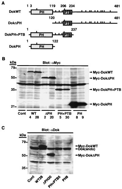
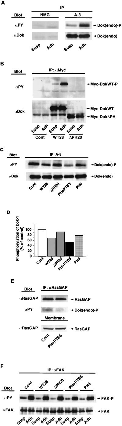
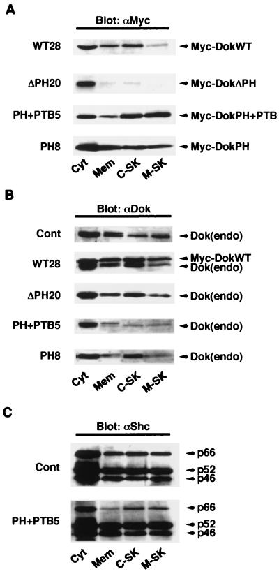
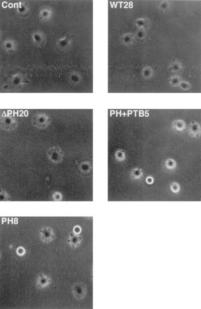
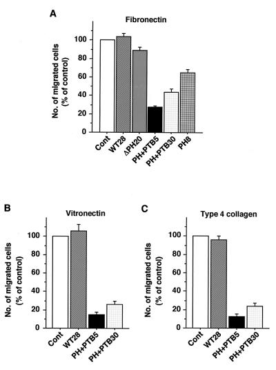
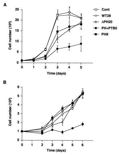
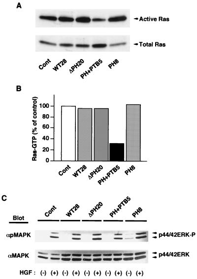
Dok-1 (p62(Dok)) is a multiple-site docking protein that acts downstream of receptor and nonreceptor tyrosine kinases. Although it has been proposed to contribute to the control of cell growth and migration through association with the Ras GTPase-activating protein and the adapter protein Nck, the role of Dok-1 remains largely unknown. The functions of Dok-1 have now been investigated by the generation of two different COOH-terminal truncation mutants of this protein: one (DokPH+PTB) containing the pleckstrin homology and phosphotyrosine-binding domains, and the other (DokPH) composed only of the pleckstrin homology domain. Both of these mutant proteins were shown to act in a dominant negative manner. Overexpression of each of the mutants in highly metastatic B16F10 mouse melanoma cells thus both inhibited the tyrosine phosphorylation of endogenous Dok-1 induced by cell adhesion as well as reduced the association of the endogenous protein with cellular membranes and the cytoskeleton. Overexpression of DokPH+PTB in these cells also markedly reduced both the rates of cell spreading, migration, and growth as well as the extent of Ras activation. The effects of DokPH on these processes were less pronounced than were those of DokPH+PTB, indicating the importance of the phosphotyrosine-binding domain. These results suggest that at least in B16F10 cells, Dok-1 positively regulates not only cell spreading and migration but also cell growth and Ras activity.


Figures

FIG. 1
FIG. 1
Generation and expression of Dok-1 mutants. (A) Schematic representation of recombinant Dok-1 proteins. The numbers correspond to amino acid positions of human Dok-1, and the locations of tyrosine residues are indicated by vertical bars. (B) Cell lysates were prepared from control B16F10 cells transfected with the empty vector (Cont) as well as from two independent clones of B16F10 cells expressing each of the recombinant Dok-1 proteins. Lysates (20 μg of protein) were subjected to immunoblot analysis with MAb 9E10 to the Myc tag (αMyc). (C) Cell lysates (20 μg of protein) prepared from the indicated cell lines were subjected to immunoblot analysis with polyclonal antibodies specific for the COOH-terminal region of Dok-1 (αDok). The positions of recombinant Dok-1 proteins, endogenous Dok-1 [Dok(endo)], and molecular size standards are indicated. The ∼40-kDa immunoreactive material in panels B and C comprises nonspecific cross-reactive polypeptides of unknown origin. Data in all figures are representative of three independent experiments.
FIG. 2
FIG. 2
Adhesion-induced tyrosine phosphorylation of endogenous Dok-1 and FAK in established B16F10 cell lines expressing recombinant Dok-1 proteins. (A) B16F10 cells were detached from culture dishes and either maintained in suspension (Susp) or replated on fibronectin-coated dishes (Adh). After incubation for 30 min at 37°C, cells were lysed and subjected to immunoprecipitation (IP) with MAb A-3 to Dok-1 or with normal mouse immunoglobulin G (NMG). The immunoprecipitates were then subjected to immunoblot analysis with HRP-conjugated MAb PY20 to phosphotyrosine (αPY). Duplicate immunoprecipitates were probed with polyclonal antibodies to Dok-1 (αDok) to verify the presence of equal amounts of Dok-1 in each sample. (B) The indicated cell lines (Cont [control], WT28, and ΔPH20) were treated as in panel A, and the resulting cell lysates were subjected to immunoprecipitation with MAb 9E10 to the Myc tag (αMyc). The phosphotyrosine content of each recombinant Dok-1 protein was assessed as in panel A. (C) The extent of adhesion-induced tyrosine phosphorylation of endogenous Dok-1 in the indicated cell lines was determined as in panel A. (D) The phosphotyrosine content of endogenous Dok-1 in the experiment shown in panel C was quantified by scanning densitometry with the NIH Image program, normalized for the amount of Dok-1 protein in each sample, and was expressed as a percentage of the value for control cells transfected with the empty vector. (E) The indicated cell lines were treated as in panel A and the resulting detargent-solubilized membrane fraction was subjected to immunoprecipitation with MAb B4F8 to RasGAP (αRasGAP). The immunoprecipitates were then subjected to immunoblot analysis with HRP-conjugated MAb PY20 to detect tyrosine-phosphorylated Dok-1 bound to RasGAP. Duplicate immunoprecipitates were probed with the MAb to RasGAP to verify the presence of equal amounts of RasGAP in each sample. Aliquots of each membrane fraction (Membrane) were also directly probed with the MAb to RasGAP. (F) The indicated cell lines were treated as in panel A and subjected to immunoprecipitation with polyclonal antibody C-20 to FAK (αFAK). The immunoprecipitates were then subjected to immunoblot analysis with HRP-conjugated MAb PY20 to phosphotyrosine. Duplicate immunoprecipitates were probed with polyclonal antibody 06-543 to FAK (αFAK) to verify the presence of equal amounts of FAK in each sample. The positions of endogenous Dok-1, exogenous Dok-1, RasGAP, FAK, and the phosphorylated forms of these various proteins [Dok(endo)-P, Myc-DokWT-P, and FAK-P] are indicated.
FIG. 3
FIG. 3
Effects of transient overexpression of DokPH+PTB and IRS-1PH+PTB on tyrosine phosphorylation of Dok-1. (A) B16F10 cells were transiently cotransfected with 0.5 μg of pRc/CMV encoding HA-tagged wild-type mouse Dok-1 (HA-DokWT) and the indicated amount of pCl-neo encoding Myc-tagged DokPH+PTB. Forty-eight hours after transfection, cell lysates were prepared and subjected to immunoprecipitation (IP) with MAb 12CA5 to the HA tag (αHA). The immunoprecipitates were then subjected to immunoblot analysis with HRP-conjugated MAb PY20 to phosphotyrosine (αPY). Duplicate immunoprecipitates were probed with polyclonal antibodies to Dok-1 (αDok) to verify the presence of equal amounts of Dok-1 in each sample. Total cell lysates (Lysate) were also probed with a MAb to the Myc tag (αMyc) to determine the amount of the mutant Dok-1 protein expressed. (B) Cells were transiently cotransfected with 1 μg of pCl-neo encoding Myc-tagged wild-type human Dok-1 (Myc-DokWT) and the indicated amount of pSRα encoding HA-tagged IRS-1PH+PTB. Cell lysates prepared as in panel A were subjected to immunoprecipitation with a MAb to the Myc tag and subsequent immunoblot analysis either with MAb PY20 or with polyclonal antibodies to Dok-1. Total cell lysates were also probed with a MAb to the HA tag to determine the amount of the mutant IRS-1 protein expressed.
FIG. 4
FIG. 4
Effects of overexpression of Dok-1 mutants on the subcellular localization of endogenous Dok-1. (A) The indicated cell lines were fractionated into cytosolic (Cyt), membrane (Mem), cytoskeletal (C-SK), and membrane-skeletal (M-SK) components, and each fraction was then subjected to immunoblot analysis with MAb 9E10 to the Myc tag (αMyc). (B and C) Subcellular fractions from each cell line were subjected to immunoblot analysis either with polyclonal antibodies to Dok-1 (αDok) (B) or with a MAb to Shc (αShc) (C) to examine the subcellular localization of endogenous Dok-1 and Shc, respectively. The positions of exogenous Dok-1 proteins, endogenous Dok-1, and three isoforms of Shc (p66, p52, and p46) are indicated. Cont, control.
FIG. 5
FIG. 5
Effects of overexpression of Dok-1 mutants on cell spreading on fibronectin. The indicated cell lines were detached from their culture dishes and replated in serum-free MEM on dishes coated with fibronectin. The cells were then allowed to adhere and spread at 37°C for 30 min, after which they were photographed in random fields with the use of phase-contrast optics. Cont, control. Original magnification, ×200.
FIG. 6
FIG. 6
Effects of overexpression of Dok-1 mutants on cell migration on ECM proteins. The indicated cell lines were seeded onto porous membranes that had been both coated with fibronectin (A), vitronectin (B), or type 4 collagen (C) and placed in Boyden multiwell chambers. After incubation at 37°C for 3 h, cells that had migrated were stained with Giemsa solution. The number of migrated cells was counted and expressed as a percentage of the value for control cells transfected with the empty vector (Cont). Data are means ± SE of triplicate determinations from three independent experiments.
FIG. 7
FIG. 7
Effects of overexpression of Dok-1 mutants on cell growth. The rate of cell growth was monitored for the indicated cell lines in the presence of 10% (A) or 0.5% (B) FBS. Data are means ± SE of triplicate determinations from three independent experiments. Cont, control.
FIG. 8
FIG. 8
Effects of overexpression of Dok-1 mutants on the activity of Ras and ERK. (A) The active (GTP-bound) form of Ras was precipitated from lysates of the indicated cell lines with a GST fusion protein containing the Ras-binding domain of c-Raf-1. The precipitates were then subjected to immunoblot analysis with a MAb to H-Ras (top). Whole-cell lysates were also subjected directly to immunoblot analysis with the same MAb to determine the total amount of Ras (bottom). Cont, control. (B) The amount of activated Ras in panel A was quantified by scanning densitometry with the NIH Image program, normalized for the amount of total Ras protein, and expressed as a percentage of the value for control cells transfected with the empty vector. (C) The indicated cell lines were incubated for 5 min in the absence (−) or presence (+) of hepatocyte growth factor (HGF; 40 ng/ml). Total cell lysates prepared were subjected to immunoblot analysis with antibodies specific for tyrosine-phosphorylated ERK (αpMAPK) or for total ERK protein (αMAPK).
FIG. 9
FIG. 9
Effects of inhibition of Dok-1 function on the activity of Rho. (A) B16F10 cells transfected with the empty vector (Cont, [control]) or expressing DokPH+PTB (PH+PTB5) were detached from their culture dishes and then either maintained in suspension (Susp) or replated on fibronectin-coated dishes (Adh). After incubation of cells for 30 min in MEM supplemented with 1% FBS, the active form of Rho was precipitated from cell lysates with a GST fusion protein containing the Rho-binding domain of Rhotekin. The resulting precipitates were then subjected to immunoblot analysis with a MAb to RhoA (top). Whole-cell lysates were also directly subjected to immunoblot analysis with the same MAb to determine the total amount Rho (bottom). (B) The same two cell lines were deprived of serum for 12 h and then incubated for 1 min in the absence (−) or presence (+) of 4 μM LPA (Sigma). The active form of Rho was then precipitated and analyzed as in panel A.

Similar articles

Cited by

References

    1. Bouton A H, Kanner S B, Vines R R, Wang H C, Gibbs J B, Parsons J T. Transformation by pp60src or stimulation of cells with epidermal growth factor induces the stable association of tyrosine-phosphorylated cellular proteins with GTPase-activating protein. Mol Cell Biol. 1991;11:945–953. - PMC - PubMed
    1. Carpino N, Wisniewski D, Strife A, Marshak D, Kobayashi R, Stillman B, Clarkson B. p62(dok): a constitutively tyrosine-phosphorylated, GAP-associated protein in chronic myelogenous leukemia progenitor cells. Cell. 1997;88:197–204. - PubMed
    1. Clark E A, King W G, Brugge J S, Symons M, Hynes R O. Integrin-mediated signals regulated by members of the Rho family of GTPases. J Cell Biol. 1998;142:573–586. - PMC - PubMed
    1. Cong F, Yuan B, Goff S P. Characterization of a novel member of the DOK family that binds and modulates Abl signaling. Mol Cell Biol. 1999;19:8314–8325. - PMC - PubMed
    1. DeClue J E, Vass W C, Johnson M R, Stacey D W, Lowy D R. Functional role of GTPase-activating protein in cell transformation by pp60v-src. Mol Cell Biol. 1993;13:6799–6809. - PMC - PubMed

Publication types

LinkOut - more resources