. 2001 Aug;21(16):5577-90.
doi: 10.1128/MCB.21.16.5577-5590.2001.

Dichotomy of AML1-ETO functions: growth arrest versus block of differentiation

Affiliations

Dichotomy of AML1-ETO functions: growth arrest versus block of differentiation

S A Burel et al. Mol Cell Biol. 2001 Aug.

Abstract

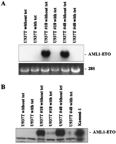
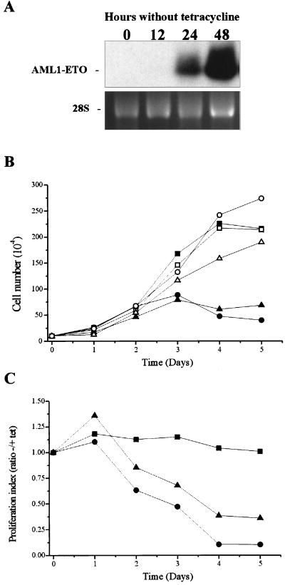
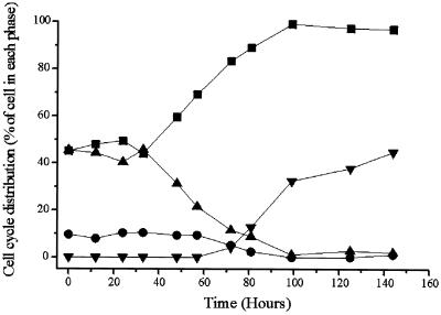
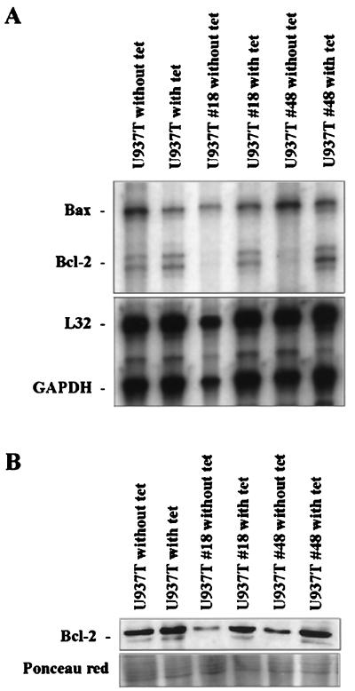
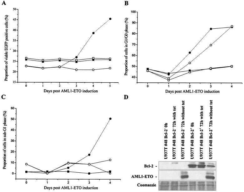
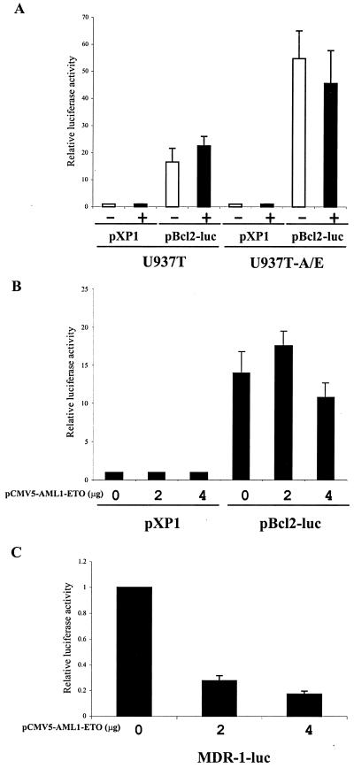
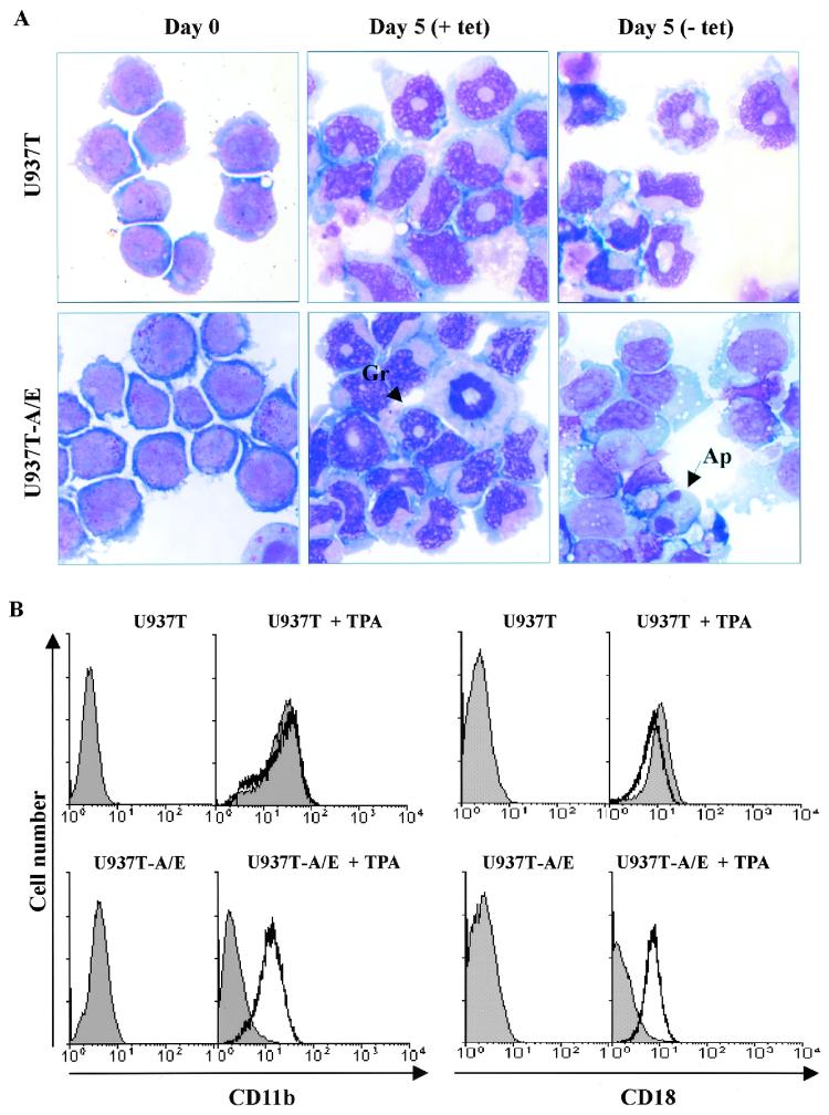
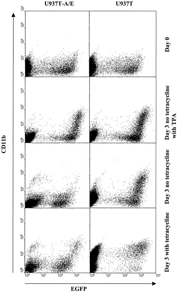
The fusion gene AML1-ETO is the product of t(8;21)(q22;q22), one of the most common chromosomal translocations associated with acute myeloid leukemia. To investigate the impact of AML1-ETO on hematopoiesis, tetracycline-inducible AML1-ETO-expressing cell lines were generated using myeloid cells. AML1-ETO is tightly and strongly induced upon tetracycline withdrawal. The proliferation of AML1-ETO(+) cells was markedly reduced, and most of the cells eventually underwent apoptosis. RNase protection assays revealed that the amount of Bcl-2 mRNA was decreased after AML1-ETO induction. Enforced expression of Bcl-2 was able to significantly delay, but not completely overcome, AML1-ETO-induced apoptosis. Prior to the onset of apoptosis, we also studied the ability of AML1-ETO to modulate differentiation. AML1-ETO expression altered granulocytic differentiation of U937T-A/E cells. More significantly, this change of differentiation was associated with the down-regulation of CCAAT/enhancer binding protein alpha (C/EBPalpha), a key regulator of granulocytic differentiation. These observations suggest a dichotomy in the functions of AML1-ETO: (i) reduction of granulocytic differentiation correlated with decreased expression of C/EBPalpha and (ii) growth arrest leading to apoptosis with decreased expression of CDK4, c-myc, and Bcl-2. We predict that the preleukemic AML1-ETO(+) cells must overcome AML1-ETO-induced growth arrest and apoptosis prior to fulfilling their leukemogenic potential.


Figures

FIG. 1
FIG. 1
Inducible AML1-ETO expression in U937T cells stably transfected with pUHD-AML1-ETO. (A) Northern blot analyses showing the expression of AML1-ETO in the presence or absence of tetracycline (with tet or without tet, respectively) in U937T parental cells and stably transfected cells. Total RNA (10 μg) for each sample was electrophoresed, blotted, and hybridized with α-32P-labeled ETO cDNA. The 28S rRNA is shown for the relative amounts of loaded RNA. (B) Western blot analyses showing the expression of AML1-ETO protein in U937T-A/E pools 18 and 48 following tetracycline withdrawal for 72 h. Proteins from U937T and U937T-A/E cell lines were analyzed by immunodetection using an anti-human ETO antibody. Protein prepared from t(8;21) Kasumi-1 cells was used as a positive control.
FIG. 2
FIG. 2
Induction of AML1-ETO expression inhibits the proliferation of U937T-A/E cells. (A) Northern blot analyses showing the time course of AML1-ETO induction following tetracycline removal in U937T-A/E pool 48 cells. Total RNA (10 μg) was prepared from U937T-A/E pool 48 cells cultured for 0, 12, 24, and 48 h after withdrawal of tetracycline, electrophoresed, blotted, and hybridized with α-32P-labeled ETO cDNA. The 28S rRNA is shown to demonstrate relative loading. (B and C) Growth curves for induced U937T-A/E cells. Cells were maintained in the medium either with or without tetracycline (1 μg/ml). The viability was measured daily by trypan blue exclusion. This experiment is representative of three independent experiments that gave similar results. (B) Only viable cells with the ability to exclude trypan blue are represented. Results for U937T cells (square), U937T-A/E pool 18 cells (triangle), and U937T-A/E pool 48 cells (circle) are shown. Solid symbols, absence of tetracycline; open symbols, presence of tetracycline. (C) The ratio of viable cells in the presence or the absence of tetracycline is shown for each cell line. Cells unaffected by the withdrawal of tetracycline exhibit a ratio close to 1. Results for U937T cells (square), U937T-A/E pool 18 cells (triangle), and U937T-A/E pool 48 cells (circle) are shown.
FIG. 3
FIG. 3
Cell cycle analysis of U937T-A/E pool 48 cells upon tetracycline withdrawal. The cell cycle distribution was determined by propidium iodide staining of the cell nuclei at different time points as indicated. The cells were analyzed by flow cytometry. Results for cells in G0/G1 phase (square), G2/M phase (circle), S phase (triangle), and sub-G1 phase (inverted triangle) are shown.
FIG. 4
FIG. 4
Inducible expression of AML1-ETO leads to apoptosis in U937T-A/E cells. Detection of apoptotic cells was performed by acridine orange and ethidium bromide double staining. U937T-A/E cells were maintained for 72 h in the medium either containing 1 μg of tetracycline/ml (A) or not containing tetracycline (B). Both live and dead cells take up acridine orange. Acridine orange intercalates into DNA and RNA, making the former appear green while the latter stains red. Thus a viable cell has bright green chromatin in its nucleus and red-orange cytoplasm. Ethidium bromide is only taken up by nonviable cells. Ethidium bromide intercalates into DNA, making it appear orange, but binds only weakly to RNA, which may appear slightly red. Thus a dead cell has bright orange chromatin (the ethidium overwhelms the acridine) and its cytoplasm, if it has any contents remaining, appears dark red. Cells that have undergone necrosis have the fluorescent features of nonviable cells but do not have apoptotic nuclear morphology. VN, viable cells with normal nuclei (bright green chromatin with organized structure); VA, viable cells with apoptotic nuclei (bright green chromatin which is highly condensed or fragmented); NVA, nonviable cells with apoptotic nuclei (bright orange chromatin which is highly condensed or fragmented).
FIG. 5
FIG. 5
The Bcl-2 apoptotic pathway is involved in apoptotic response of induced U937T-A/E cells. (A) RPA analyses showing the reduced expression of Bcl-2 mRNA. Total RNA (10 μg) was prepared from U937T and derived clones cultured for 72 h after withdrawal of tetracycline and hybridized with α-32P-labeled in vitro-transcribed probes from the Bcl-2 family (only Bcl-2 and Bax are represented along with L32 and GAPDH [glyceraldehyde-3-phosphate dehydrogenase] as the internal control) and resolved on a denaturing polyacrylamide gel. (B) Western blot analyses showing the expression of Bcl-2 proteins in U937T cells and U937T-A/E pool 18 and 48 cells following tetracycline withdrawal for 72 h. Ponceau red staining is shown to indicate the relative amount of protein in each lane.
FIG. 6
FIG. 6
Ectopic expression of Bcl-2 delays apoptosis in U937T-A/E cells. (A) U937T-A/E cells were infected with either MSCV–Bcl-2–IRES–EGFP (circle) or MSCV-IRES-EGFP (square) and cultured in the presence (open symbol) or the absence (closed symbol) of tetracycline (1 μg/ml). Retrovirus-infected cells were confirmed based on EGFP fluorescence. Apoptotic and necrotic cells were discriminated from viable cells by double annexin V-PE and 7-AAD staining. (B and C) Cell cycle analysis of U937T-A/E pool 48 cells infected with MSCV–Bcl-2–IRES–EGFP upon tetracycline withdrawal. The cell cycle distribution was determined by propidium iodide staining of the cell nuclei at different time points as indicated. The cells were initially sorted on the basis of EGFP expression. Open symbols, EGFP+ cells; solid symbols, EGFP cells. The cells were cultured in the presence (square) or absence (circle) of tetracycline and were analyzed by flow cytometry. (B) Cells in G0/G1 phase; the percentage does not reflect the proportion of cells in sub-G1 phase. (C) Proportion of cells in sub-G1 phase (apoptotic) compared to the total population. (D) Western blot analyses showing the expression of Bcl-2 and AML1-ETO proteins in EGFP+ and EGFP U937T-A/E pool 48 cells infected with MSCV–Bcl-2–IRES–EGFP following tetracycline withdrawal for 72 h. Coomassie staining is shown to indicate the relative amount of protein in each lane.
FIG. 7
FIG. 7
Transactivation analysis of the Bcl-2 promoter with AML1-ETO. (A) U937T and U937T-A/E cells were transfected with either promoterless firefly luciferase reporter pXP1 or the 3.7-kb Bcl-2 gene upstream sequence–firefly luciferase reporter pBcl2-luc after they had been cultured in the absence or presence of tetracycline for 12 h. The pRL-Null Renilla luciferase construct was cotransfected to normalize transfection efficiency. (B) U937T cells were transfected with pXP1 or pBcl2-luc and various amounts of pCMV5 or pCMV5-AML1-ETO, along with pRL-Null to normalize transfection efficiency. The activity of the promoter was calculated as the ratio of the firefly luciferase activity and the Renilla luciferase activity. The transactivation was calculated as the ratio between pXP1 and pBcl2-luc with 0, 2, and 4 μg of pCMV5-AML1-ETO, assuming a value of 1 for each in the presence of pXP1. (C) U937T cells were transfected with MDR-1-luc and various amounts of pCMV5 or pCMV5-AML1-ETO, along with pRL- Null to normalize transfection efficiency. The activity of the promoter was calculated as the ratio of the firefly luciferase activity and the Renilla luciferase activity. The transactivation was calculated as the ratio between 0 μg and 2 or 4 μg of pCMV5-AML1-ETO. The results are the means of three independent experiments (standard errors of the means are shown when measurable).
FIG. 8
FIG. 8
AML1-ETO blocks granulocytic differentiation of U937T-A/E cells. TPA (65 nM) was added to the cell culture medium after U937T and U937T-A/E cells were cultured in the presence or absence of tetracycline (1 μg/ml) for 24 h. Cell culture was continued in the same condition after the addition of TPA. (A) Wright-Giemsa staining of cytospin preparation of U937T and U937T-A/E cells without (+ tet) or with (− tet) AML1-ETO expression upon TPA treatment. Gr, granulocyte; Ap, apoptotic cell. (B) Flow cytometry profile of U937T and U937T-A/E cells treated for 48 h with 65 nM TPA 24 h after AML1-ETO induction. The cells were stained with anti-human CD11b-PE or anti-human CD18-FITC. Profiles of cells in the presence (unshaded) or the absence (shaded) of tetracycline are presented. Undifferentiated cells appeared as CD11blow and CD18low. Differentiated cells appeared as CD11bhigh and CD18high.
FIG. 9
FIG. 9
AML1-ETO down-regulates the expression of C/EBPα, CDK4, and c-myc. (A) Total RNA (10 μg) was prepared from Kasumi-1 cells and U937T cells and their derived clones after they had been cultured in the presence or the absence of tetracycline for 72 h, electrophoresed, blotted, and hybridized with α-32P-labeled C/EBPα cDNA. (B) Time course of the expression of C/EBPα, CDK4, and c-myc upon tetracycline withdrawal. (C) Effects of TPA on the level of C/EBPα mRNA. The cells were treated with 65 nM TPA for 48 h starting 24 h after induction of AML1-ETO. The 28S rRNA is shown to show relative RNA loading in the gel.
FIG. 10
FIG. 10
AML1-ETO inhibits spontaneous U937T differentiation caused by ectopic expression of C/EBPα. Flow cytometry profile of U937T and U937T-A/E cells 2 days (day 0 for tetracycline withdrawal) and 5 days (3 days after tetracycline withdrawal) after infection with MSCV-C/EBPα-IRES-EGFP. The cells were also treated with 65 nM TPA 24 h after AML1-ETO induction. The cells were stained with anti-human CD11b-PE. Profiles of cells in the presence (unshaded) or absence (shaded) of tetracycline are presented. Undifferentiated cells appeared as CD11blow. Differentiated cells appeared as CD11bhigh.

References

    1. Ahn M Y, Huang G, Bae S C, Wee H J, Kim W Y, Ito Y. Negative regulation of granulocytic differentiation in the myeloid precursor cell line 32Dcl3 by ear-2, a mammalian homolog of Drosophila seven-up, and a chimeric leukemogenic gene, AML1/ETO. Proc Natl Acad Sci USA. 1998;95:1812–1817. - PMC - PubMed
    1. Banker D E, Radich J, Becker A, Kerkof K, Norwood T, Willman C, Appelbaum F R. The t(8;21) translocation is not consistently associated with high Bcl-2 expression in de novo acute myeloid leukemias of adults. Clin Cancer Res. 1998;4:3051–3062. - PubMed
    1. Boer J, Bonten-Surtel J, Grosveld G. Overexpression of the nucleoporin CAN/NUP214 induces growth arrest, nucleocytoplasmic transport defects, and apoptosis. Mol Cell Biol. 1998;18:1236–1247. - PMC - PubMed
    1. Borner C. Diminished cell proliferation associated with the death-protective activity of Bcl-2. J Biol Chem. 1996;271:12695–12698. - PubMed
    1. Buchholz F, Refaeli Y, Trumpp A, Bishop J M. Inducible chromosomal translocation of AML1 and ETO genes through Cre/loxP-mediated recombination in the mouse. EMBO Rep. 2000;1:133–139. - PMC - PubMed

Publication types

MeSH terms

Substances

LinkOut - more resources