. 2001 Aug;183(16):4848-59.
doi: 10.1128/JB.183.16.4848-4859.2001.

Structure-function analysis of BfpB, a secretin-like protein encoded by the bundle-forming-pilus operon of enteropathogenic Escherichia coli

Affiliations

Structure-function analysis of BfpB, a secretin-like protein encoded by the bundle-forming-pilus operon of enteropathogenic Escherichia coli

S A Schmidt et al. J Bacteriol. 2001 Aug.

Abstract

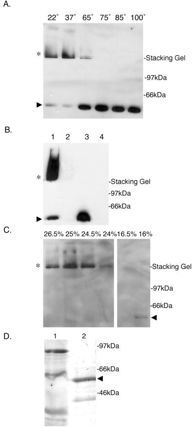
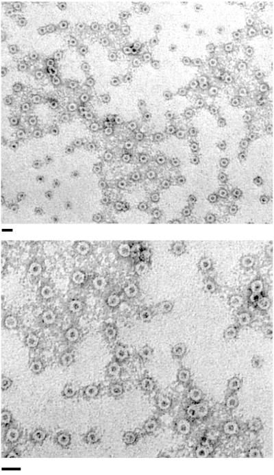
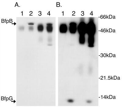
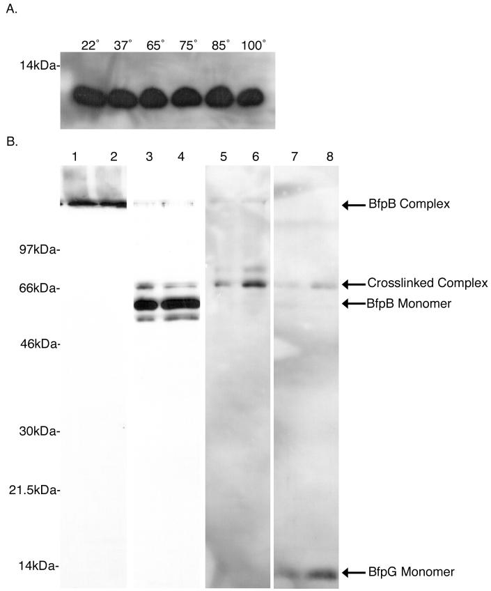
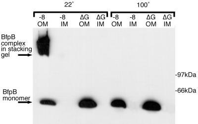
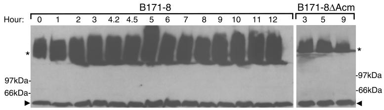
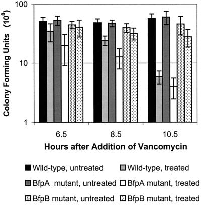
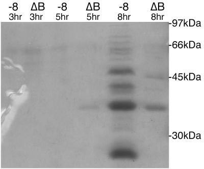
Production of type IV bundle-forming pili by enteropathogenic Escherichia coli (EPEC) requires BfpB, an outer-membrane lipoprotein and member of the secretin protein superfamily. BfpB was found to compose a ring-shaped, high-molecular-weight outer-membrane complex that is stable in 4% sodium dodecyl sulfate at temperatures of < or = 65 degrees C. Chemical cross-linking and immunoprecipitation experiments disclosed that the BfpB multimeric complex interacts with BfpG, and mutational studies showed that BfpG is required for the formation and/or stability of the multimer but not for the outer-membrane localization of BfpB. Formation of the BfpB multimer also does not require BfpA, the repeating subunit of the pilus filament. Functional studies of the BfpB-BfpG complex revealed that its presence confers vancomycin sensitivity, indicating that it may form an incompletely gated channel through the outer membrane. BfpB expression is also associated with accumulation of EPEC proteins in growth medium, suggesting that it may support both pilus biogenesis and protein secretion.


Figures

FIG. 1
FIG. 1
BfpB forms a HMW outer-membrane complex. (A) Temperature-dependent stability of the BfpB multimer in SDS. Purified outer membranes (3 μg of protein), prepared from the wild-type strain B171-8, were incubated in SDS-PAGE sample buffer at the indicated temperatures (in centrograde). Following SDS-PAGE, Western blot analysis with anti-BfpB antiserum was performed. When incubated in PAGE sample buffer at temperatures of 22 and 37°C, BfpB migrates principally as an HMW complex (∗) with some monomeric species (▸). At temperatures greater than 65°C, the predominant species is the monomer of BfpB (▸). (B) HMW complexes reactive with BfpB antisera (∗) are not detected in the bfpB deletion strain, B171-8ΔB2. Whole cells were sonicated and incubated in SDS-PAGE sample buffer for 7 min at the indicated temperature, and equal protein amounts were loaded onto a 12% polyacrylamide gel. Western blot analysis with anti-BfpB antiserum was performed. Lane 1, B171-8 whole-cell sonicate incubated at room temperature; lane 2, B171-8ΔB2 whole-cell sonicate incubated at room temperature; lane 3, B171-8 whole-cell sonicate incubated at 100°C; lane 4, B171-8ΔB2 whole-cell sonicate incubated at 100°C. (C) Isolation of the BfpB multimer by sucrose gradient centrifugation. Outer membranes isolated from B171-8 bacteria were solubilized at 37°C in a buffer containing 4% SDS and loaded onto a linear sucrose gradient. Following centrifugation, 20-μl aliquots from individual sucrose gradient fractions were mixed with an equal volume of SDS-PAGE sample buffer and loaded onto a 12% polyacrylamide gel without prior heating of the sample. Immunoblotting with anti-BfpB antiserum was used to detect BfpB. The BfpB HMW complex (∗) localized to 25% sucrose, while the BfpB monomer localized to 16% sucrose (◂). (D) Characterization of the isolated BfpB multimer. Sucrose gradient fractions containing the BfpB multimer were boiled in sample buffer and separated on a 12% polyacrylamide gel, and the proteins were visualized by silver staining. Lane 1, 30 μg of the outer-membrane starting material, heated for 7 min at 100°C in SDS-PAGE sample buffer; lane 2, 30 μg of sucrose gradient fractions containing the BfpB HMW complex, heated for 7 min at 100°C in SDS-PAGE sample buffer. The primary protein identified by silver staining corresponds to the size of the BfpB monomer (◂). Immunoblotting with anti-BfpB antiserum was used to confirm the identity of the two silver-stained bands and to conclude that the smaller 39-kDa band is a BfpB breakdown product. A band corresponding to BfpB cannot be detected in the crude outer-membrane fraction, most likely because it is a minor component of the outer membrane.
FIG. 2
FIG. 2
The isolated BfpB HMW complex adopts a ring-like configuration. Negatively stained BfpB complexes, prepared by SDS treatment at 37°C and sucrose gradient centrifugation, were visualized with transmission EM. Bar, 30 nm.
FIG. 3
FIG. 3
BfpB and BfpG coimmunoprecipitate. (A) An antiserum to BfpG coimmunoprecipitates BfpB. Immunoprecipitates were heated to 80°C in SDS-PAGE sample buffer, separated by PAGE, transferred to a nylon membrane for Western analysis, and probed with anti-BfpB antiserum. Lane 1, B171-8ΔG immunoprecipitated with anti-BfpG; lane 2, B171-8 immunoprecipitated with anti-BfpG; lane 3, B171-8ΔB immunoprecipitated with anti-BfpB; lane 4, B171-8 immunoprecipitated with anti-BfpB. Both the anti-BfpB and the anti-BfpG antibodies were able to immunoprecipitate BfpB protein. The dark abundant bands between BfpB and BfpG are rabbit immunoglobulins, Western reactive with the HRP-labeled secondary donkey anti-rabbit immunoglobulin G antibody used in Western detection. (B) An antiserum to BfpB coimmunoprecipitates BfpG. The membrane shown in panel A was sequentially probed with anti-BfpG antiserum without prior stripping of the BfpB antibodies. Both the anti-BfpB and anti-BfpG antibodies were able to immunoprecipitate BfpG protein.
FIG. 4
FIG. 4
The BfpB complex can be chemically cross-linked with BfpG. (A) BfpG is a monomer under conditions that BfpB is a multimer. Three micrograms of outer-membrane protein from the B171-8 wild-type strain was incubated in sample buffer containing 4% SDS for 7 min at various temperatures. Following SDS-PAGE, Western blot analysis using αBfpG antiserum was performed. BfpG runs as a monomer at every incubation temperature. (B) DTSSP cross-links BfpG to the BfpB multimer. One milligram of outer membranes from the wild-type B171-8 strain was treated with a 0.2 mM solution of the reducible cross-linker DTSSP. After cross-linking, BfpB complexes were purified. The same two adjacent sucrose gradient fractions, containing BfpB complexes, were used in each of the following immunoblots. Lanes 1 and 2, anti-BfpB immunoblot of purified complexes incubated at 22°C in sample buffer with no 2-ME; lanes 3 and 4, anti-BfpB immunoblot of purified complexes incubated at 100°C in sample buffer with no 2-ME; lanes 5 and 6, anti-BfpG immunoblot of purified complexes incubated at 100°C in sample buffer with no 2-ME; lanes 7 and 8, anti-BfpG immunoblot of purified complexes incubated at 100°C in sample buffer with 2-ME. The intensity of the cross-linked complex decreases and BfpG monomer appears.
FIG. 5
FIG. 5
BfpB requires BfpG for multimerization but not localization to the outer membrane. Three micrograms of outer- and inner-membrane proteins from B171-8 and B171-8ΔG was loaded onto a 15% polyacrylamide gel. Western blot analysis with anti-BfpB antiserum was performed. Samples on the left half were incubated at 100°C in sample buffer for 7 min, while samples on the right were incubated at 22°C for 7 min.
FIG. 6
FIG. 6
BfpB forms a HMW complex in the absence of BfpA expression. Cells were collected from cultures of either the wild-type strain B171-8 or the BfpA knockout mutant, B171-8ΔAcm, at the indicated times and sonicated, and equal amounts of protein were loaded onto 12% polyarcylamide gels. Western analysis with anti-BfpB antiserum was performed with B171-8 (left) and B171-8ΔAcm (right). Arrowheads, BfpB monomers; stars, BfpB complexes.
FIG. 7
FIG. 7
The BfpB complex renders cells susceptible to vancomycin. Vancomycin at a final concentration of 8 μg/ml was added to shaking DMEM cultures at 37°C, 1.5 h after inoculation from an overnight culture. Bacteria were collected from vancomycin-treated cultures and untreated control cultures, diluted, and plated 6.5, 8.5, and 10.5 h after addition of the antibiotic. Surviving colonies were counted the following day. Each data point on the graph is the average of at least six separate experiments, and error bars represent the standard deviations. The wild-type strain is B171-8, the BfpA mutant is B171-8ΔAcm, and the BfpB mutant is B171-8ΔB. B171-8 and B171-8ΔAcm, both of which express the outer-membrane BfpB multimer, were significantly killed by vancomycin 10.5 h after the addition of the antibiotic. By contrast, the viability of B171-8ΔB, which does not express the outer-membrane BfpB multimer, was not significantly affected by antibiotic addition.
FIG. 8
FIG. 8
Comparative analysis of proteins accumulating in tissue culture medium during growth of the wild-type strain, B171-8, and the BfpB deletion strain, B171-8ΔB. Tissue culture medium was collected 3, 5, and 8 h after a 1:100 dilution of an overnight culture into DMEM, and the cells were pelleted by centrifugation. The supernatants were filter sterilized, concentrated 300-fold by ultrafiltration, and analyzed by SDS-PAGE and Coomassie blue staining. In the absence of the BfpB HMW complex, the secretion of proteins into the media is reduced at the 8-h time point. Each lane contains 25 μl of concentrated supernatant from either B171-8 or B171-8ΔB culture media collected at the indicated time point.

References

    1. Anantha R P, Stone K D, Donnenberg M S. Effects of bfp mutations on biogenesis of functional enteropathogenic Escherichia coli type IV pili. J Bacteriol. 2000;182:2498–2506. - PMC - PubMed
    1. Anantha R P, Stone K D, Donnenberg M S. Role of BfpF, a member of the PilT family of putative nucleotide-binding proteins, in type IV pilus biogenesis and in interactions between enteropathogenic Escherichia coli and host cells. Infect Immun. 1998;66:122–131. - PMC - PubMed
    1. Bieber D, Ramer S W, Wu C Y, Murray W J, Tobe T, Fernandez R, Schoolnik G K. Type IV pili, transient bacterial aggregates, and virulence of enteropathogenic Escherichia coli. Science. 1998;280:2114–2118. - PubMed
    1. Bitter W, Koster M, Latijnhouwers M, de Cock H, Tommassen J. Formation of oligomeric rings by XcpQ and PilQ, which are involved in protein transport across the outer membrane of Pseudomonas aeruginosa. Mol Microbiol. 1998;27:209–219. - PubMed
    1. Bitter W, Tommassen J. Ushers and other doorkeepers. Trends Microbiol. 1999;7:4–6. , 6–7. - PubMed

Publication types

MeSH terms

LinkOut - more resources