. 2001 Aug 1;21(15):5473-83.
doi: 10.1523/JNEUROSCI.21-15-05473.2001.

Physiological modulation of rabphilin phosphorylation

Affiliations

Physiological modulation of rabphilin phosphorylation

D L Foletti et al. J Neurosci. .

Abstract

The dynamic modulation of protein function by phosphorylation plays an important role in regulating synaptic plasticity. Several proteins involved in synaptic transmission have been shown to be targets of protein kinases and phosphatases. A thorough analysis of the physiological role of these modifications has been hampered by the lack of reagents that specifically recognize the phosphorylated states of these proteins. In this study we analyze the physiological modulation of rabphilin using phosphospecific antibodies. We show that phosphorylation on serine-234 and serine-274 of rabphilin is dynamically regulated both under basal and stimulated conditions by the activity of kinases and phosphatases. The two sites are differentially phosphorylated by the stimulation of various kinases, suggesting a possible convergence of different pathways to modulate the function of the protein. Maximal stimulation was observed under plasma membrane-depolarizing conditions that trigger synaptic vesicle exocytosis. The increase in phosphorylation was critically dependent on external Ca(2+) and on the presence of Rab3a, a small GTPase that recruits rabphilin to synaptic vesicles. The rapid phosphorylation and dephosphorylation during and after stimulation demonstrates the transient nature of the modification. Our results indicate that rabphilin is phosphorylated on synaptic vesicles by Ca(2+)-dependent kinases that become active in synaptic terminals during exocytosis. We have found that phosphorabphilin has a reduced affinity for membranes; we therefore propose that the modulation of the membrane association of rabphilin has a role in the synaptic vesicle life cycle, perhaps in vesicle mobilization in preparation for subsequent rounds of neurotransmission.


Figures

Fig. 1.
Fig. 1.
Stimulation of phosphorylation on S234 and S274 of rabphilin. Acute slices prepared from 6- to 8-week-old rats were incubated in normal Ringer's solution supplied with the indicated pharmacological agents. At the end of the incubation, the slices were flash-frozen in liquid nitrogen and processed for quantitative Western blotting with the phosphospecific antibodies against rabphilin S234-P (A) and S274-P (B). Each panel shows a representative Western blot result and summarizes the quantitative analysis of four to eight experiments (mean and SEM).PDBu, Phorbol-12,13-dibutyrate used at 1 μm; FO, forskolin used at 50 μm; 8-CPT-cAMP/IBMX, adenosine 3′,5′-cyclic monophosphate, 8-(4-chlorophenylthio)/3-isobutyl-1-methylxanthine used at 500 or 50 μm, respectively; 4-AP, 4-aminopyridine used at 100 μm; sphi, sphingosine used at 30 μm; TEA, tetraethylammonium chloride used at 25 mm. All the above incubations, as well as the unstimulated condition, were for 30 min.2′K+, After 28 min in Ringer's solution, the slices were incubated for 2 min in Ringer's solution containing 56 mm K+.
Fig. 2.
Fig. 2.
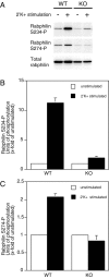
The high K+-induced increase in rabphilin phosphorylation is severely reduced (S234-P) or completely abolished (S274-P) in slices prepared from Rab3a knock-out mice. Acute slices prepared from wild-type (WT) and Rab3a knock-out (KO) mice were incubated in normal Ringer's solution. Unstimulated slices and slices that were subjected to a 2 min 56 mm K+ stimulation were flash-frozen in liquid nitrogen and processed for quantitative Western blotting to detect changes in rabphilin phosphorylation at S234 and S274.A, Representative Western blot results. B, C, Quantitative analysis of four independent experiments (mean and SEM). In both WT and KO, the level of phosphorylation after stimulation is expressed relative to the level in unstimulated slices.
Fig. 3.
Fig. 3.
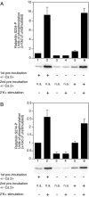
The high K+-induced increase in rabphilin phosphorylation at S234 and S274 is strictly dependent on extracellular Ca2+. Acute slices from 6- to 8-week-old rats were preincubated for 15 min in Ringer's solution with or without Ca2+, followed by incubation for 2 min in the presence or absence of 56 mm K+ and subsequent flash freezing. Some slices were first preincubated for 15 min in Ringer's solution without Ca2+ and then supplemented with Ca2+ for 15 min before a 2 min incubation in the presence or absence of 56 mmK+ and subsequent flash freezing. Slices were processed for quantitative Western blotting to detect changes in rabphilin phosphorylation at S234 (A) and S274 (B). Each panel shows a representative Western blot result and summarizes the analysis of four to seven independent experiments (mean and SEM, for each condition the level of phosphorylation is expressed relative to the level observed in unstimulated slices under normal Ca2+conditions).
Fig. 4.
Fig. 4.
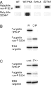
A large proportion of rabphilin is phosphorylated on S234 after a 2 min high K+ stimulation.A, Specificity of the antibody specific for the form of rabphilin that is not phosphorylated at S234 (αnon-P S234). Equal amounts of a recombinant fragment of rabphilin (aa 1–361), wild-type (WT), or serine to alanine mutants at the phosphorylation sites (S234A and S274A), together with the recombinant WT fragment phosphorylated in vitro with purified PKA (WT/PKA), were resolved by SDS-PAGE and transferred to nitrocellulose membranes. The blots were probed with αnon-P S234 or the antibody specific for the form of rabphilin that is phosphorylated at S234 (αS234-P). B, Estimate of the proportion of rabphilin phosphorylated at S234 after a 2 min high K+ stimulation. Equivalent pools of acute slices prepared from 6- to 8-week-old rats were stimulated for 2 min in Ringer's solution with 56 mm K+. The slices were flash-frozen in liquid nitrogen, and the two pools were separately homogenized in the presence or absence of phosphatase inhibitors (PI). The slices homogenized in the absence of phosphatase inhibitors were additionally treated with calf intestinal phosphatase (CIP). Equal amounts of total protein were subjected to Western blotting. The blots were probed with an antibody that recognizes total rabphilin (irrespective of its phosphorylation state) to confirm equal loading, as well as with the antibody that recognizes only S234-phosphorylated rabphilin to confirm the complete dephosphorylation in the CIP-treated sample. In the blot probed with the αnon-P S234 the increase in signal after dephosphorylation (CIP), compared with the signal obtained from the sample prepared in the presence of phosphatase inhibitors (PI), reflects the proportion of rabphilin phosphorylated on S234 after a 2 min high K+ stimulation. C, After a high K+ stimulation the increase in rabphilin S234-P is mirrored by a decrease in rabphilin non-P S234. Equal amounts of total protein from unstimulated slices (unst) and from slices stimulated for 2 min in Ringer's solution with 56 mmK+ (2′K+) were subjected to Western blotting. The blots were probed with an antibody that recognizes total rabphilin to confirm equal loading, as well as with the antibodies that specifically recognize rabphilin phosphorylated or not phosphorylated at S234. The increase in rabphilin phosphorylated at S234 after high K+ stimulation is matched by an equivalent decrease in the signal obtained with the antibody that recognizes only S234 nonphosphorylated rabphilin.
Fig. 5.
Fig. 5.
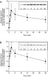
The high K+ induced increase in rabphilin phosphorylation is maximal within 2 min. Acute slices prepared from 6- to 8-week-old rats were incubated in Ringer's solution containing 56 mm K+. At the indicated time points, slices were flash-frozen in liquid nitrogen and subsequently processed for quantitative Western blotting to detect changes in rabphilin phosphorylation at S234 (A) and S274 (B). The insets show representative Western blot results of the time course of phosphorylation; the graphs summarize the analysis of six (S234-P) and five (S274-P) independent experiments (mean and SEM).
Fig. 6.
Fig. 6.
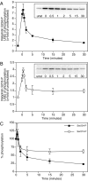
A rapid phosphatase activity dephosphorylates rabphilin S234-P and S274-P. Acute slices prepared from 6- to 8-week-old rats were first subjected to a 2 min incubation in Ringer's solution containing 56 mmK+ and then rapidly transferred to normal Ringer's solution. Slices were flash-frozen in liquid nitrogen before stimulation (unst), at the end of the 2′ high K+ stimulation (t = 0), and at the indicated time points after transfer to normal Ringer's solution.A (rabphilin S234-P) and B (rabphilin S274-P), Insets show representative Western blot results of the time course of dephosphorylation, and the graphs summarize the analysis of four independent experiments (mean and SEM).C, The graph shows the extent of dephosphorylation over time relative to the level of phosphorylation at the end of the stimulation (4 independent experiments, mean and SEM).
Fig. 7.
Fig. 7.
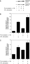
Inhibition of phosphatase activity increases both the basal and the high K+ stimulated level of rabphilin phosphorylation at S234 and S274. Acute slices prepared from 6- to 8-week-old rats were preincubated for 30 min in normal Ringer's solution with or without the addition of 1 μm OA. Subsequently, slices were either flash-frozen in liquid nitrogen or first stimulated for 2 min in Ringer's solution with 56 mmK+ and then flash-frozen. Finally, slices were processed for quantitative Western blotting to detect changes in rabphilin phosphorylation. A, Representative Western blot results.B, C, Quantitative analysis of rabphilin phosphorylation on S234 (B) and S274 (C) of four to eight independent experiments (mean and SEM).
Fig. 8.
Fig. 8.
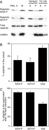
Phosphorabphilin has reduced affinity for membranes. Acute slices were prepared from 6- to 8-week-old rats and stimulated for 2 min in Ringer's solution with 56 mmK+. The slices were homogenized, and the homogenate was spun at low speed (1000 × g) to make postnuclear supernatant (PNS). The PNS was further centrifuged at high speed (100,000 × g) to separate the cytosol (C) and membrane (M) fractions. Equal amounts of total protein for PNS, cytosol, and membranes were used for Western blotting. The membrane fraction was resuspended and extracted with either 1m NaCl or 1% Triton X-100 (TX-100). After a high-speed spin to separate the supernatant (S, extracted proteins) from the pellet (P, not extracted proteins), equal volumes of the two samples were subjected to Western blotting. A, Representative Western blot results with samples probed with the two antibodies specific for the S234 and S274 phosphorylated forms of rabphilin, the antibody to detect total rabphilin, and an antibody against the integral synaptic vesicle protein VAMP-2 to control for the fractionation and membrane extraction protocols. B, Quantitative analysis of cytosol and membrane distributions of phosphorabphilin compared with total rabphilin (4–6 independent experiments, mean and SEM).C, Quantitative analysis of the relative amounts of phosphorabphilin and total rabphilin released into the supernatants after membrane extraction with 1 m NaCl (4–6 independent experiments, mean and SEM).

References

    1. Alger BE, Dhanjal SS, Dingledine R, Garthwaite J, Henderson G, King GL, Lipton P, North A, Schwartzkroin PA, Sears TA, Segal M, Whittingham TS, Williams J. In: Brain slices (Dingledine R, ed), pp 381–437. New York; Plenum: 1984.
    1. Arribas M, Regazzi R, Garcia E, Wollheim CB, De Camilli P. The stimulatory effect of rabphilin 3a on regulated exocytosis from insulin-secreting cells does not require an association-dissociation cycle with membranes mediated by Rab 3. Eur J Cell Biol. 1997;74:209–216. - PubMed
    1. Bock JB, Matern HT, Peden AA, Scheller RH. A genomic perspective on membrane compartment organization. Nature. 2001;409:839–841. - PubMed
    1. Burns ME, Sasaki T, Takai Y, Augustine GJ. Rabphilin-3A: a multifunctional regulator of synaptic vesicle traffic. J Gen Physiol. 1998;111:243–255. - PMC - PubMed
    1. Chung SH, Takai Y, Holz RW. Evidence that the Rab3a-binding protein, rabphilin3a, enhances regulated secretion. Studies in adrenal chromaffin cells. J Biol Chem. 1995;270:16714–16718. - PubMed

MeSH terms

LinkOut - more resources