. 2001 Aug 1;21(15):5546-58.
doi: 10.1523/JNEUROSCI.21-15-05546.2001.

Dopamine D1 receptor-dependent trafficking of striatal NMDA glutamate receptors to the postsynaptic membrane

Affiliations

Dopamine D1 receptor-dependent trafficking of striatal NMDA glutamate receptors to the postsynaptic membrane

A W Dunah et al. J Neurosci. .

Abstract

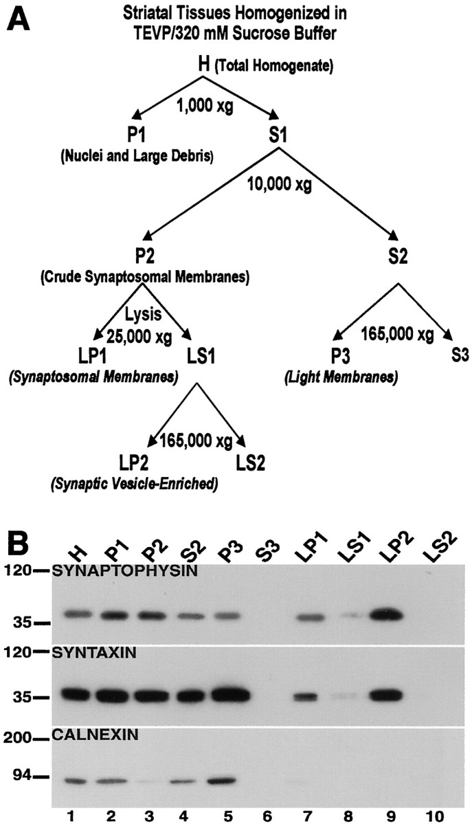
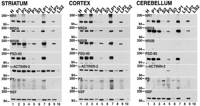
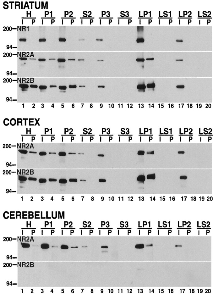
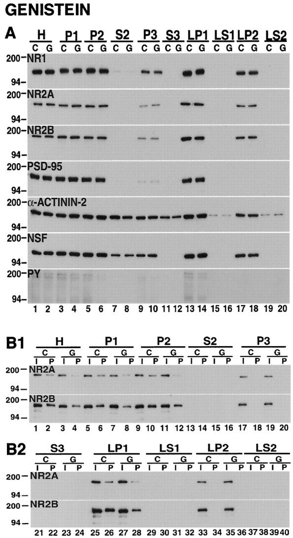
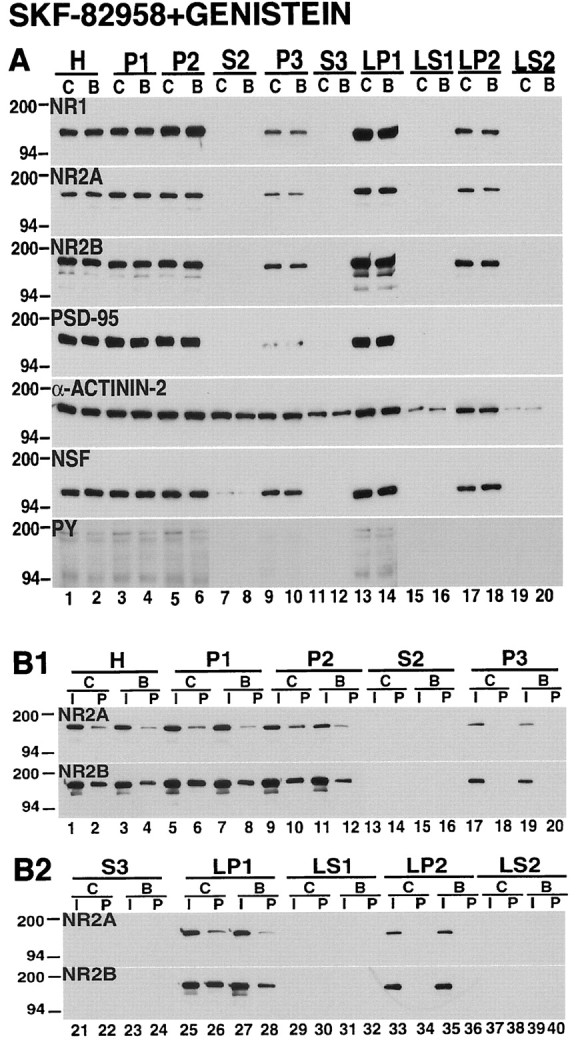
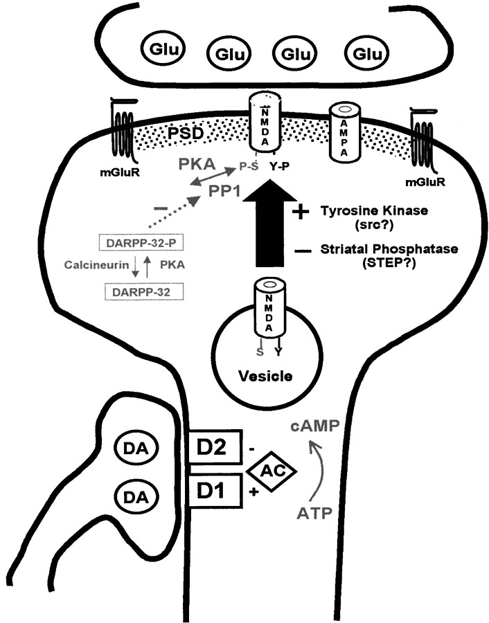
Recent work has shown substantial alterations in NMDA receptor subunit expression, assembly, and phosphorylation in the dopamine-depleted striatum of a rodent 6-hydroxydopamine model of Parkinson's disease. These modifications are hypothesized to result from the trafficking of NMDA receptors between subcellular compartments. Here we show that in rat striatal tissues the NR2A and NR2B subunits in the synaptosomal membrane, and not those in the light membrane and synaptic vesicle-enriched compartments, are tyrosine phosphorylated. The dopamine D1 receptor agonist SKF-82958 produces (1) an increase in NR1, NR2A, and NR2B proteins in the synaptosomal membrane fraction; (2) a decrease in NR1, NR2A, and NR2B proteins in the light membrane and synaptic vesicle-enriched fractions; and (3) an increase in the tyrosine phosphorylation of NR2A and NR2B in the synaptosomal membrane compartment. The protein phosphatase inhibitor pervanadate reproduces the alterations in subcellular distribution and phosphorylation, whereas the effects of the dopamine D1 receptor agonist are blocked by genistein, a protein tyrosine kinase inhibitor. Dopamine D1 receptor agonist treatment does not change the subcellular distribution of the AMPA receptor subunits GluR1 or GluR2/3 in the striatum and has no effect on cortical or cerebellar NMDA receptor subunits. These data reveal a rapid dopamine D1 receptor- and tyrosine kinase-dependent trafficking of striatal NMDA receptors between intracellular and postsynaptic sites. The subcellular trafficking of striatal NMDA receptors may play a significant role both in the pathogenesis of Parkinson's disease and in the development of adverse effects of chronic dopaminergic therapy in parkinsonian patients.


Figures

Fig. 1.
Fig. 1.
Characterization of fractionated subcellular compartments of the striatum. A, Schematic for the biochemical fractionation. The procedure for the subcellular separation of proteins as depicted in this schematic is described in Materials and Methods. B, Characterization of subcellular compartments. The isolated biochemical fractions from striatal tissues were separated by SDS-PAGE, and the blots were probed with antibodies against synaptophysin (top), syntaxin (middle), and calnexin (bottom). Synaptophysin is highly concentrated in the synaptic vesicle-enriched fraction (LP2, lane 9); syntaxin is enriched in the light membrane (P3, lane 5) and synaptic vesicle-enriched (LP2,lane 9) fractions compared with the synaptosomal membrane (LP1, lane 7); and calnexin is found in the light membrane fraction (P3,lane 5), but not in the synaptosomal membrane (LP1, lane 7) nor synaptic vesicle-enriched (LP2, lane 9) fractions.
Fig. 2.
Fig. 2.
NMDA receptor subunits are distributed differentially between subcellular compartments in the rat brain. The tissue samples from the striatum, cortex, and cerebellum were homogenized immediately after dissection, separated into different biochemical fractions as described in Materials and Methods, and resolved on SDS-polyacrylamide gels. Total protein (10 μg) from each fraction was loaded in each lane. The blots were probed with anti-NR1, anti-NR2A, anti-NR2B, anti-PSD-95, anti-α-actinin-2, anti-phosphotyrosine (PY), and anti-NSF antibodies. H, Total homogenate; P1, nuclei and large debris; P2, crude synaptosomal fraction; P3, light membrane fraction;LP1, synaptosomal membrane fraction; LP2, synaptic vesicle-enriched fraction. S2, S3, LS1, andLS2 are supernatants from P2, P3, LP1, and LP2, respectively. The positions and sizes of molecular weight markers are indicated in kilodaltons.
Fig. 3.
Fig. 3.
NMDA receptors present in the synaptosomal membrane fraction, but not those in the light membrane and synaptic vesicle-enriched compartments, are tyrosine phosphorylated. Samples from rat striatum (top), cortex (middle), and cerebellum (bottom) were homogenized immediately after dissection, fractionated, solubilized, and immunoprecipitated with anti-phosphotyrosine antibody. The inputs (I;lanes 1, 3, 5, 7, 9, 11, 13, 15, 17, 19; 5 μg) and pellets (P; lanes 2, 4, 6, 8, 10, 12, 14, 16, 18, 20; 40 μg) were separated on SDS-PAGE gels, and the blots were probed with anti-NR1, anti-NR2A, and anti-NR2B antibodies. Tyrosine-phosphorylated NR2A and NR2B subunits were detected in LP1 (lane 13), but not in P3 (lane 10) and LP2 (lane 18) fractions.
Fig. 4.
Fig. 4.
The dopamine D1 receptor agonist SKF-82958 produces subcellular redistribution and an increase in the tyrosine phosphorylation of striatal NMDA receptors. A, Subcellular distribution. Samples from tissues that were incubated for 10 min under control conditions (C) or with 50 μm SKF-82958 (S) were subjected to biochemical fractionation. The subcellular fractions were resolved by SDS-PAGE, and the blots were probed with NR1, NR2A, NR2B, PSD-95, α-actinin-2, NSF, and anti-phosphotyrosine (PY) antibodies. SKF-82958 decreased NR1, NR2A, and NR2B in P3 (lane 10) and LP2 (lane 18) fractions; it increased these subunits in LP1 (lane 14), and it increased phosphotyrosine proteins (PY). B1, B2, Tyrosine phosphorylation. The subcellular-fractionated samples from control (C) and SKF-82958-treated (S) striatal tissues were immunoprecipitated with anti-phosphotyrosine antibody and immunoblotted with anti-NR2A and anti-NR2B. The inputs (I) and pellets (P) for each biochemical fraction are indicated across the top of the figure. SKF-82958 increased tyrosine-phosphorylated NR2A and NR2B in H (lane 4), P1 (lane 8), P2 (lane 12), and LP1 (lane 28) fractions.
Fig. 5.
Fig. 5.
Densitometric quantification of NMDA receptor subunit NR1, NR2A, and NR2B proteins in striatal tissues that were treated with the dopamine D1 receptor agonist SKF-82958 (left panels) and with a combination of the dopamine D1 receptor agonist SKF-82958 and a protein tyrosine kinase inhibitor genistein (right panels). The exposed films from the experiments depicted in Figures 4A and 10A were scanned and analyzed as described in Materials and Methods. Values on the ordinate represent the relative levels of NR1, NR2A, and NR2B proteins given as a percentage of the control samples. Data are means ± SEM obtained from three rats. Asterisks indicate significant differences between treatment and control samples (p < 0.05, ANOVA). The alteration in the subcellular distribution of NR1, NR2A, and NR2B subunits produced by SKF-82958 was blocked by genistein.
Fig. 6.
Fig. 6.
The dopamine D2 receptor agonist quinpirole does not alter the subcellular distribution and tyrosine phosphorylation of striatal NMDA receptors. A, Subcellular distribution. Samples from tissues that were incubated for 10 min under control conditions (C) or with 100 μmquinpirole (a dopamine D2 receptor agonist; Q) were separated into subcellular fractions and subjected to electrophoresis on SDS-polyacrylamide gels. The blots were probed with NR1, NR2A, NR2B, PSD-95, α-actinin-2, NSF, and anti-phosphotyrosine (PY) antibodies. There were no significant differences in the subcellular distribution of the analyzed proteins between control and quinpirole-treated samples. B1, B2, Tyrosine phosphorylation. Striatal tissues from control (C) and quinpirole-treated (Q) samples were solubilized and precipitated with anti-phosphotyrosine antibody. The inputs (I) and pellets (P) are indicated across the top of the figure. The resulting blots were immunoblotted for the NR2A and NR2B subunits. Quinpirole had no apparent effect on the tyrosine phosphorylation of NR2A and NR2B subunits.
Fig. 7.
Fig. 7.
The dopamine D1 receptor agonist SKF-82958 had no effect on the subcellular distribution and tyrosine phosphorylation of striatal AMPA receptors. A, Subcellular distribution. Fractionated striatal tissues that were incubated for 10 min under control conditions (C) or with 50 μm SKF-82958 (S) were resolved by SDS-PAGE, and the blots were probed with antibodies specific for GluR1 and GluR2/3. SKF-82958 had no apparent effect on the subcellular distribution of GluR1 and GluR2/3 subunits. B1, B2, Tyrosine phosphorylation. The fractionated striatal samples were precipitated with anti-phosphotyrosine antibody and immunoblotted for GluR1 and GluR2/3. The inputs (I) and pellets (P) are indicated across thetop of the figure. SKF-82958 produced no changes in the tyrosine phosphorylation of GluR1 or GluR2/3.
Fig. 8.
Fig. 8.
The protein phosphatase inhibitor pervanadate induces subcellular redistribution and an increase in tyrosine phosphorylation of striatal NMDA receptors. A, Subcellular distribution. Striatal samples from tissues that were incubated for 10 min under control conditions (C) or with 200 μm pervanadate (P) were fractionated and resolved on SDS-PAGE gels. The blots were probed with NR1, NR2A, NR2B, PSD-95, α-actinin, NSF, and anti-phosphotyrosine (PY) antibodies. Similar to the effects produced by SKF-82958, pervanadate reduced the levels of NR1, NR2A, and NR2B in P3 (lane 10) and LP2 (lane 18); it increased the levels of these NMDA subunits in LP1 (lane 14) and markedly increased phosphotyrosine proteins (PY). B1, B2, Tyrosine phosphorylation. Samples from the subcellular-fractionated control (C) and pervanadate-treated (P) striatal tissues were immunoprecipitated with anti-phosphotyrosine antibody. The inputs (I) and pellets (P) were electrophoresed and probed for NR2A and NR2B subunits. Tyrosine phosphorylation of NR2A and NR2B was increased in H (lane 4), P1 (lane 8), P2 (lane 12), and LP1 (lane 28).
Fig. 9.
Fig. 9.
The protein tyrosine kinase inhibitor genistein does not change the subcellular distribution of striatal NMDA receptors but decreases the tyrosine phosphorylation of NR2A and NR2B.A, Subcellular distribution. Fractions from striatal tissues that were incubated for 10 min under control conditions (C) or with 100 μm genistein (G) were electrophoresed on SDS-polyacrylamide gels, and the blots were probed with NR1, NR2A, NR2B, PSD-95, α-actinin-2, NSF, and anti-phosphotyrosine (PY) antibodies. The subcellular localizations of NMDA receptor subunits and the other proteins that were studied were not changed by genistein, but there were reductions in total phosphotyrosine proteins (PY). B1, B2, Tyrosine phosphorylation. The fractionated samples from control (C) and genistein-treated (G) striatal tissues were immunoprecipitated by using anti-phosphotyrosine antibody and were immunoblotted with anti-NR2A and anti-NR2B antibodies. The inputs (I) and pellets (P) are indicated across the top of the figure. Genistein decreased the tyrosine phosphorylation of NR2A and NR2B in H (lane 4), P1 (lane 8), P2 (lane 12), and LP1 (lane 28).
Fig. 10.
Fig. 10.
The dopamine D1 receptor agonist-induced alterations in subcellular distribution and tyrosine phosphorylation of striatal NMDA receptors are inhibited by the protein tyrosine kinase inhibitor genistein. A, Subcellular distribution. Samples from tissues that were incubated for 10 min under control conditions (C) or with 50 μmSKF-82958 and 100 μm genistein (B) were subjected to subcellular fractionation. The proteins were separated on SDS-PAGE, and the blots were probed with antibodies against NR1, NR2A, NR2B, PSD-95, α-actinin-2, NSF, and phosphotyrosine proteins (PY). In the presence of genistein, SKF-82958 produced no alteration in the distribution of NMDA receptor subunits. B1, B2, Tyrosine phosphorylation. Solubilized control (C) and treated samples (B) were immunoprecipitated with anti-phosphotyrosine antibody, and the blots were probed with NR2A and NR2B antibodies. The inputs (I) and pellets (P) are indicated across thetop of the figure. Genistein inhibited the increase in tyrosine phosphorylation of NR2A and NR2B produced by the dopamine D1 receptor agonist SKF-82958.
Fig. 11.
Fig. 11.
Model for the trafficking of NMDA receptors at the corticostriatal synapse. Illustrated is a dendritic spine of a striatal projection neuron receiving input from a cortical axon, using glutamate as a transmitter (Glu) at the head of the spine, and from a nigrostriatal axon, using dopamine (DA) as a transmitter and forming a synapse on the shaft of the spine. The effects of glutamate are mediated by NMDA, AMPA, and metabotropic glutamate receptors (mGluR). These are linked to each other and to additional signaling molecules via the proteins of the postsynaptic density (PSD). Dopaminergic inputs are mediated by the dopamine D1 and D2 receptors. These receptors have reciprocal effects on the formation of cAMP by adenylyl cyclase (AC), but most striatal neurons express a preponderance of a single dopamine receptor type, depending on whether they contribute to the direct or indirect pathways. In addition to the NMDA receptors that are present at the excitatory synapse, our data suggest that there is a pool of assembled receptors within intracellular vesicular compartments. The vesicle-associated receptors are characterized by the absence of tyrosine (Y) phosphorylation of the NR2A and NR2B subunits. Activation of dopamine D1 receptors leads (via mechanisms that are not well defined) to tyrosine phosphorylation of the vesicular NMDA receptor subunits by a tyrosine kinase [perhaps a member of the src family reported to phosphorylate NMDA subunits at tyrosine residues (Suzuki and Okumura-Noji, 1995)] and insertion of the receptors into the synaptic membrane. There is also a tonically active tyrosine phosphatase, which may be a member of the STEP family. The diagram also illustrates the serine (S) phosphorylation of the NR1 subunit, which is regulated both by the direct action of cAMP on protein kinase A (PKA) as well as by the DARPP-32 pathway; phosphorylation of DARPP-32 by PKA inhibits the dephosphorylation of NR1 by protein phosphatase-1 (PP-1; Snyder et al., 1998). Although protein kinase C has been reported to modulate the trafficking of NMDA receptors (Lan et al., 2001), the precise role of cAMP in the phosphorylation of NMDA subunits at tyrosine residues is not known. At present it is also uncertain whether serine phosphorylation plays a role in the trafficking of NMDA receptors at the corticostriatal synapse.

References

    1. Barinaga M. Secrets of secretion revealed. Science. 1993;260:487–489. - PubMed
    1. Bennett MK, Calakos N, Scheller RH. Syntaxin: a synaptic protein implicated in docking of synaptic vesicles at presynaptic active zones. Science. 1992;257:255–259. - PubMed
    1. Blanchet PJ, Papa SM, Metman LV, Mouradian MM, Chase TN. Modulation of levodopa-induced motor response complications by NMDA antagonists in Parkinson's disease. Neurosci Biobehav Rev. 1997;21:447–453. - PubMed
    1. Boulanger LM, Lombroso PJ, Raghunathan A, During MJ, Wahle P, Naegele JR. Cellular and molecular characterization of a brain-enriched protein tyrosine phosphatase. J Neurosci. 1995;15:1532–1544. - PMC - PubMed
    1. Cepeda C, Levine MS. Dopamine and N-methyl-d-aspartate receptor interactions in the neostriatum. Dev Neurosci. 1998;20:1–18. - PubMed

Publication types

MeSH terms

LinkOut - more resources