. 2001 Aug 1;21(15):5643-51.
doi: 10.1523/JNEUROSCI.21-15-05643.2001.

Asymmetric segregation of Numb in retinal development and the influence of the pigmented epithelium

Affiliations

Asymmetric segregation of Numb in retinal development and the influence of the pigmented epithelium

M Cayouette et al. J Neurosci. .

Abstract

Asymmetric segregation of cell-fate determinants during cytokinesis plays an important part in controlling cell-fate choice in invertebrates. During Drosophila neurogenesis, for example, asymmetric segregation of the Numb protein, which inhibits Notch signaling, is necessary for the two daughter cells of a division to have different fates. In vertebrates, the role of asymmetric segregation of cell-fate determinants is uncertain, and the way the process might be regulated is unknown. We have studied the orientation of cell divisions and the distribution of Numb in the developing rat retina. We show that, whereas most retinal neuroepithelial cells divide with their mitotic spindles oriented parallel to the plane of the neuroepithelium, a substantial minority divides with their spindles oriented perpendicularly. The proportion of these vertically dividing cells changes during development, peaking around the day of birth. Numb appears to be inherited only by the apical daughter cell when a neuroepithelial cell divides vertically. Similarly, in dissociated cell cultures, some retinal neuroepithelial cells divide asymmetrically and distribute Numb to only one of the two daughter cells, suggesting that the dissociated cells can retain their polarity in vitro. Using retinal explant cultures, we find that the retinal pigment epithelium apparently promotes vertical divisions in the neural retina. To our knowledge, this is the first evidence that asymmetric segregation of cell-fate determinants may contribute to cell diversification in the mammalian retina and that an epithelium controls this process by influencing the plane of division in the adjacent neural retina.


Figures

Fig. 1.
Fig. 1.
Mitotic spindle orientations in dividing retinal neuroepithelial cells. P0 retinal sections were stained with both anti-γ-tubulin antibodies to visualize the centrosomes (green) and propidium iodide to visualize DNA (red). Both metaphase (A–C) and anaphase–telophase (D–F) mitotic cells were analyzed. Most dividing cells had the mitotic spindle aligned horizontally relative to the plane of the tissue (A, D), but in some cells the spindle was aligned perpendicular to the plane of the tissue (C, F) or in an intermediate orientation (B, E). The retinal pigment epithelium isdown in all figures.
Fig. 2.
Fig. 2.
The distribution of mitotic spindle orientations at different developmental ages. The graphs show frequency histograms of the orientations of mitotic spindles in embryonic and postnatal rat retinas at E18 (A), P0 (B), and P4 (C). Four animals were analyzed at E18 (n = 153 cells) and P0 (n = 135 cells), whereas three animals were analyzed at P4 (n = 104 cells). The distributions at different times were compared using the Kolmogorov–Smirnov test and found to be significantly different for embryonic and postnatal retina: E18 versus P0, p = 0.0001; E18 versus P4,p = 0.005. The pie charts show the proportions of vertical (light gray) and horizontal (dark gray) spindles at the different times, where vertical was defined as an angle ≥45 degrees. The differences in the proportions are highly significant when compared using the χ2 test: E18 versus P0, p < 0.0001; E18 versus P4,p = 0.03.
Fig. 3.
Fig. 3.
The effect of the retinal pigment epithelium (RPE) on the orientation of mitotic spindles.A, B, Cryostat sections through P0 retinal explants cultured for 24 hr either with (A) or without (B) the RPE in place. C, D, Frequency histograms of the orientations of mitotic spindles in comparable retinal explants cultured for 24 hr either with (C) or without (D) the RPE. The results were pooled from two separate experiments that involved a total of 11 rats (+RPE explants, n = 193 cells; −RPE explants, n = 176 cells). Pie charts show the proportion of vertical (light gray) and horizontal (dark gray) spindles in each condition. The Kolmogorov–Smirnov test shows that the difference between the frequency distributions in A and B is highly significant (p = 0.0088). Comparison of the proportions of vertical and horizontal spindles using the χ2 test also shows a significant difference (p = 0.0342).
Fig. 4.
Fig. 4.
Apical localization of Numb in P0 retinal neuroepithelial cells. Retinal sections were stained with monoclonal anti-Numb antibody (green) and propidium iodide (red). A, Low magnification view showing that Numb staining is mainly concentrated on the apical side of the neuroepithelium. B, High magnification view showing the finger-like staining of Numb associated with the apical pole of interphase neuroepithelial cells (arrowheads point to two examples of finger-like staining). C–F, Numb distribution in mitotic neuroepithelial cells (arrowheads). Metaphase (C) and anaphase–telophase (D) cells dividing with their spindles aligned horizontally, relative to the plane of the tissue, show an apical crescent of Numb, suggesting that both daughter cells of these divisions will inherit Numb. In cells dividing with their mitotic spindles aligned more perpendicularly to the plane of the tissue (E and F show two examples of cells in anaphase–telophase), Numb is likely to be inherited preferentially by the apical daughter cell.
Fig. 5.
Fig. 5.
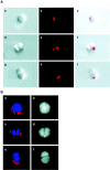
Asymmetric distribution of Numb and symmetric distribution of Notch-1 in dissociated retinal cell cultures.A, Differential-interference contrast images of dissociated retinal cells (a, d,g), immunofluorescence images of Numb staining (red) of the corresponding cells (b,e, h), and overlaid images of both (c, f, i). Some single cells show Numb confined to one pole (a–c). In most cell doublets, Numb is found at the same pole in both cells, consistent with the cells being produced by a symmetric division in culture (d–f). In some doublets, however, Numb is found only in one of the cells, with a distribution consistent with the two cells being produced by an asymmetric division in which only one daughter cell inherited Numb (g–i).B, Numb (a, c,e) and Notch (b, d,f) immunofluorescence staining in dissociated retinal cells. The DNA is stained in blue with Hoescht dye in a, c, and e. In a single cell in which Numb is segregated to one pole (a), Notch-1 is evenly distributed on the cell surface (b). In cell doublets (c–f), Notch-1 is always observed evenly distributed on the surface of both cells (d, f), irrespective of whether Numb is found on both cells (c) or only on one (e).
Fig. 6.
Fig. 6.
A model for how asymmetric cell division in the retina may influence cell-fate. Some retinal neuroepithelial cells reorient their mitotic spindles and divide vertically. Because Numb is concentrated at the apical pole of the cells, it is likely to be inherited preferentially by the apical daughter cell of such divisions, where it inhibits Notch signaling. The basal daughter cell inherits little Numb, allowing unopposed Notch signaling and differentiation into a Müller cell. The apical cell might divide again or develop into another cell type.
Fig. 7.
Fig. 7.
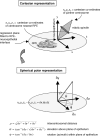
Calculation of the orientation of mitotic spindles, using three-dimensional coordinate geometry. To determine the spindle orientation, a regression fit is first made to the interface between the RPE and the neural retina to provide a reference plane or axis. The x, y, and zCartesian coordinates of the two centrosomes of the spindle are transformed such that they are now expressed relative to the fitted plane or line, as illustrated in the topdiagram, which shows a schematic mitotic spindle. The RPE is shown directly beneath the mitotic cell in thex–z plane, and the positions of the two centrosomes are indicated by the triplet coordinate pairsx1,y1,z1andx2,y2,z2. To calculate the elevation of the spindle above thex–z plane, it is necessary to represent this information in spherical polar coordinate form. This is done as follows: the difference between the triplets is calculated (shown in the bottomdiagram) and represented by the values δx, δy, and δz. These values are then substituted into the equations (derived from trigonometry) shown at thebottom of the figure to yield the values ρ (the distance between the centrosomes), θ (the elevation of the spindle out of the xz plane), and ϕ (the rotation of the spindle about the y-axis, also termed the azimuth). These values are also illustrated on theright side of the bottom diagram.

Similar articles

Cited by

References

    1. Carmena A, Murugasu-Oei B, Menon D, Jimenez F, Chia W. Inscuteable and numb mediate asymmetric muscle progenitor cell divisions during Drosophila myogenesis. Genes Dev 12 1998. 304 315 [Erratum (1998) 12:1241] - PMC - PubMed
    1. Cepko CL. The roles of intrinsic and extrinsic cues and bHLH genes in the determination of retinal cell fates. Curr Opin Neurobiol. 1999;9:37–46. - PubMed
    1. Cepko CL, Austin CP, Yang X, Alexiades M, Ezzeddine D. Cell fate determination in the vertebrate retina. Proc Natl Acad Sci USA. 1996;93:589–595. - PMC - PubMed
    1. Chang JT, Esumi N, Moore K, Li Y, Zhang S, Chew C, Goodman B, Rattner A, Moody S, Stetten G, Campochiaro PA, Zack DJ. Cloning and characterization of a secreted frizzled-related protein that is expressed by the retinal pigment epithelium. Hum Mol Genet. 1999;8:575–583. - PubMed
    1. Chenn A, McConnell SK. Cleavage orientation and the asymmetric inheritance of Notch1 immunoreactivity in mammalian neurogenesis. Cell. 1995;82:631–641. - PubMed

Publication types

LinkOut - more resources