. 2001 Aug 1;21(15):5703-14.
doi: 10.1523/JNEUROSCI.21-15-05703.2001.

Generation of aggregated beta-amyloid in the rat hippocampus impairs synaptic transmission and plasticity and causes memory deficits

Affiliations

Generation of aggregated beta-amyloid in the rat hippocampus impairs synaptic transmission and plasticity and causes memory deficits

A Stéphan et al. J Neurosci. .

Abstract

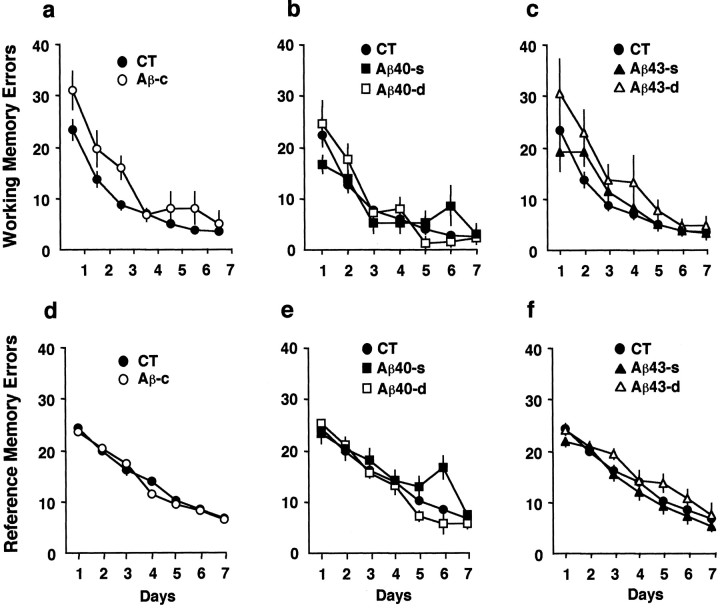
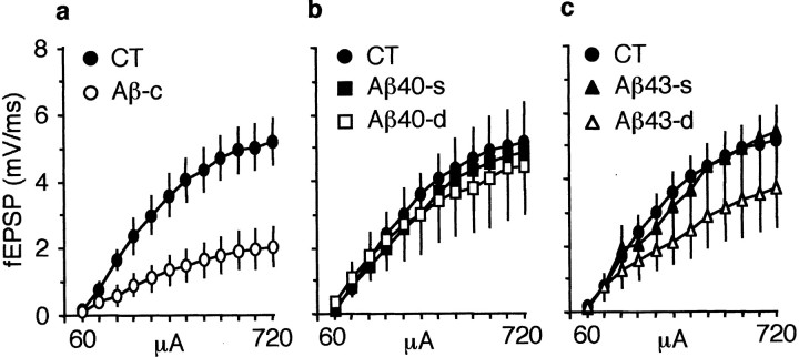
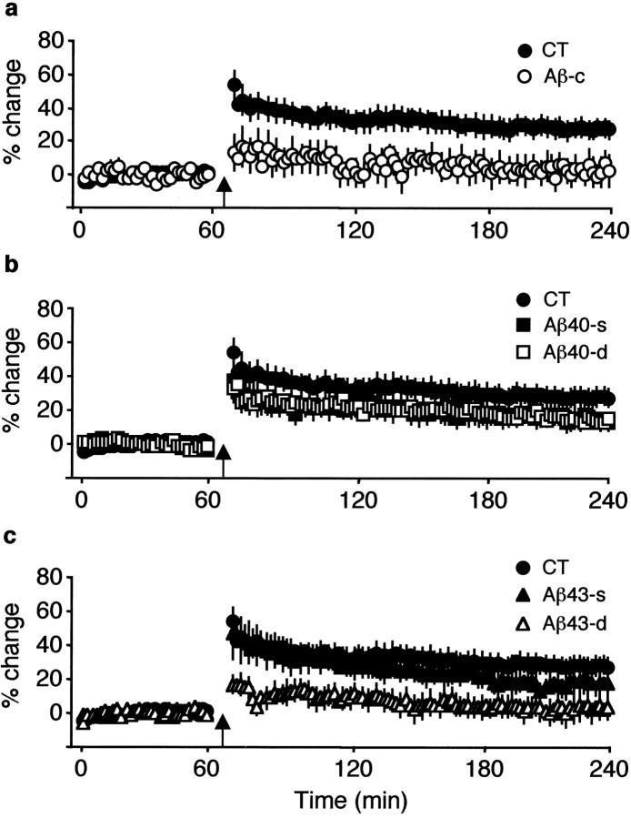
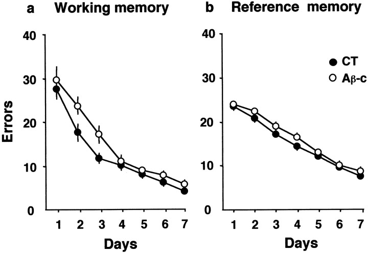
We injected a combination of the beta-amyloids (Abetas) Abeta40 and Abeta43 to "seed" formation of amyloid deposits in the dorsal dentate gyrus of rats in vivo, on the basis of a theory of Jarrett and Landsbury (1993). Rats were tested on several different learning tasks, and synaptic transmission and plasticity were assessed in vivo. Between 7 and 16 weeks after injection, we found aggregated amyloid material, reactive astrocytosis, microgliosis, and cell loss around the sites of injection. Rats were impaired specifically in working memory type tasks in accordance with the type of memory deficit observed in the early stages of Alzheimer's disease. Synaptic transmission and long-term potentiation, a candidate cellular mechanism for memory, were severely impaired in vivo. Injections of the same dose of fragments individually did not induce these effects. These findings suggest that aggregated amyloid material induces cognitive deficits similar to those observed in the early phases of Alzheimer's disease via an alteration in neuronal transmission and plasticity.


Figures

Fig. 1.
Fig. 1.
Spatial learning on the radial arm maze, 4 weeks after focal injections of amyloid peptides, shows the ability of rats to learn the working memory element (a–c) or the reference memory element (d–f).a, Rats injected with combined fragments (Aβ-c) showed impairment in the working memory aspect in the first few days of training (blocks of 8 trials/d) compared with control rats (CT). This difference was modest but significant. b, Rats injected with either dose of Aβ40 [single dose (Aβ40-s); double dose (Aβ40-d)] were not significantly impaired in working memory. c, Whereas rats injected with the low dose of Aβ43 were not impaired in learning, those rats injected with the high dose of Aβ43 showed an overall impairment. d–f, All rats were able to learn the reference memory aspect of the task to the same level as the control rats. The y-axis denotes the total number of reentries made in the working memory element (a–c) and entries into nonbaited arms in the reference memory element (d–f) of the task.
Fig. 2.
Fig. 2.
I–O curves of the field EPSP (fEPSP) in rats with focal injections of amyloid fragments. a, Rats injected with the combined fragments (Aβ-c) show a significant reduction in the size of the fEPSP at all intensities compared with control (CT) rats. b, In rats injected with a single dose (Aβ40-s) or a double dose (Aβ40-d) of Aβ40, there was no effect of the peptide at any intensity compared with theCT group. c, The single dose of Aβ43 had no effect on I–O curves, but rats injected with the double dose showed a significant reduction in fEPSP, although not as severe as that of rats injected with the combined peptides. The y-axis denotes the slope of the fEPSP measured in millivolts per millisecond, and the x-axis indicates the lowest and highest intensities used in microamperes. Increments were an average of 60 μA.
Fig. 3.
Fig. 3.
LTP in the dentate gyrus measured for 3 hr after the induction of LTP in rats injected with amyloid peptides.a, In rats injected with the combined fragments (Aβ-c), no LTP was induced after the tetanus. b, Aβ40 injected at either a single dose (Aβ40-s) or double dose (Aβ40-d) had no significant effect on either induction or maintenance of LTP compared with control rats (CT), although there was a slight reduction throughout the recording period. c, The single dose of Aβ43 had no significant effect on induction or maintenance of LTP, but at double the dose, there was a blockade of induction of LTP in a manner similar to that of the Aβ-cgroup, and this was also significantly reduced compared with the control group. The y-axis denotes the percentage of change in slope of the fEPSP after induction of LTP (indicated byarrow), and the x-axis denotes the time of recording.
Fig. 4.
Fig. 4.
Nissl-stained sections (a–f) show the amount of damage to the dentate gyrus and cell loss in sample rats in each group, and Thioflavin S staining (g–j) shows where amyloid material has aggregated in rats in which focal injections were made.a, Nissl staining in a control rat injected with the vehicle solution shows a small amount of damage and loss of cells that is likely to be caused by mechanical damage. g, Thioflavin S staining around the site of injection in vehicle-injected rats does not fluoresce, indicating the absence of aggregate amyloid.b, c, Nissl staining in rats injected with Aβ40-s (b) and Aβ43-s (c) shows that there is no greater level of cell loss than that observed with the vehicle-injected rats. d, h, Nissl staining in a rat injected with Aβ40-d (d) shows a small lesion that is likely to be caused by the increased volume injected, because there is no accompanying presence of aggregated amyloid detected with Thioflavin S staining (h). e, f, In rats injected with Aβ43-d (e) or the combined injections (f), there is the presence of a lesion that is exacerbated by aggregated material. The level of cell loss and damage made in the dentate gyrus and the amount of aggregated material observed in and around the lesion were similar in these two groups. i, An example of Thioflavin S positively stained aggregated material outside of the lesion in a rat injected with Aβ43-d is shown. j, An example of aggregated material in and around the site of the lesion in a rat injected with the combined peptides is shown. Scale bars: a–f, 500 μm;g–j, 200 μm.
Fig. 5.
Fig. 5.
The performance of rats with distributed injections of the combined fragments (Aβ-c) or vehicle solution in the radial arm maze task. a, Rats injected with the fragments were impaired in the working memory aspect of the task, in much the same manner as were the rats with focal injections of the combined peptides. b, Also similar to the rats with focal injection, those with distributed injections of the combined fragments were not impaired in the reference memory task. They-axis denotes the total number of reentries made in the working memory element (a) and entries into nonbaited arms in the reference memory element (b) of the task. CT, Control.
Fig. 6.
Fig. 6.
Spatial navigation in the water maze. No impairment was observed in rats injected with combined fragments (Aβ-c; open circles orbars) compared with the control group receiving vehicle (VH; closed circles orbars) in either the acquisition phase (a) or the probe trial measured 24 hr later (b). Both groups spent significantly (double asterisks) more time in the training quadrant than in the others. Ar, Adjacent right;Tg, target; Al, adjacent left; Opp, opposite.
Fig. 7.
Fig. 7.
Working memory in the eight-arm elimination task. Rats injected with the combined fragments (Aβ-c) were impaired (asterisks) at the beginning of training compared with control rats (CT) but attained a similar level of performance with overtraining. Thex-axis denotes the number of trials, and they-axis denotes the number of reentries.
Fig. 8.
Fig. 8.
Delayed match-to-place on the radial arm maze. Rats injected with combined fragments (Aβ-c) were impaired at a 0 and 10 min delay (asterisks) compared with the control group (CT). When the delay was extended (20–180 min), a similar increase in the number of errors was observed in both the CT group and rats injected with the combined peptides.
Fig. 9.
Fig. 9.
Synaptic transmission and LTP in rats with distributed injections of vehicle (Vh) or combined peptides (Aβ-c). a, I–O curves of the fEPSP show that rats injected withAβ-c have a significantly smaller increase in fEPSP when compared with Vh-injected rats. This effect is similar to that observed with focal injections. They-axis denotes the change in the slope of the fEPSP in millivolts per millisecond, and the x-axis denotes increasing intensities (increments of 60 μA). b, Sample responses from rats injected with the vehicle solution and combined peptides before and after (arrowhead) induction of LTP are shown. The left-hand panel shows potentiation 30 min after induction of LTP, and the right-hand panelshows potentiation 3 hr after induction of LTP. c, Rats injected with the combined peptides show normal induction of LTP of the fEPSP, but this declined back to basal levels 3 hr after the tetanus. The y-axis denotes the percentage of change in fEPSP slope. d, In contrast, the spike amplitude was potentiated, although significantly less than in the vehicle-injected rats, and the level of potentiation was maintained for the duration of recording. The y-axis denotes the change in spike amplitude in millivolts, and the arrow indicates when the tetanus was delivered.
Fig. 10.
Fig. 10.
Representative examples of pathological markers of AD in the dentate gyrus from a rat injected with the vehicle solution (a–c) or with the combined peptides (d–j). a–c, Serial sections from a vehicle-injected rat show the site of injection (arrowhead) marked with Nissl staining (a), minimal cell loss restricted to the site of injection (arrowhead) with NeuN (b), and no Aβ immunoreactivity (c). d, e, Serial sections from a rat injected with the combined fragments and stained with Nissl (d) or NeuN (e) show cell loss near the injection site (arrowhead) that is more extensive than that observed with vehicle-injected rats. Note the thinning of the cell body layer in the upper blade of the dentate gyrus, indicating additional cell loss away from the site of injection.f, h, Aβ immunoreactivity shows the presence of amyloid material in the dentate gyrus corresponding with the site of injection observed with Nissl and NeuN (f) and shows amyloid material further from the site of injection (h). g, i, j, Adjacent sections tof show Thioflavin S-positive staining, suggesting that the amyloid material is aggregated (g), and inflammation reactivity around the site of injection (i, j). i, Extensive GFAP-positive staining forming a virtual wall around the site of the injection is shown.Inset, An astrocyte with swollen processes, magnified 1000×, is shown. j, OX-42 immunostaining in the adjacent section shows strong microglial reactivity in the site of the injection. Inset, An example of a microglia magnified 1000× is shown. Scale bars: h, 100 μm; a–g, i, j, 200 μm.

References

    1. Abraham WC, Bliss TV, Goddard GV. Heterosynaptic changes accompany long-term but not short-term potentiation of the perforant path in the anaesthetized rat. J Physiol (Lond) 1985;363:335–349. - PMC - PubMed
    1. Akiyama H, Barger S, Barnum S, Bradt B, Bauer J, Cole GM, Cooper NR, Eikelenboom P, Emmerling M, Fiebich BL, Finch CE, Frautschy S, Griffin WS, Hampel H, Hull M, Landreth G, Lue L, Mrak R, Mackenzie IR, McGeer PL, O'Banion MK, Pachter J, Pasinetti G, Plata-Salaman C, Rogers J, Rydel R, Shen Y, Streit W, Strohmeyer R, Tooyoma I, Van Muiswinkel FL, Veerhuis R, Walker D, Webster S, Wegrzyniak B, Wenk G, Wyss-Coray T. Inflammation and Alzheimer's disease. Neurobiol Aging. 2000;21:383–421. - PMC - PubMed
    1. Annaert WG, Levesque L, Craessaerts K, Dierinck I, Snellings G, Westaway D, George-Hyslop PS, Cordell B, Fraser P, De Strooper B. Presenilin 1 controls gamma-secretase processing of amyloid precursor protein in pre-Golgi compartments of hippocampal neurons. J Cell Biol. 1999;147:277–294. - PMC - PubMed
    1. Ball MJ, Fisman M, Hachinski V, Blume W, Fox A, Kral VA, Kirshen AJ, Fox H, Mersky H. A new definition of Alzheimer's disease: a hippocampal dementia. Lancet. 1985;1:14–16. - PubMed
    1. Barone S, Tandon P, McGinty JF, Tilson HA. The effects of NGF and fetal cell transplants on spatial learning after intradentate administration of colchicine. Exp Neurol. 1991;114:351–363. - PubMed

Publication types

MeSH terms