. 2001 Aug 1;21(15):5715-22.
doi: 10.1523/JNEUROSCI.21-15-05715.2001.

Acquisition of eyeblink conditioning is critically dependent on normal function in cerebellar cortical lobule HVI

Affiliations

Acquisition of eyeblink conditioning is critically dependent on normal function in cerebellar cortical lobule HVI

P J Attwell et al. J Neurosci. .

Abstract

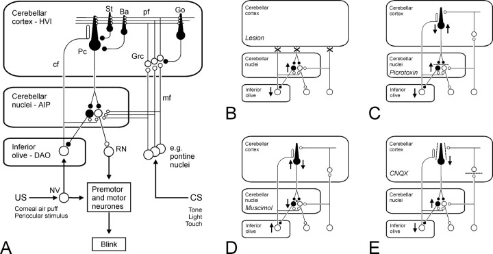
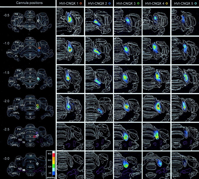
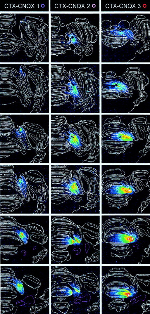
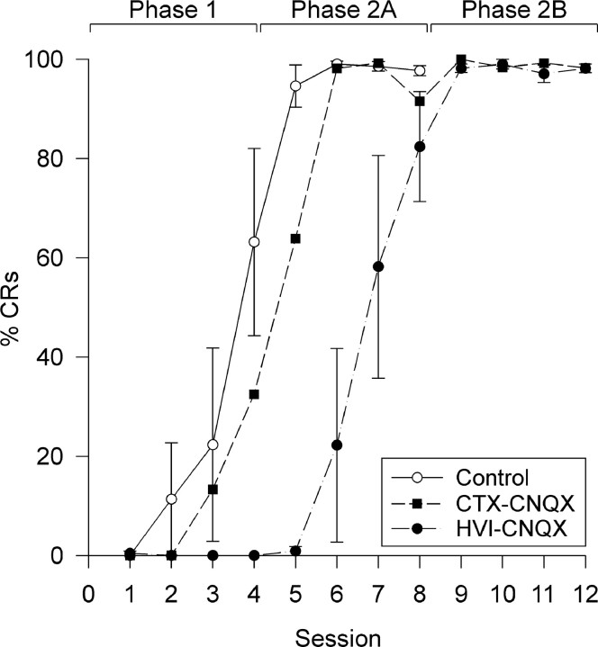
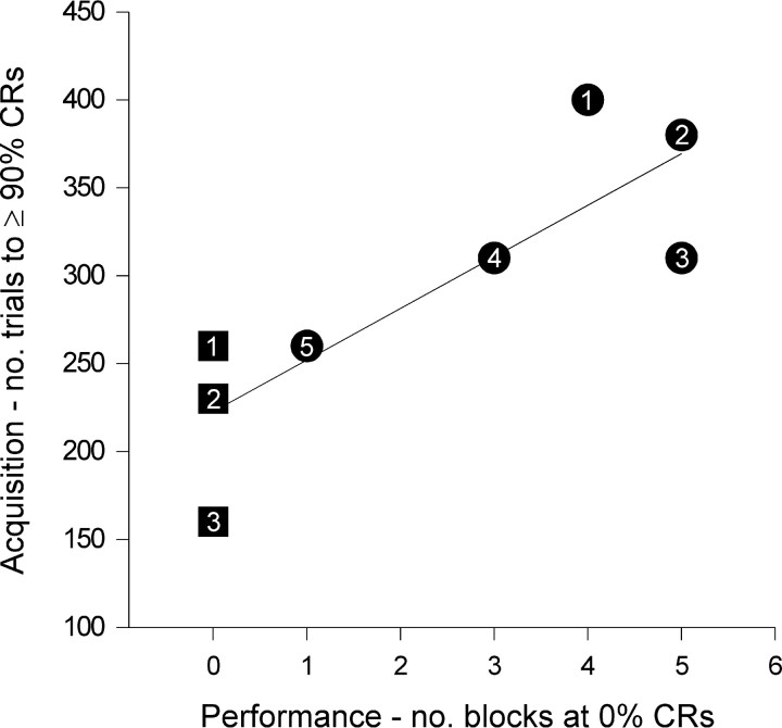
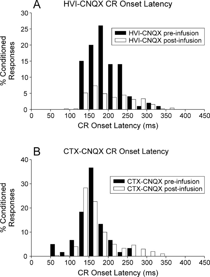
Classical conditioning of the nictitating membrane response (NMR)/eyeblink response of rabbits is a simple form of cerebellar-dependent, associative motor learning. Reversible inactivations of the cerebellar nuclei and inferior olive have implicated the olivo-cortico-nuclear loop in the acquisition of nictitating membrane conditioning, but the role of the cerebellar cortex in acquisition has not been tested directly. Here we have used local infusions of the water-soluble, disodium salt of 6-cyano-7-nitroquinoxaline-2,3-dione reversibly to block cerebellar cortical AMPA/kainate receptors in lobule HVI during acquisition training. After the drug effects dissipated, there was no evidence that acquisition had taken place; the subjects behaved as if naive. Further training without inactivation then allowed normal acquisition, and further inactivations during performance of conditioned responses abolished these established responses. There was a strong correlation between the inactivation effects on acquisition and subsequent inactivation effects on performance, indicating that the same eyeblink-control cortical microzones are engaged in learning and expressing this behavior. The cortical component of the olivo-cortico-nuclear loop is essential for acquisition of classically conditioned nictitating membrane response learning, and eyeblink control areas in HVI are critical. Our findings are consistent with models of cerebellar learning that assign essential plasticity to the cortex or to a distribution between levels in olivo-cortico-nuclear modules.


Figures

Fig. 1.
Fig. 1.
Olivo-cortico-nuclear loops: lesions and inactivations. A, A model of the cerebellum as a mediator of eyeblink conditioning. CS- and US-related information converges within the cerebellar cortex and within the cerebellar nuclei through mossy fiber and climbing fiber inputs, respectively (for review, see Yeo and Hesslow, 1998). Excitatory neurons and synapses are shown in white; inhibitory neurons and synapses are shown in black. AIP, Anterior interpositus nucleus; Ba, basket cell;cf, climbing fiber; DAO, dorsal accessory olive; Go, Golgi cell; Grc, granule cell;HVI, cortical lobule HVI; NV, trigeminal nucleus; Pc, Purkinje cell; pf, parallel fibers; RN, red nucleus; St, stellate cell. BE, Simplified views of the circuitry shown in A, with cortical interneurons, multiple mossy fiber inputs, and some brainstem circuits omitted for clarity. Each panel shows how information transmission and excitabilities within the olivo-cortico-nuclear loop may change after a different intervention. Excitability increases (↑) and decreases (↓) are indicated. B, After a cortical lesion, loss of Purkinje cell inhibition leads to increased excitabilities in the cerebellar nuclei and their efferent targets, consistent with enhanced unconditioned reflex eyeblinks (Yeo and Hardiman, 1992; Gruart and Yeo, 1995). In previously conditioned subjects, this lesion abolishes conditioned NM responses (Yeo et al., 1985a; Yeo and Hardiman, 1992) but can unmask short-latency, CS-driven eyelid responses (Perrett et al., 1993), consistent with the suggestion that conditioning induces plasticity within the cerebellar nuclei. C, Blockade of GABAA receptors in the cerebellar nuclei (indicated bybarred synapses) by local picrotoxin infusions disinhibits excitatory output neurons and the inhibitory nucleo-olivary projection. Increased inhibition in the inferior olive has effects similar to cutting or reversibly cooling climbing fiber inputs to the cortex (Colin et al., 1980; Montarolo et al., 1982) to produce a loss of complex spikes but a significant increase in Purkinje cell, simple spike frequencies. This blockade can also unmask short-latency, CS-driven eyelid responses (Garcia and Mauk, 1998). D, Muscimol infusions in the cerebellar nuclei (active at synapses marked with an asterisk) agonize GABAA receptors and strongly depress nuclear excitabilities. Nucleo-olivary inhibition is depressed so olivary excitability will be increased. Increased climbing fiber activity increases complex spike activity with a corollary reduction in simple spike activity (Andersson and Hesslow 1987); this dual excitability change is indicated by ↑ and ↓. Nuclear muscimol infusions prevent acquisition and extinction of NMR conditioning (Krupa et al., 1993; Hardiman et al., 1996; Ramnani and Yeo, 1996; Yeo et al., 1997). The disruption of acquisition can relate to loss of normal function at any level in the olivo-cortico-nuclear loop. E, CNQX infusions in the cerebellar cortex block ionotropic, non-NMDA receptor-mediated transmission. The main targets (shown as barred synapses) are parallel fiber inputs to Purkinje cells (and cortical interneurons; data not shown), climbing fiber inputs to Purkinje cells, and mossy fiber to granule cell synapses. The block of parallel fiber synapses would reduce simple spike activity in Purkinje cells but may not abolish spontaneous activity. Cerebellar nuclear neurons would be partially disinhibited. Cortical CNQX infusions block performance of established NM conditioned responses (Attwell et al., 1999). In the present study, CNQX infusions reveal no short-latency, CS-driven responses but they do prevent acquisition in naive subjects.
Fig. 2.
Fig. 2.
Cannula tip positions and 3H-CNQX distributions after localized infusions into cerebellar cortex. Incolumn 1, cannula tip locations are shown for all subjects on a series of six, standard transverse sections at levels from 0.5 mm anterior to 3.0 mm posterior to skull lambda. White rings indicate locations for the vehicle-infused subjects in the HVI–control group. Colored rings indicate locations for subjects in the HVI–CNQX and CTX–CNQX groups and are identified against each of these subjects in the autoradiography column headings. The cerebellar nuclei are shown with pink boundaries.Columns 2–6 and 7–9show a series of actual transverse sections for all CNQX-infused subjects at levels corresponding to the standards. Subjects HVI-CNQX 1–5 are ranked by CNQX effects on behavior; HVI-CNQX 1 is most affected. Lobule and granule cell boundaries are shown in white. The density of 3H-CNQX binding is color-coded. Densitometry calibration: picomoles of CNQX per milligram tissue equivalent.
Fig. 2.
Fig. 2.
Cannula tip positions and 3H-CNQX distributions after localized infusions into cerebellar cortex. Incolumn 1, cannula tip locations are shown for all subjects on a series of six, standard transverse sections at levels from 0.5 mm anterior to 3.0 mm posterior to skull lambda. White rings indicate locations for the vehicle-infused subjects in the HVI–control group. Colored rings indicate locations for subjects in the HVI–CNQX and CTX–CNQX groups and are identified against each of these subjects in the autoradiography column headings. The cerebellar nuclei are shown with pink boundaries.Columns 2–6 and 7–9show a series of actual transverse sections for all CNQX-infused subjects at levels corresponding to the standards. Subjects HVI-CNQX 1–5 are ranked by CNQX effects on behavior; HVI-CNQX 1 is most affected. Lobule and granule cell boundaries are shown in white. The density of 3H-CNQX binding is color-coded. Densitometry calibration: picomoles of CNQX per milligram tissue equivalent.
Fig. 3.
Fig. 3.
Effects of cerebellar cortical CNQX on acquisition of NMR conditioning. Daily, mean session % CR (±SEM) for control, CTX–CNQX, and HVI–CNQX groups. Subjects in control and CTX–CNQX groups developed CRs during phase 1 and reached asymptotic CR frequencies in phase 2A. Subjects in the HVI–CNQX group showed no CRs in phase 1 and reached asymptotic CR frequencies in phase 2B.
Fig. 4.
Fig. 4.
Correlation of CNQX effects on acquisition and subsequent performance. Acquisition and performance measures for subjects CTX–CNQX 1–3 (each subject numbered in a square symbol) and HVI–CNQX 1–5 (each subject numbered in acircle symbol) groups. Subject identifiers correspond to those in Figure 2. y-axis: CNQX effects on acquisition (expressed as number of trials in phases 1, 2A, and 2B needed to reach a criterion of 90% CR within a 10-trial block).x-axis: CNQX effects on performance (expressed as total number of 10-trial blocks in which CR frequency was 0%, during the phase 3 performance testing session).
Fig. 5.
Fig. 5.
Distribution of CR onset latencies during administration of, and recovery from, cortical CNQX infusions.A, Frequency histogram (25 msec bin widths) of CR onset latencies for all subjects in the HVI–CNQX group before and after CNQX infusions. % CRs are derived from 20 preinfusion trials (all subjects) and 50 (1 subject) or 100 (4 subjects) post-infusion trials. There is an overall reduction of CR frequency but little effect on the distribution of CR onset latencies. B, Frequency histogram (25 msec bin widths) of CR onset latencies for all subjects in the HVI–CTX group before (20 trials) and after (100 trials) CNQX infusions. There is a mild extension of some CR onset latencies to between 250 and 350 msec and no evidence of CR onset latency reductions.

References

    1. Albus J. A theory of cerebellar function. Math Biosci. 1971;10:25–61.
    1. Andersson G, Hesslow G. Inferior olive excitability after high frequency climbing fibre activation in the cat. Exp Brain Res. 1987;67:523–532. - PubMed
    1. Andersson G, Garwicz M, Hesslow G. Evidence for a GABA-mediated cerebellar inhibition of the inferior olive in the cat. Exp Brain Res. 1988;72:450–456. - PubMed
    1. Attwell PJ, Rahman S, Ivarsson M, Yeo CH. Cerebellar cortical AMPA-kainate receptor blockade prevents performance of classically conditioned nictitating membrane responses. J Neurosci. 1999;19:RC45. - PMC - PubMed
    1. Colin P, Manil J, Desclin JC. The olivocerebellar system. 1. Delayed and slow inhibitory effects: an overlooked salient feature of cerebellar climbing fibers. Brain Res. 1980;187:3–27. - PubMed

Publication types

MeSH terms

LinkOut - more resources