. 2001 Aug 1;21(15):5723-9.
doi: 10.1523/JNEUROSCI.21-15-05723.2001.

Progesterone blockade of estrogen activation of mu-opioid receptors regulates reproductive behavior

Affiliations

Progesterone blockade of estrogen activation of mu-opioid receptors regulates reproductive behavior

K Sinchak et al. J Neurosci. .

Abstract

The mu-opioid receptor (MOR), a G-protein-coupled receptor, is internalized after endogenous agonist binding. Although receptor activation and internalization are separate events, internalization is a good assay for activation because endogenous opioid peptides all induce internalization. Estrogen treatment of ovariectomized rats induces MOR internalization, providing a neurochemical signature of estrogen activation of the medial preoptic nucleus. MOR activation appears to be the mechanism via which estrogen acts in the medial preoptic area to prevent the display of female reproductive behavior during the first 20-24 hr after estrogen treatment. Naltrexone, an alkaloid universal opioid receptor antagonist, prevented MOR internalization, suggesting that estrogen induces the release of endogenous opioid peptides that in turn activate the MOR. Enkephalins and beta-endorphin are nonselective endogenous MOR ligands. The most selective endogenous MOR ligands are the endomorphins. Infusions of selective MOR agonists, H-Tyr-d-Ala-Gly-N-Met-Phe-glycinol-enkephalin (DAMGO) or endomorphin-1, into the medial preoptic nucleus attenuated lordosis, and their effects were blocked with the MOR antagonist H-d-Phe-Cys-Tyr-d-Trp-Orn-Thr-Pen-Thr-NH(2) (CTOP). Infusion of endomorphin-1 internalized MOR. To determine whether progestin also acts via the MOR system to facilitate reproductive behavior, ovariectomized rats were primed with 17beta-estradiol and progesterone. Progestin facilitation of lordosis was correlated with a reduction of estrogen-induced MOR internalization. Progestin reversed estrogen-induced MOR internalization, suggesting that progesterone blocked estrogen-induced endogenous opioid release, relieving estrogen inhibition and facilitating lordosis. These results indicate a central role of MOR in the mediation of sex steroid activation of the CNS to regulate female reproductive behavior.


Figures

Fig. 1.
Fig. 1.
Time course of responsiveness of estrogen-treated females to progesterone facilitation of sexual receptivity as measured by lordosis quotient. Females were tested at various times after 2 μg of EB and 500 μg of progesterone or 4 hr after oil vehicle (−) treatment. Similar symbols (+, *) signify equivalent groups within the EB + progesterone-treated groups only (SNKp < 0.05). EB + oil vehicle and EB + progesterone groups of females were not compared statistically.
Fig. 2.
Fig. 2.
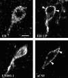
Confocal projections of MOR immunoreactivity in the MPN of ovariectomized rats that were treated with 2 μg of estradiol benzoate followed by either oil vehicle (EB) or 500 μg of progesterone (EB + P) 26 hr later and were killed 30 hr after the initial treatment (Experiment II) or received a microinfusion of either ENDO-1 (50 nmol) or aCSF into the lateral ventricle (Experiment V). The resulting images illustrate that both EB- and ENDO-1-treated animals had an internalized pattern of MOR immunoreactivity (arrows). In EB + P- and aCSF-treated rats, MOR immunoreactivity was associated with the plasma membrane (arrowheads), indicating that these receptors were not activated. Scale bar, 10 μm.
Fig. 3.
Fig. 3.
Bright-field photomicrographs of 3,3′-diaminobenzidine tetrahydrochloride-stained MOR immunoreactivity localization in the medial MPN dorsal to the central MPN in the region quantified in ovariectomized rats treated with 2 μg of estradiol benzoate and either oil vehicle (EB) or 500 μg of progesterone (EB + P) 26 hr later or oil vehicle initially and oil vehicle 26 hr later (CONTROL) and killed 30 hr after the initial injection. CONTROL rats had a lower density of distinct MOR-immunoreactive processes compared with EB-treated rats. Although the density of distinct processes was low in the CONTROL rats, distinct processes suggested that a basal, estrogen-independent internalization of MOR occurs. TheEB + P-treated rats exhibited a reversal of the estrogen-induced increase in MOR-immunoreactive fiber density toCONTROL levels. Distinct varicose MOR-immunoreactive fibers (arrowheads) were increased in EB-treated rats compared with the levels in the CONTROL and EB + P groups that had MOR immunoreactivity that was mainly diffuse (arrows).
Fig. 4.
Fig. 4.
Effects of estrogen and progesterone treatment on the density of μ-opioid receptor-immunoreactive (MORi) fibers stained with 3,3′-diaminobenzidine tetrahydrochloride in the MPN. Ovariectomized rats were treated with 2 μg of estradiol benzoate and either oil vehicle (EB) or 500 μg of progesterone (EB + P) 26 hr later or oil vehicle initially and 26 hr later oil vehicle (CONTROL) again. Animals were killed 4 hr after the final injection and processed for MOR immunocytochemistry (*, significantly greater MOR-immunoreactive fiber density than was found with other treatment groups; p < 0.05, SNK).
Fig. 5.
Fig. 5.
Effects of bilateral ENDO-1 (1, 5, 25 nmol/side) or aCSF microinjections (0.5 μl of total volume/side) into the medial preoptic area on the lordosis quotient 10 and 60 min after microinjection. Ovariectomized rats were maximally receptive after sequential treatment with 2 μg of EB + 500 μg of progesterone 26 hr after EB. Animals were tested 30 hr after EB treatment. Values are means ± SEM (*, significantly less than aCSF and 1 nmol treatments at 10 min and less than within group treatments in the 60 min test; p < 0.05, SNK).
Fig. 6.
Fig. 6.
Effects of bilateral infusions of either the MOR-specific antagonist CTOP (25 nmol/0.5 μl volume) or aCSF 10 min before bilateral infusion of the MOR-specific agonist DAMGO (0.2 nmol/0.5 μl volume) into the medial preoptic area on lordosis quotient 10 min after the last injection. Ovariectomized rats were maximally receptive after treatment with 2 μg of EB + 500 μg of progesterone 26 hr after EB. Animals were tested 30 hr after EB treatment. Values are means ± SEM (*, significantly less than all other treatments; p < 0.05, SNK).
Fig. 7.
Fig. 7.
Effects of bilateral infusions of either the MOR-specific antagonist CTOP (25 nmol/0.5 μl volume) or aCSF 10 min before bilateral infusion of ENDO-1 (25 nmol/0.5 μl volume) or aCSF on lordosis quotient 10 min after the last injection. Ovariectomized rats were maximally receptive after sequential treatment with 2 μg of EB + 500 μg of progesterone 26 hr after EB. Animals were tested 30 hr after EB treatment. Values are means ± SEM (*, significantly less than all other treatments; p < 0.05, SNK).
Fig. 8.
Fig. 8.
MORi fiber density in the MPN of ovariectomized rats that received either ENDO-1 (50 nmol) or aCSF microinfusion into the lateral ventricle (*, significantly higher density of MOR-immunoreactive fibers compared with that in aCSF animals;p < 0.001). Each value is the mean ± SEM (n = 3; df = 4).

Similar articles

Cited by

References

    1. Allen BJ, Rogers SD, Ghilardi JR, Menning PM, Kuskowski MA, Basbaum AI, Simone DA, Mantyh PW. Noxious cutaneous thermal stimuli induce a graded release of endogenous substance P in the spinal cord: imaging peptide action in vivo. J Neurosci. 1997;17:5921–5927. - PMC - PubMed
    1. Babcock AM, Block GJ, Micevych PE. Injections of cholecystokinin into the ventromedial hypothalamic nucleus inhibit lordosis behavior in the rat. Physiol Behav. 1988;43:195–199. - PubMed
    1. Beach F. Hormones and behavior. Hoeber; New York: 1948.
    1. Dournaud P, Boudin H, Schonbrunn A, Tannenbaum GS, Beaudet A. Interrelationships between somatostatin sst2A receptors and somatostatin-containing axons in rat brain: evidence for regulation of cell surface receptors by endogenous somatostatin. J Neurosci. 1998;18:1056–1071. - PMC - PubMed
    1. Eckersell CB, Micevych PE. Opiate receptors modulate estrogen-induced cholecystokinin and tachykinin but not enkephalin mRNA levels in the limbic system and hypothalamus. Neuroscience. 1997;80:473–485. - PubMed

Publication types

MeSH terms