. 2001 Aug 1;21(15):5752-63.
doi: 10.1523/JNEUROSCI.21-15-05752.2001.

Behavioral properties of the trigeminal somatosensory system in rats performing whisker-dependent tactile discriminations

Affiliations

Behavioral properties of the trigeminal somatosensory system in rats performing whisker-dependent tactile discriminations

D J Krupa et al. J Neurosci. .

Abstract

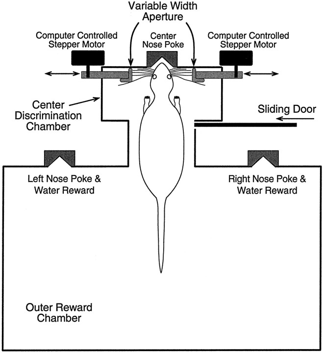
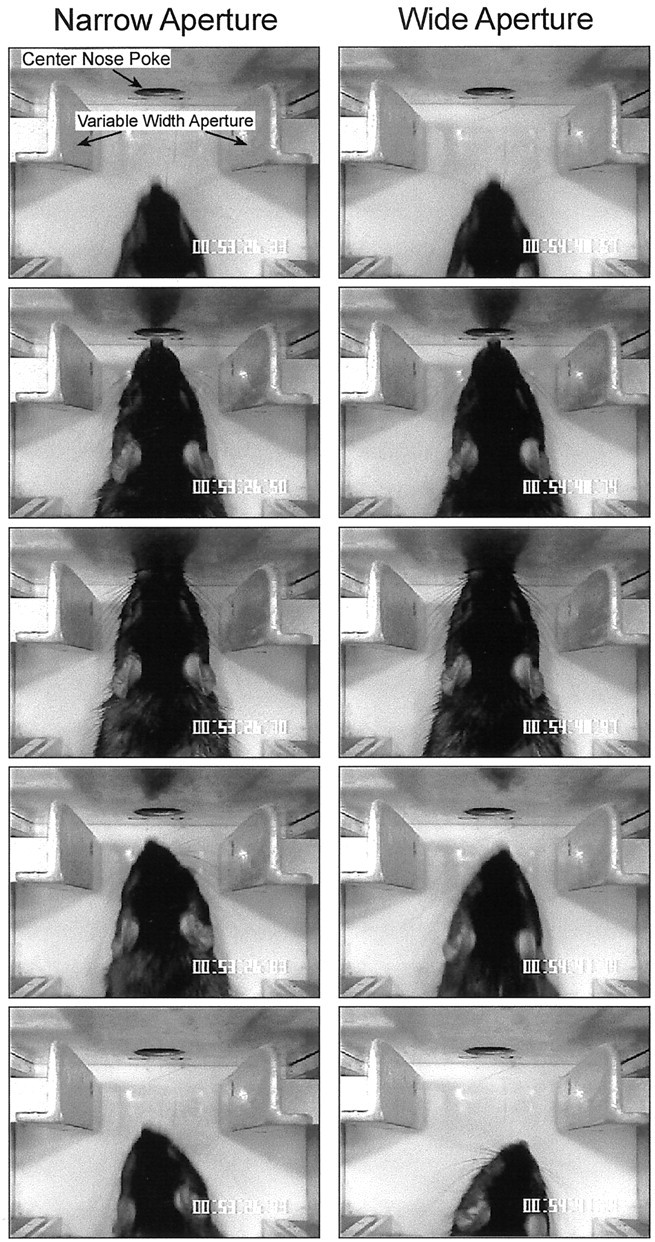
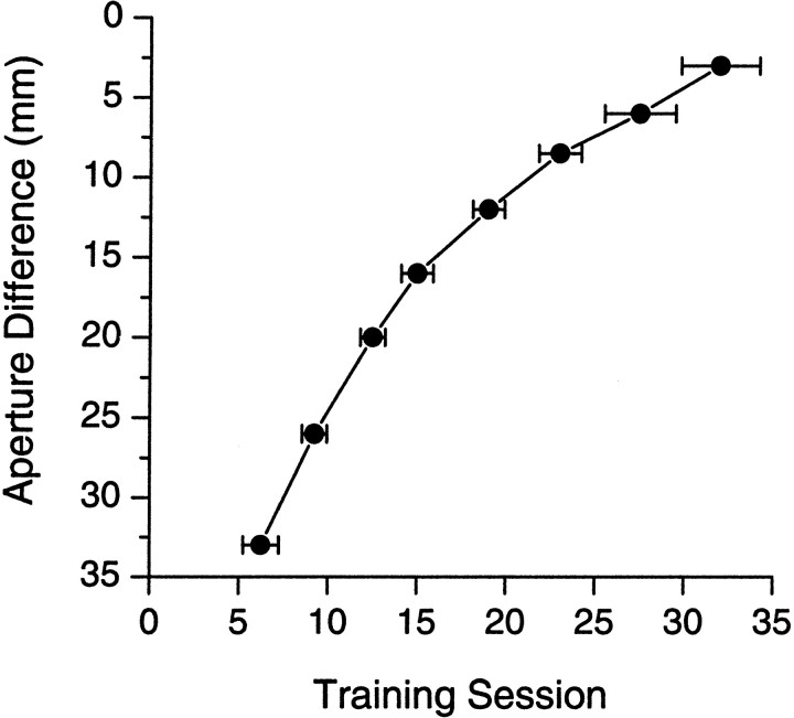
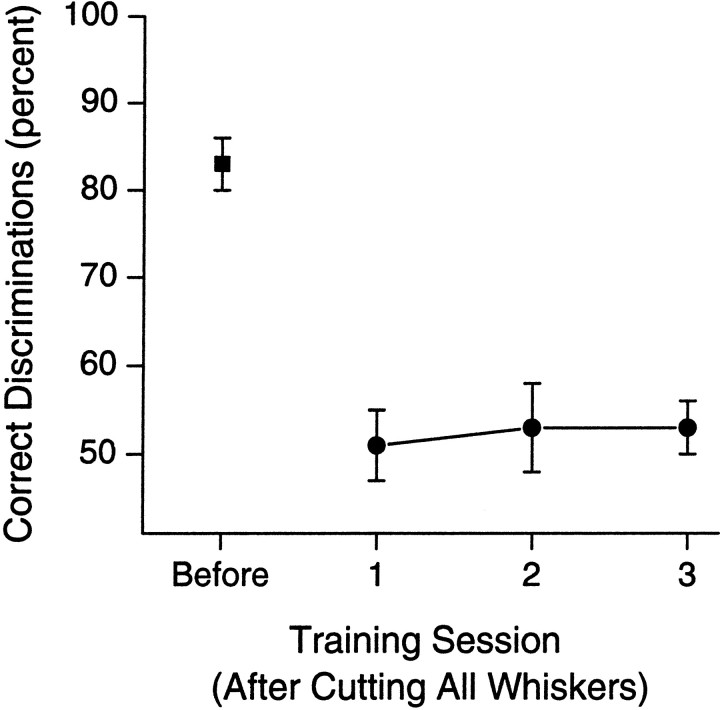
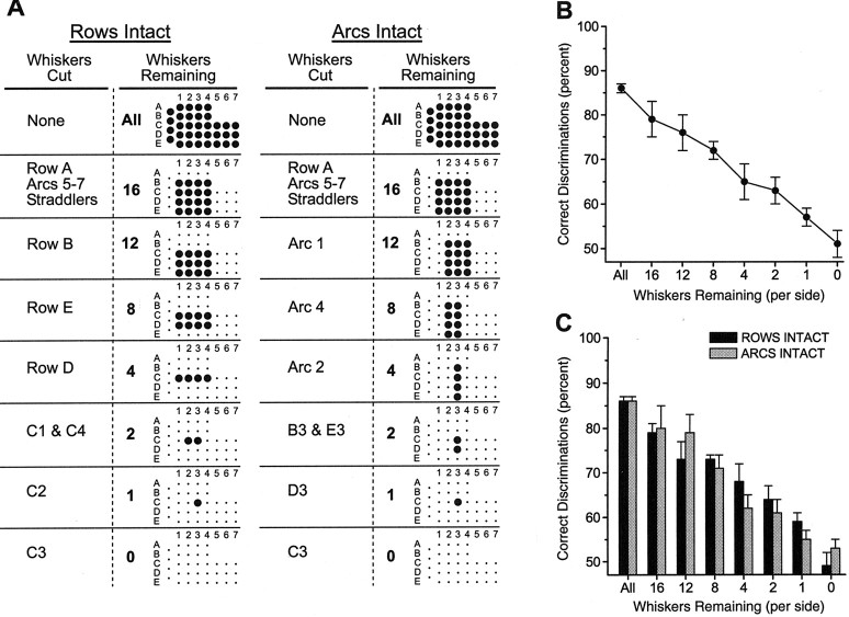
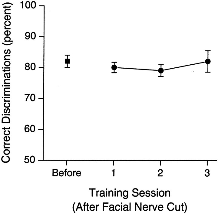
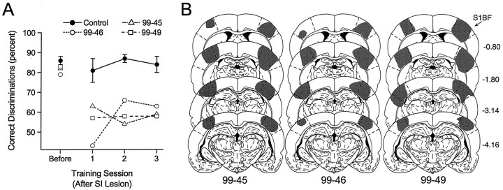
To address several fundamental questions regarding how multiwhisker tactile stimuli are integrated and processed by the trigeminal somatosensory system, a novel behavioral task was developed that required rats to discriminate the width of either a wide or narrow aperture using only their large mystacial vibrissae. Rats quickly acquired this task and could accurately discriminate between apertures of very similar width. Accurate discriminations required a large number of intact facial whiskers. Systematic removal of individual whiskers caused a decrease in performance that was directly proportional to the number of whiskers removed, indicating that tactile information from multiple whiskers is integrated as rats gauge aperture width. In different groups of rats, different sets of whiskers were removed in patterns that preferentially left whisker rows or whisker arcs intact. These different whisker removals caused similar decreases in performance, indicating that individual whiskers within the vibrissal array are functionally equivalent during performance of this task. Lesions of the barrel cortex abolished the ability of rats to discriminate, demonstrating that this region is critically involved in this tactile behavior. Interestingly, sectioning the facial nerve, which abolished whisker movements, did not affect the ability to perform accurate discriminations, indicating that active whisker movements are not necessary for accurate performance of the task. Collectively, these results indicate that the trigeminal somatosensory system forms internal representations of external stimuli (in this case, aperture width) by integrating tactile input from many functionally equivalent facial whiskers and that the vibrissal array can function as a fine-grained distance detector without active whisker movements.


Figures

Fig. 1.
Fig. 1.
Schematic diagram of the behavioral training apparatus. The width of the variable-width aperture is set by rotating the computer-controlled stepper motors. Moving each aperture inward, toward the center, results in a narrower aperture; moving them outward results in a wider aperture. At the start of each training session, rats are placed in the outer reward chamber with the sliding door closed. When the door is opened, rats enter the center discrimination chamber and poke their nose into the center nose poke and sample the variable-width aperture with their facial whiskers. After poking their nose into the center nose poke (detected by interrupting an infrared photobeam), rats then back out into the outer reward chamber and poke their nose into either the left or right reward nose poke to receive a water reward, a left nose poke if the aperture is narrow and a right nose poke if the aperture is wide. Immediately after rats poke their nose into either the left or right nose poke, the sliding door between the outer reward chamber and the center discrimination chamber is closed. A new trial begins when the sliding door is again opened.
Fig. 2.
Fig. 2.
Each column represents a series of still frames (captured from videotape) showing a rat performing a correct discrimination of a narrow (62 mm; left column) or wide (68 mm; right column) aperture. The video sequence progresses from top to bottom. The topmost frame in each column shows the rat approaching the center nose poke and variable-width aperture. The second frame shows the whiskers in contact with the aperture. The third frame shows the rat completing the center poke. The fourth and fifth framesshow the rat withdrawing from the center poke and aperture. In these two frames it can be seen that the rat has begun to turn left (narrow) or right (wide), indicating that the rat has correctly determined the relative width of the aperture and is turning toward the appropriate reward poke. Also, only the large facial whiskers contact the aperture walls. The smaller whiskers around the nose and lips do not contact the aperture. Finally, notice the similarity between the wide and narrow apertures. Even when shown side-by-side, it is difficult to distinguish between the two visually. Rats were capable of discriminating apertures even more similar in width (62 and 65 mm). The time display in the lower right-hand corner of eachframe (hours:minutes:seconds:hundredths of a second) shows the elapsed time between each frame. The chamber is illuminated with an infrared light source so that the rat cannot use visual cues.
Fig. 3.
Fig. 3.
Mean (±SEM) cumulative number of training sessions (during phase 3 training) necessary for a group of rats (n = 5) to reach criterion at each progressively more difficult aperture setting. Each data point represents a different step in the sequence (see Materials and Methods, Fine-grained distance detection). The first data point (Aperture Difference of 33 mm) corresponds to aperture settings of 85 mm (wide) and 52 mm (narrow). The final data point (Aperture Difference of 3 mm) corresponds to aperture settings of 62 and 65 mm. Rats rapidly learn to perform the discrimination, and they are capable of accurately discriminating between apertures that are very similar in width.
Fig. 4.
Fig. 4.
Mean (±SEM) percentage of correct discriminations for a group of rats (n = 5) in the session before cutting all of the large facial whiskers on both sides of the face (Before) and for each of the three training sessions (1–3) after the whisker cuts. Cutting all large facial whiskers completely abolished the ability of the rats to perform the discrimination. There was no recovery of performance over the three sessions after whisker cuts.
Fig. 5.
Fig. 5.
A, Graphical depiction of the whiskers removed at each stage of the experiment. The leftmost two columns refer to the ROWS INTACT group, and therightmost two columns refer to the ARCS INTACT group. The Whiskers Cut columns describe which whiskers were cut at each stage. The Whiskers Remaining columns show how many total whiskers were remaining and a depiction of which whiskers. Each grid represents the vibrissal array.Large dots represent intact whiskers; small dots are cut whiskers. Thus, after the first set of whisker cuts, a grid of 16 intact whiskers remained (Rows B–E, Arcs 1–4). Subsequent cuts further reduced the total number of intact whiskers. As seen in each group, different sets of whiskers were removed at each step. B, Mean (±SEM) percentage of correct discriminations for all rats (n = 8) in which whiskers were systematically removed. There is a gradual decrease in performance that is correlated with the total number of whiskers removed. Each data point represents the mean percentage of correct discriminations of all three training sessions for both groups of rats (ROWS INTACT and ARCS INTACT) after each set of whisker cuts.C, Discrimination performance for each separate group of rats in which different sets of whiskers were cut. There is no difference between groups, indicating that different whiskers in the vibrissal array are functionally equivalent.
Fig. 6.
Fig. 6.
Mean (±SEM) percentage of correct discriminations for a group of rats (n = 3) in the session before bilaterally cutting the facial nerve (Before) and for each of the three training sessions (1–3) after the nerve cut. Cutting the facial nerve did not affect performance.
Fig. 7.
Fig. 7.
A, Mean (±SEM) percentage of correct discriminations in the session before aspiration of the barrel region of the SI cortex (Before) and for the three sessions (1–3) after cortical lesions for each of the rats with SI lesions (n = 3; open symbols). Cortical lesions significantly impaired the ability of the rats to perform the discrimination. The performance of control rats (n = 3; Control) did not differ before or after surgery. B, Extent of the cortical lesions (gray regions, shown on standard sections) for each cortically lesioned rat. Numerals on the right represent the distance (millimeters) of each section posterior from the bregma skull suture. Regions demarcated bydashed lines represent the barrel region of the primary SI cortex. Atlas templates are modified from Paxinos and Watson (1986).S1BF, Barrel region of the SI cortex.Numerals below each column identify individual rats.

References

    1. Armstrong-James M, Fox K. Spatiotemporal convergence and divergence in the rat S1 “barrel” cortex. J Comp Neurol. 1987;263:265–281. - PubMed
    1. Arvidsson J. Somatotopic organization of vibrissae afferents in the trigeminal sensory nuclei of the rat studied by transganglionic transport of HRP. J Comp Neurol. 1982;211:84–92. - PubMed
    1. Belford GR, Killackey HP. Vibrissae representation in subcortical trigeminal centers of the neonatal rat. J Comp Neurol. 1979;183:305–321. - PubMed
    1. Brecht M, Preilowski B, Merzenich MM. Functional architecture of the mystacial vibrissae. Behav Brain Res. 1997;84:81–97. - PubMed
    1. Carvell GE, Simons DJ. Biometric analyses of vibrissal tactile discrimination in the rat. J Neurosci. 1990;10:2638–2648. - PMC - PubMed

Publication types

MeSH terms

LinkOut - more resources